Vol.3, No.12, 1022-1028 (2011) Natural Science
http://dx.doi.org/10.4236/ns.2011.312127
Copyright © 2011 SciRes. OPEN ACCESS
Pathogenicity characterization with implicit and explicit
molecular dynamics simulation
Sigit Jaya Herlambang*, Rosari Saleh
Department of Physics, Mathematics and Science Faculty, Universitas Indonesia, Depok, Indonesia;
*Corresponding Author: sigit.jaya.herlambang@gmail.com
Received 3 October 2011; revised 8 November 2011; accepted 19 November 2011.
ABSTRACT
The contribution of water molecules in molecu-
lar dynamics simulation (MDS) is unquestiona-
bly high, particularly for enzymatic interaction
which occurred in the cytoplasmic environment.
The addition of water molecules to the system
will surely influence different direct interaction
between active site residues and substrate. We
try to theoretically investigate to what extent the
pathogenicity characterization will varies in dif-
ferent neuraminidase-sialic acid complex sys-
tems. The heating dynamics simulations were
produced with and without TIP3P water mole-
cules. The periodic boundary system was made
for explicitly added TIP3P water molecules and
generalized born molecular volume (GBMV) en-
ergy contribution was added for implicit solvent
system. Both complexes, neuraminidase-sialic
acid of A/Tokyo/3/67 and A/Pennsylvania/10218/
84, which have a different pathogenicity levels
were minimized and simulated. The result shows
more residues produced hydrogen bonds with
substrate when water molecules were not added
to the system. The binding free energies also
show differences. Overall, even the values of
energy is different, but an implicit solvent pro-
vides the similar result (HPAI complex has
higher activity than LPAI for both systems) in
characterization of pathogenic virus neuramini-
dase activity.
Keywords: Implicit; Explicit; Solvation; Molecular
Dynamics Simulation; Neuraminidase; Sialic Acid
1. INTRODUCTION
The correlation between neuraminidase (NA) catalytic
activity with the pathogenicity level has been studied in
several experiments [1-11]. The results shows the same
similarity that the high pathogenic avian influenza
(HPAI) virus have NA’s activity higher than the low
pathogenic avian influenza (LPAI). Further investigation
even revealed specific NA’s residue which increase the
virulence, viral pathogenicity, and cause a resistance into
NA inhibitors (NAI).
The NA catalytic process, which cleavage matured
virion that attached in the cell wall, occurred in the
aqueous environment. To produce molecular dynamics
simulation of NA-SA complex, the contribution of water
molecules to the interactions need to be added into the
system. Since, the addition of water molecules explicitly
into the system requires high computational costs, many
alternative methods were achieved to add aqueous envi-
ronment implicitly to suppress the time of simulation.
One of many methods widely used is GBMV [12].
In this study, the NA-SA complexes were compared
in explicit and implicit solvent to get insight on how the
results still have a capability to characterizing the patho-
genicity level of avian influenza virus. The comparison
was build in the subject of the structural changes through
root mean square deviation (RMSD), the number of NA
residues formed hydrogen bond with SA directly and
indirectly, percentage of occupation, and free binding
energy.
2. MATERIALS AND METHODS
2.1. Structure Preparation
The NA sequence of A/Tokyo/3/67 avian influenza
virus (AIV), that was isolated in 1967 [13] during a time
of prevalent infection, obtained from NCBI [14] with
accession code AAB05621. The sequence was being
aligned with the NA sequence of A/Pennsylvania/10218/
84 (accession code BAF48360) which is a non-patho-
genic AIV [15]. These AIV’s were chosen to delegated
two different level of pathogenicity. Sequences align-
ment was executed with BLOSUM 30 scoring matrix in
fast pairwise alignment method.
S. J. Herlambang et al. / Natural Science 3 (2011) 1022-1028
Copyright © 2011 SciRes. OPEN ACCESS
102
1023
The result of sequences alignment was being used in
homology modeling to generate LPAI NA molecule. The
template molecule, which is A/Tokyo/3/67 NA-SA
complex [16], was obtained from RCSB protein data
bank [17]. The CHARMm forcefield utilized to adding
missing hydrogen atoms [18,19]. The original LPAI and
HPAI NA-SA complex molecules were copied and the
explicit water molecules were added into duplicated mole-
cules as TIP3P waterbox [20].
2.2. Minimization and Heating MDS
The minimization and heating MDS were done with
periodic boundary implicit and explicit solvation sys-
tems. The GBMV implicit solvation was included in
original and unsolvated HPAI and LPAI NA-SA com-
plexes minimization and heating MDS. Both HPAI and
LPAI complexes in both systems minimized within two
steps, 1,000,000-steps maximum with gradient energy
0.5 kcal/mol steepest descents and 1,000,000-steps maxi-
mum with gradient energy 0.1 kcal/mol conjugate gra-
dient. Heating MDS was successfully completed with 20
picoseconds (ps) time simulation. All systems tempera-
ture rose from 0 into 300 K with the parameters used
were 20,000 steps, 0.001 time step, non-bond list radius
14 Å, switching function 10-12 Å [21,22], the SHAKE
algorithm [23] was applied (only in the implicit solva-
tion) to fix lengths of all bonds involving hydrogen at-
oms, and the trajectory data were stored every 0.1 ps. All
phases of the study described in this section from struc-
ture preparation to molecular dynamics simulation were
conducted with Discovery Studio 2.1 (Accelrys).
3. RESULTS
3.1. All Atoms, Backbone, and SA RMSD
Figures 1(a), 1(b), and 1(c) visualize the structural
changes during the simulation. The movement of sub-
jected atoms could be monitored through its fluctuative
RMSD. The higher fluctuate indicates higher movement.
The structural stability of each molecule and the whole
atoms in the complexes could also be analyzed from
those figures. Overall, the explicitly solvated complexes
show much better stability than the implicit.
Figure 1(a) depicts the RMSD of all atoms in the
complex (without water molecules). Both solvation shows
progressive increasement around 0 - 5 ps, but in the im-
plicit solvation systems, the movements of all atoms are
higher than in the explicit. This difference must be caused
by the water molecules that surrounded the NA-SA
complexes. Although the stability of implicit solvation
systems is lower than the other, it has an advantage. A
significant result in pathogenicity characterization is more
(a)
(b)
(c)
Figure 1. The RMSD vs simulation time of: (a)
NA-SA complexes all atoms; (b) NA backbone
atoms; (c) SA atoms.
obvious since the LPAI shows a higher movement dur-
ing the simulation. Similar result in the pathogenicity
characterization showed in Figure 1(b) which differed
only on the value of the RMSD. The value of all atoms
RMSD is higher than the NA backbone.
Figure 1(c) shows SA movement during the simula-
S. J. Herlambang et al. / Natural Science 3 (2011) 1022-1028
Copyright © 2011 SciRes. OPEN ACCESS
1024
tion. With explicit solvation, SA which is bound in the
HPAI NA binding pocket RMSD fluctuate more than in
LPAI NA. The fluctuation noticed around 10-15 ps in
simulation time. The result shows differences in the im-
plicit solvation which have higher fluctuation of SA
RMSD when bound the LPAI NA than in the HPAI NA.
Different interaction in the explicit solvation between
SA-water molecules may cause the different SA move-
ment in the NA binding pocket during the simulation.
3.2. The Hydrogen Bonds between NA
Residues and SA
The comparison of hydrogen bonds formed between
functional residues (residues which interact directly with
substrate, in this case is SA) and SA has been done. This
is needed since hydrogen bonds cluster bind tightly. Ta-
ble 1 listed all NA residues that formed hydrogen bonds
with SA complemented with its interaction energy and
distance. This section also provided the percentage of
hydrogen bonds occupation and the figure of water mo-
lecules in explicit solvation which linkage framework
residues to bind SA indirectly.
In Table 1 the results shows that the HPAI NA-SA
complex produce larger number of hydrogen bonds
formed, compared with LPAI NA-SA complex, in the
last conformation. This is appears in both implicit and
explicit solvation systems. In implicit HPAI NA-SA
complex there are 15 hydrogen bonds formed while
LPAI only have 8. The best interaction (lowest interac-
tion energy) and the shortest distance of NA residues-SA
in an implicit solvation was made by hydrogen bond
formed between Arg152 HH21 atom and SA O7 atom in
the HPAI complex with –41.61 kcal/mol and 1.81 Å,
respectively.
In explicit LPAI NA-SA complex there are only 4 hy-
drogen bonds formed while in HPAI produced 7 hydro-
gen bonds. The lowest interaction energy was made by
hydrogen bond formed between Glu276 OE1 atom and
SA H5 atom with –41.11 kcal/mol. The shortest distance
was made by hydrogen bond formed between Asp151
OD1 atom and SA H3 atom with 1.73 Å. In the last
conformation of simulation also revealed several water
molecules formed hydrogen bond with the SA. Fasci-
natingly, the water molecules which linkage several
residues appears only in HPAI (look Figures 2(a) and
2(b)). Those residues are Glu119, Asp151, Ser179,
Arg224, and Glu227.
The percentage of occupation offers a complementary
observation into the stability of hydrogen bonds formed
during the simulation. From Figure 3 could be seen that
several functional residues formed hydrogen bonds with
SA which have percentage of occupation larger than
80%. This is indicated that those residues have strong
hydrogen bonds [23]. In the explicit HPAI NA-SA com-
plex observed that there are four NA residues formed a
strong hydrogen bond with SA. Those residues are
Asp151, Arg152, Glu276, and Arg371. In other hand,
the explicit LPAI NA-SA complex produce three strong
hydrogen bonds that formed by Asp151, Arg224, and
Glu276 with SA atoms.
In the implicit HPAI NA-SA complex there are pro-
duced five strong hydrogen bonds formed by SA with
Arg118, Arg152, Glu276, Arg292, and Arg371. Three of
those five residues are known as triad Arg 118-292-371
which occupy S1 active site. This is not occurred in the
implicit LPAI NA-SA complex which formed four strong
hydrogen bonds involving Asp151, Glu276, Arg292, and
Tyr406. In spite of that, the results shows similarity that
the numbers of strong hydrogen bonds formed in HPAI
is larger than LPAI in both solvation applied in NA-SA
complexes for the simulation. For both solvation also
revealed that strong hydrogen bonds was made by
Glu276 for all complexes simulated. This indicates that
the Glu276 has played a role in NA-SA binding.
3.3. End-Point Free Binding Energy
Table 2 shows free binding energies of all NA-SA
complex molecules in the last conformation based on
Amaro et al. [24]. This method improves the time of
calculation than if all of trajectories saved were calcu-
lated. The results are known similar to other studies that
calculated the binding free energy of NA-inhibitor com-
plexes with different approaches [25,26]. In the explicit
solvation systems, complex molecules calculated with-
out any implicit solvation energetic contribution while in
the implicit solvation, the calculation executed with the
addition of GBMV implicit solvation energy. The NA-
SA complexes in HPAI is lower than LPAI for both sol-
vation which in other way tells that the HPAI NA have a
better interaction with SA than in the LPAI [27,28]. The
similarity of the results could be seen as the indicator
that pathogenicity characterization is available to per-
form either with implicit solvation or explicit solvation.
4. DISCUSSION
The main focus in this study is to compare the implicit
solvation GBMV with explicit solvation and both possi-
bility to determine the difference of pathogenicity of
AIV through the interaction of its NA-SA. Overall, al-
most all of the results show similarities. Except in the
SA RMSD that shown different stability between HPAI
and LPAI in both implicit and explicit solvation. In the
implicit solvation the HPAI is more stable than LPAI
while in explicit solvation the LPAI is the more stable
than HPAI. Even in the explicit solvation the differences
S. J. Herlambang et al. / Natural Science 3 (2011) 1022-1028
Copyright © 2011 SciRes. OPEN ACCESS
102
1025
Table 1. The hydrogen bonds formed in the last of simulation completed with its energy of
interaction and distance.
Hydrogen Bonds
Atom 1 Atom 2 E (kcal/mol) Distance (Å)
Arg152 HH21 Sialic Acid O10 –36.42 2.45 Explicit HPAI
Arg292 HH22 Sialic Acid O1B–28.52 2.13
Arg371 HH12 Sialic Acid O1A–32.84 2.22
Arg371 HH12 Sialic Acid O1B–31.47 2.03
Arg371 HH22 Sialic Acid O1B–34.45 2.33
Asp151 OD1 Sialic Acid H3 –34.24 1.73
Glu276 OE1 Sialic Acid H5 –41.11 1.90
Asp151 OD1 Sialic Acid H15 –39.43 2.29 Explicit LPAI
Arg224 HH11 Sialic Acid O9 –36.62 2.44
Arg371 HH22 Sialic Acid O1A–30.54 2.07
Glu276 OE1 Sialic Acid H17 –32.79 2.01
Arg118 HH12 Sialic Acid O4 –37.87 2.00 Implicit HPAI
Arg152 HE Sialic Acid O10 –24.59 2.24
Arg152 HH21 Sialic Acid O7 –41.61 1.81
Arg224 HH11 Sialic Acid O9 –32.64 2.32
Arg292 HH22 Sialic Acid O1B–35.73 1.95
Arg292 HH22 Sialic Acid O1A–28.50 2.45
Arg371 HH12 Sialic Acid O1B–35.91 1.94
Arg371 HH22 Sialic Acid O1A–28.82 2.43
Arg371 HH22 Sialic Acid O1B–37.08 1.86
Tyr406 OH Sialic Acid H1 –27.82 1.91
Glu119 OE2 Sialic Acid H3 –40.74 1.92
Glu276 OE1 Sialic Acid H5 –41.52 1.87
Glu276 OE2 Sialic Acid H5 –33.75 2.37
Glu276 OE1 Sialic Acid H6 –34.78 2.30
Glu276 OE2 Sialic Acid H6 –34.57 2.31
Arg292 HH12 Sialic Acid O1A–31.79 2.20 Implicit LPAI
Arg292 HH12 Sialic Acid O1B–36.55 1.90
Arg292 HH21 Sialic Acid O8 –33.60 2.25
Arg292 HH22 Sialic Acid O1A–36.09 1.93
Tyr406 HH Sialic Acid O1B–41.15 1.90
Asp151 OD1 Sialic Acid H15 –39.57 2.00
Asp151 OD2 Sialic Acid H15 –37.91 2.10
Glu276 OE2 Sialic Acid H17 –41.31 1.88
S. J. Herlambang et al. / Natural Science 3 (2011) 1022-1028
Copyright © 2011 SciRes. OPEN ACCESS
1026
(a)
(b)
Figure 2. The water molecules which produce hydro-
gen bond with SA in: (a) HPAI NA-SA complex; (b)
LPAI NA-SA complex.
Figure 3. The percentage occupation of hydrogen
bonds formed during the simulation.
Table 2. Free binding energy.
Free Energy (kcal/mol)
HPAI Explicit
Solvation
LPAI Explicit
Solvation
HPAI Implicit
Solvation
LPAI Implicit
Solvation
–361.55 –273.64 –108.60 2.85
is subtle, it could still be an indicator that there is may be
another explanation for this confusing results. It is
probably be caused by the interaction of SA with water
molecules in explicit solvation simulation. As indirect
interaction was made in HPAI from the residues that
formed a hydrogen bonds with water molecules that also
produces a hydrogen bonds with SA, which did not oc-
curred in LPAI NA-SA complex. However, deep inves-
tigation is still needed to look further in this issue.
The NA-SA binding with and without water mole-
cules influences different positional fluctuations of com-
plex molecules. It appears in our RMSD graphics that
the molecules in the explicit solvation system have lower
RMSD values. Non-bond interactions between complex
molecules and water molecules stabilize the system that
cause the movement of the complex molecules is mini-
mum. In both explicit and implicit solvation, HPAI has
more stable movement which can be seen from its mini-
mum fluctuation RMSD. This RMSD comparison is ame-
nable with multiple studies that infer that a higher sub-
strate RMSD suggests the superior neuraminidase ability
to reject an inhibitor [23,29,30]. While in this study, SA
high RMSD related with neuraminidase acceptance
abilities when cleavage the virion from the edge of the
cell.
The specific residues that formed hydrogen bonds
with SA in our experiment agree with the results from
other studies [1-11]. The key role of Asp151, Glu276,
Arg292, and Arg371 are important that the changes in
one of those residues could followed by the resistance of
inhibitors. In both explicit and implicit solvation systems,
their importance is obviously observed. It could be seen
from its interaction energy, distance and percentage of
occupation. Those three results provide the evidence that
they have a strong interaction in almost all NA-SA com-
plexes simulated.
The end point free binding energy calculation results
for explicit simulation is very low. It seems because the
calculation for the explicit solvation systems, the im-
plicit solvation contribution was not added. The GBMV
free binding energy calculation then also executed for
explicit complex molecules. The results show the values
of –1.37 kcal/mol and 17.97 kcal/mol for HPAI and
LPAI, respectively. Compared by other studies [25,26,
31], the calculation with GBMV is at their results range
(–21.7 kcal/mol to 7.3 kcal/mol). This is indicate that our
S. J. Herlambang et al. / Natural Science 3 (2011) 1022-1028
Copyright © 2011 SciRes. OPEN ACCESS
102
1027
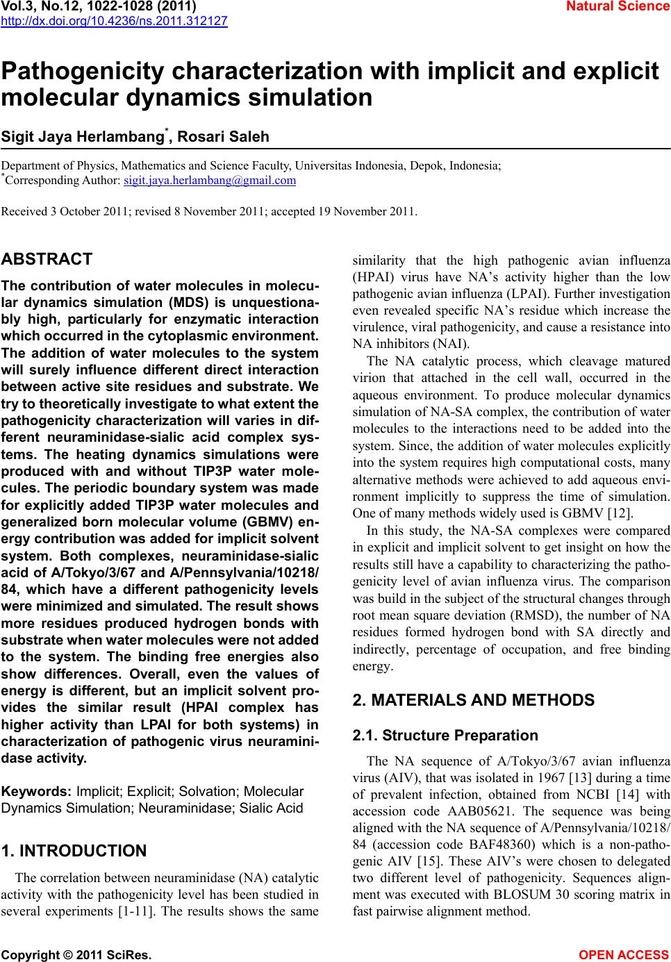
results could be used to characterize virus pathogenicity.
5. CONCLUSION
Differences in pathogenicity level can be assessed by
implicit or explicit solvation heating dynamics simulation.
Even the structural changes and energetic analysis show
significant value, the implicit solvation systems results
are similar to with the explicit solvation systems. The
HPAI complexes molecules in both systems show higher
activity than the LPAI complexes molecules. The use of
explicit solvation is suggested for better results and
systems stability, but the use of implicit solvation is still
resembles the explicit solvation.
6. ACKNOWLEDGEMENTS
We would like to express gratitude towards Ding Ming Chee of Ac-
celrys Singapore for the Accelrys Discovery Studio 2.1 trial sent to us.
REFERENCES
[1] Russell, R.J., Haire, L.F., Stevens, D.J., Collins, P.J., Lin,
Y.P., Blackburn, G.M., Hay, A.J., Gamblin, S.J. and Ske-
hel, J.J. (2006) The structure of H5N1 avian influenza
neuraminidase suggests new opportunities for drug de-
sign. Nature, 443, 45-49. doi:10.1038/nature05114
[2] Chachra, R. and Rizzo, R.C. (2008) Origins of resistance
conferred by the R292K neuraminidase mutation via
molecular dynamics and free energy calculations. Jour-
nal of Chemical Theory and Computation, 4, 1526-1540.
doi:10.1021/ct800068v
[3] McKimm-Breschkin, J.L., Sahasrabudhe, A., Blick, T.J.,
McDonald, M., Colman, P.M., Hart, G.J., Bethell, R.C.
and Varghese, J.N. (1998) Mutations in a conserved
residue in the influenza virus neuraminidase active site
decreases sensitivity to Neu5Ac2en derivatives. Journal
of Virology, 72, 2456-2462.
[4] Mishin, V.P., Hayden, F.G. and Gubareva, L.V. (2005)
Susceptibilities of antiviral-resistant influenza viruses to
novel neuraminidase inhibitors. Antimicrobial Agents
and Chemotherapy, 49, 4515-4520.
doi:10.1128/AAC.49.11.4515-4520.2005
[5] Sheu, T.G., Deyde, V.M., Okomo-Adhiambo, M., Garten,
R.J., Xu, X., Bright, R.A., Butler, E.N., Wallis, T.R.,
Klimov, A.I. and Gubareva, L.V. (2008) Surveillance for
neuraminidase inhibitor resistance among human influ-
enza A and B viruses circulating worldwide from 2004 to
2008. Antimicrobial Agents and Chemotherapy, 52, 3284-
3292.
[6] Wetherall, N.T., Trivedi, T., Zeller, J., Hodges-Savola, C.,
McKimm-Breschkin, J.L., Zambon, M. and Hayden, F.G.
(2003) Evaluation of neuraminidase enzyme assays using
different substrates to measure susceptibility of influenza
clinical isolates to neuraminidase inhibitors: Report of
the Neuraminidase Inhibitor Susceptibility Network.
Journal of Clinical Microbiology, 41, 742-750.
doi:10.1128/JCM.41.2.742-750.2003
[7] McKimm-Breschkin, J.L., Trivedi, T., Hampson, A., Hay,
A., Klimov, A., Tashiro, M., Hayden, F.G. and Zambon,
M. (2003) Neuraminidase sequence analysis and suscep-
tibilities of influenza virus clinical isolates to zanamivir
and oseltamivir. Antimicrobial Agents and Chemotherapy,
47, 2264-2272. doi:10.1128/AAC.47.7.2264-2272.2003
[8] Yen, H., Ilyushina, N.A., Salomon, R., Hoffmann, E.,
Webster, R.G. and Govorkova, E.A. (2007) Neuramini-
dase inhibitor-resistant recombinant a/vietnam/1203/04
(H5N1) influenza viruses retain their replication effi-
ciency and pathogenicity in vitro and in vivo. Journal of
Virology, 81, 12418-12426. doi:10.1128/JVI.01067-07
[9] Meijer, A., Lackenby, A., Hungnes, O., Lina, B., van der
Werf, S., Schweiger, B., Opp, M., Paget, J., van de
Kassteele, J., Hay, J. and Zambon. M. (2009) Osel-
tamivir-resistant influenza virus A (H1N1), Europe, 2007-
08 Season. Emerging Infectious Diseases, 15, 552-560.
doi:10.3201/eid1504.081280
[10] Monto, A.S., McKimm-Breschkin, J.L., Macken, C.,
Hampson, A.W., Hay, A., Klimov, A., Tashiro, M., Web-
ster, R.G., Aymard, M., Hayden, F.G. and Zambon, M.
(2006) Detection of influenza viruses resistant to neural-
minidase inhibitors in global surveillance during the first
3 years of their use. Antimicrobial Agents and Chemo-
therapy, 50, 2395-2402. doi:10.1128/AAC.01339-05
[11] Tamura, D., Mitamura, K., Yamazaki, M., Fujino, M.,
Nirasawa, M., Kimura, K., Kiso, M., Shimizu, H., Ka-
wakami, C., Hiroi, S., Takahashi, S., Hata, M., Minagawa,
H., Kimura, Y., Kaneda, S., Sugita, S., Horimoto, T.,
Sugaya, N. and Kawaoka, Y. (2009) Oseltamivir-resistant
influenza A viruses circulating in Japan. Journal of
Clinical Microbiology, 47, 1424-1427.
doi:10.1128/JCM.02396-08
[12] Lee, M.S., Feig, M., Salsbury, F. R. and Brooks, C.L.III.
(2003) New analytic approximation to the standard mo-
lecular volume definition and its application to genera-
lized born calculations. Journal of Computational Che-
mistry, 24, 1348-1356. doi:10.1002/jcc.10272
[13] Pereira, M.S., Chakraverty, P. and Pane, A.R. (1969) The
influence of antigenic variation on influenza A2 epidem-
ics. Journal of Hygiene, 67, 551-557.
[14] http://www.ncbi.nlm.nih.gov/genomes/FLU/Database/np
h-select.cgi?go=database
[15] Kaverin, N.V., Rudneva, I.A., Ilyushina, N.A., Varich,
N.L., Lipatov, A.S., Smirnov, Y.A., Govorkova, E.A.,
Gitelman, A.K., Lvov, D.K. and Webster, R.G. (2002)
Structure of antigenic sites on the haemagglutinin mole-
cule of H5 avian influenza virus and phenotypic variation
of escape mutants. Journal of General Virology, 83,
2497-2505.
[16] Varghese, J.N., Mc-Kimm-Breschkin, J.L., Caldwell, J.B.,
Kortt, A.A. and Colman, P.M., (1992) The structure of
the complex between influenza virus neuraminidase and
sialic acid, the viral receptor. Proteins, 14, 327-332.
doi:10.1002/prot.340140302
[17] http://www.rcsb.org/pdb/home/home.do
[18] Brooks, B.R., Bruccoleri, R.E., Olafson, B.D., States,
D.J., Swaminathan, S. and Karplus, M. (1983) CHARMM:
A program for macromolecular energy minimization and
dynamics calculations. Journal of Computational Chem-
istry, 4, 187-217. doi:10.1002/jcc.540040211
[19] Brooks, B.R., et al., (2009) CHARMM: The biomolecu-
lar simulation program. Journal of Computational Che-
mistry, 30, 1545-1614. doi:10.1002/jcc.21287
S. J. Herlambang et al. / Natural Science 3 (2011) 1022-1028
Copyright © 2011 SciRes. OPEN ACCESS
1028
[20] Jorgensen, W.L., Chandrasekhar, J., Madura, J.D., Impey,
R.W. and Klein, M.L. (1983) Comparison of simple po-
tential functions for simulating liquid water. Journal of
Chemical Physics, 79, 926-935. doi:10.1063/1.445869
[21] Brooks, C.L.III, Pettit, B.M. and Karplus, M. (1985)
Structural and energetic effects of truncating long ranged
interactions in ionic and polar fluids. Journal of Chemi-
cal Physics, 83, 5897-5908. doi:10.1063/1.449621
[22] Steinbach, P.J. and Brooks, B.R. (1994) New sphere-
cal-cutoff methods for long-range forces in macromo-
lecular simulation. Journal of Computational Chemistry,
15, 667-683. doi:10.1002/jcc.540150702
[23] Shu, M., Lin, Z., Zhang, Y., Wu, Y., Mei, H. and Jiang, Y.
(2010) Molecular dynamics simulation of oseltamivir re-
sistance in neuraminidase of avian influennza H5N1 vi-
rus. Journal of Molecular Modeling, 17, 587-592.
[24] Amaro, R.E., Cheng, X., Ivanov, I., Xu, D. and
McCammon, J.A. (2009) Characterizing loop dynamics
and ligand recognition in human- and avian-type influ-
enza neuraminidases via generalized born molecular dy-
namics and end-point free energy calculations. Journal of
the American Chemical Society, 13, 4702-4709.
doi:10.1021/ja8085643
[25] Masukawa, K.M., Kollman, P.A. and Kuntz, I.D. (2003)
Investigation of neuraminidase-substrate recognition us-
ing molecular dynamics and free energy calculations.
Journal of Medicinal Chemistry, 46, 5628-5637.
doi:10.1021/jm030060q
[26] Smith, B.J., Colman, P.M., von Itzstein, M., Danylec, B.
and Varghese, J.N. (2000) Analysis of inhibitor binding
in influenza virus neuraminidase. Protein Science, 10,
689-696. doi:10.1110/ps.41801
[27] Guo, X.L., Wei, D.Q., Zhul, Y.S. and Chou, K.C. (2008)
Cleavage mechanism of the H5N1 hemagglutinin by
trypsin and furin. Amino Acids, 35, 375-382.
[28] Decha, P., Rungrotmongkol, T., Intharathep, P., Malaisree,
M., Aruksakunwong, O., Laohpongspaisan, C., Parasuk,
V., Sompornpisut, P., Pianwanit, S., Kokpol, S. and
Hannongbua, S. (2008) Source of high pathogenicity of
an avian influenza virus H5N1: Why H5 is better cleaved
by furin. Biophysical Journal, 95, 128-134.
doi:10.1529/biophysj.107.127456
[29] Aruksakunwong, O., Malisree, M., Decha, P., Somporn-
pisut, P., Parasuk, V., Pianwanit, S. and Hannongbua, S.
(2007) On the lower susceptibility of oseltamivir to in-
fluenza neuraminidase subtype than those in N2 and N9.
Biophysical Journal, 92, 798-807.
doi:10.1529/biophysj.106.092528
[30] Le, L., Lee, E., Schulten, K. and Truong, T.N. (2009)
Molecular modeling of swine influenza A/H1N1, Spanish
H1N1, and avian H5N1 flu N1 neuraminidases bound to
tamiflu and relenza. PloS Current.
http://www.ncbi.nlm.nih.gov/pmc/articles/PMC2762416.
4/
[31] von Itzstein, M., Wu, W.Y., Kok, G.B., Pegg, M.S.,
Dyason, J.C., Jin, B., Phan, T.V., Smythe, M.L., White,
H.F., Oliver, S.W., Colman, P.M., Varghese, J.N., Ryan,
D.M., Woods, J.M., Bethell, R.C., Hotham, V.J., Cam-
eron, J.M. and Penn, C.R. (1993) Rational design of po-
tent sialidase-based inhibitors of influenza virus replica-
tion. Nature, 363, 418-423. doi:10.1038/363418a0
ABBREVIATION
Gly Glycine Met Methionine Trp Tryphtophan
Ala Alanine Cys Cysteine Glu Glutamic Acid
Ile Isolucine Asn Asparagine His Histidine
Leu Leucine Gln Glutamine Lys Lysine
Val Valine Ser Serine Phe Phenylalanine
Pro Proline Thr Threonine Asp Aspartat Acid
Tyr Tyrosine Arg Arginine