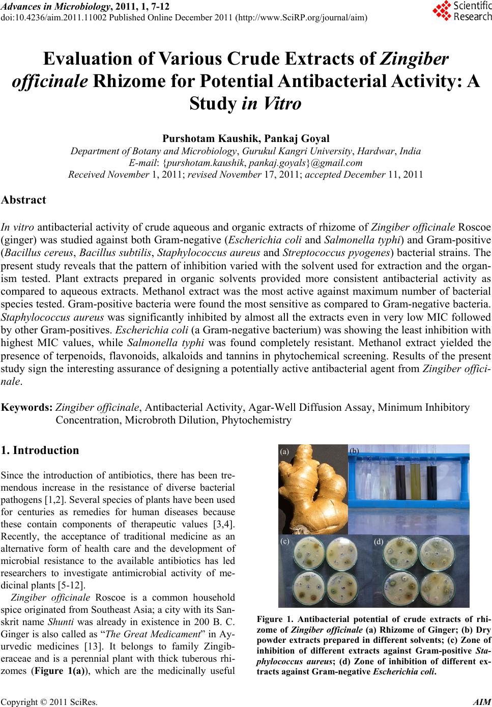
Advances in Microbiology, 2011, 1, 7-12
doi:10.4236/aim.2011.11002 Published Online December 2011 (http://www.SciRP.org/journal/aim)
Copyright © 2011 SciRes. AIM
7
Evaluation of Various Crude Extracts of Zingiber
officinale Rhizome for Potential Antibacterial Activity: A
Study in Vitro
Purshotam Kaushik, Pankaj Goyal
Department of Botany and Microbiology, Gurukul Kangri University, Hardwar, India
E-mail: {purshotam.kaushik, pankaj.goyals}@gmail.com
Received November 1, 2011; revised November 17, 2011; accepted December 11, 2011
Abstract
In vitro antibacterial activity of crude aqueous and organic extracts of rhizome of Zingiber officinale Roscoe
(ginger) was studied against both Gram-negative (Escherichia coli and Salmonella typhi) and Gram-positive
(Bacillus cereus, Bacillus subtilis, Staphylococcus aureus and Streptococcus pyogenes) bacterial strains. The
present study reveals that the pattern of inhibition varied with the solvent used for extraction and the organ-
ism tested. Plant extracts prepared in organic solvents provided more consistent antibacterial activity as
compared to aqueous extracts. Methanol extract was the most active against maximum number of bacterial
species tested. Gram-positive bacteria were found the most sensitive as compared to Gram-negative bacteria.
Staphylococcus aureus was significantly inhibited by almost all the extracts even in very low MIC followed
by other Gram-positives. Escherichia coli (a Gram-negative bacterium) was showing the least inhibition with
highest MIC values, while Salmonella typhi was found completely resistant. Methanol extract yielded the
presence of terpenoids, flavonoids, alkaloids and tannins in phytochemical screening. Results of the present
study sign the interesting assurance of designing a potentially active antibacterial agent from Zingiber offici-
nale.
Keywords: Zingiber officinale, Antibacterial Activity, Agar-Well Diffusion Assay, Minimum Inhibitory
Concentration, Microbroth Dilution, Phytochemistry
1. Introduction (b)
(a)
Since the introduction of antibiotics, there has been tre-
mendous increase in the resistance of diverse bacterial
pathogens [1,2]. Several species of plants have been used
for centuries as remedies for human diseases because
these contain components of therapeutic values [3,4].
Recently, the acceptance of traditional medicine as an
alternative form of health care and the development of
microbial resistance to the available antibiotics has led
researchers to investigate antimicrobial activity of me-
dicinal plants [5-12].
(c) (d)
Zingiber officinale Roscoe is a common household
spice originated from Southeast Asia; a city with its San-
skrit name Shunti was already in existence in 200 B. C.
Ginger is also called as “The Great Medicament” in Ay-
urvedic medicines [13]. It belongs to family Zingib-
eraceae and is a perennial plant with thick tuberous rhi-
zomes (Figure 1(a)), which are the medicinally useful
Figure 1. Antibacterial potential of crude extracts of rhi-
zome of Zingiber officinale (a) Rhizome of Ginger; (b) Dry
powder extracts prepared in different solvents; (c) Zone of
inhibition of different extracts against Gram-positive Sta-
phylococcus aureus; (d) Zone of inhibition of different ex-
tracts against Gram-negative Escherichia coli.
P. KAUSHIK ET AL.
8
part of this plant. The medicinal history of ginger has
been extensively searched throughout the world and
found to possess anti-inflammatory, cholesterol-lowering,
and antithrombotic properties [13,14]. Important second-
dary metabolites present in the rhizome are curcumene,
non-volatile hydroxyaryl compounds e.g. zingerone, gin-
geroles and shogaoles (phenylalkanones), volatile ses-
quiterpenes (e.g. zingiberene and bisabolene) and mono-
terpenoids (e.g. citral) [15]. Although, the antimicrobial
activity and chemical analysis of essential oil and olio-
resins of this plant has been investigated [16], the present
study was focussed to investigate the antibacterial poten-
tial of crude extracts of rhizome of Zingiber officinale.
Furthermore, active extracts were evaluated for their
minimum inhibitory concentrations (MICs) and phyto-
chemical screening.
2. Materials and Methods
2.1. Collection of Plant Part
Plant part (rhizome) of Zingiber officinale was collected
during winter between September 2006 and January 2007.
Rhizome was first washed under running tap water fol-
lowed by sterilized distilled water, air-dried and then
powdered with the help of sterilized pestle and mortar.
This powder was stored in airtight bottles and subjected
to various extraction procedures.
2.2. Preparation of Crude Extracts of Ginger
Rhizome
Following methods were applied in preparation of crude
extracts of rhizome of Zingiber officinale (Figure 1(b)).
2.2.1. Aqueo u s Ext r action
To make aqueous decoction, air-dried powder of plant
part (10 g) was boiled in 400 ml distilled water till one
fourth of the extract initially taken was left behind after
evaporation. The solution was then filtered using muslin
cloth. Filtrate was centrifuged at 5000 rpm for 15 min.
The supernatant was again filtered using Whatman Filter
No. 1 under strict aseptic conditions and the filtrate was
collected in fresh sterilized bottles and stored at 4˚C until
further use.
2.2.2. Organic Solvent Extraction
Air-dried powder (10 g) was thoroughly mixed with 100
ml organic solvent (viz. ethanol, methanol, hexane and
ethyl acetate). The mixture was placed at room tempera-
ture for 24 h on shaker with 150 rpm. Solution was fil-
tered through muslin cloth and then re-filtered by passing
through Whatman Filter No. 1. The filtrate thus obtained
was concentrated by complete evaporation of solvent at
room temperature to yield the pure extract. Stock solu-
tions of crude extracts for each type of organic solvent
were prepared by mixing well the appropriate amount of
dried extracts with respective solvent to obtain a final
concentration of 100 mg/ml. Each solution was stored at
4˚C after collecting in sterilized bottles until further use.
2.3. Bacterial Strains Selected for Susceptibility
Assay
A total of six bacteria namely Escherichia coli MTCC-
739, Salmonella typhi MTCC-531 (all Gram-negative
bacteria) and Bacillus cereus MTCC-430, Bacillus sub-
tilis MTCC-736, Staphylococcus aureus MTCC-740 and
Streptococcus pyogenes MTCC-442 (all Gram-positive
bacteria) were screened for present investigation. All the
above mentioned bacterial strains were collected from
Microbial Type Culture Collection (MTCC), India. These
bacterial cultures were maintained in nutrient agar slants
at 37˚C. Each of the microorganisms was reactivated
prior to susceptibility testing by transferring them into a
separate test tube containing nutrient broth and incubated
overnight at 37˚C.
2.4. Antibacterial assay
Antibacterial activities of all aqueous and organic ex-
tracts of rhizome of Zingiber officinale were determined
by standard agar well diffusion assay [17]. Petri dishes
(100 mm) containing 18 ml of Mueller Hinton Agar
(MHA) seeded with 100 µl inoculum of bacterial strain
(inoculum size was adjusted so as to deliver a final in-
oculum of approximately 108 CFU/ml). Media was al-
lowed to solidify and then individual Petri dishes were
marked for the bacteria inoculated. Wells of 6 mm di-
ameter were cut into solidified agar media with the help
of sterilized cup-borer. 50 µl of each extract was poured
in the respective well and the plates were incubated at
37˚C for overnight. Organic solvents were used as nega-
tive control while tetracycline antibiotic (5 µg·ml–1) was
used as positive control. The experiment was performed
in triplicate under strict aseptic conditions and the anti-
bacterial activity of each extract was expressed in terms
of the mean of diameter of zone of inhibition (in mm)
produced by the respective extract at the end of incuba-
tion period.
2.5. Determination of Minimum Inhibitory
Concentration
Extracts producing an inhibition zone 12 mm in diame-
ter were screened to determine minimum inhibitory con-
Copyright © 2011 SciRes. AIM
P. KAUSHIK ET AL.
Copyright © 2011 SciRes. AIM
9
centrations (MICs) by standard two-fold microbroth di-
lution methodology given by NCCLS [18]. A stock solu-
tion of each extract (viz. ethanol, methanol, ethyl acetate
and aqueous) was serially diluted in 96-wells microtiter
plate with Mueller Hinton broth to obtain a concentration
ranging from 8 µg/ml to 4096 µg/ml. A standardized
inoculum for each bacterial strain was prepared so as to
give inoculum size of approximately 5 × 10⁵ CFU/ml in
each well. Microtiter plates were then kept at 37˚C for an
overnight incubation. Following incubation, the MIC
was calculated as the lowest concentration of the extract
inhibiting the visible growth of bacterial strain.
All the chemical ingredients used in present study
were of analytical grade, and were purchased from Hi
Media, India.
2.6. Phytochemical Analysis of Active Crude
Extract
Methanol extract of ginger rhizome was evaluated for its
phytochemistry by standard methodology as given by
Harborne [19].
3. Results and Discussion
Table 1 represents the antibacterial activity of various
crude extracts prepared from the rhizome of ginger. Data
indicated that extracts prepared in organic solvents con-
sistently displayed better antibacterial activity than that
of aqueous extracts. Extracts prepared in methanol was
observed most inhibitory (diameter of zone of inhibition
ranging from 12.83 to 18.67 mm) followed by those pre-
pared in ethyl acetate (zone of inhibition 10.33 to 14.00
mm). Ethanol and hexane extract was found mild inhibi-
tory only against Staphylo coccus aureus with only 13.66
and 10.33 mm diameter of zone of inhibition, respec-
tively. The zone of inhibition observed with aqueous
extract was 15.67 mm against Gram-positive staphylo-
cocci.
All antimicrobial activities observed varied with the
type of test organism. Primary screening indicated that
extracts were more effective in inhibiting Gram-positive
bacteria when compared to Gram-negative bacteria. Sta-
phylococcus aureus (a Gram-positive bacterium) was
significantly inhibited by almost all the extracts and
found most susceptible among all the bacterial species
examined in this study (Figure 1(c)). Bacillus cereus and
Bacillus subtilis were also inhibited but comparatively
smaller zone of inhibitions were obtained.
Ginger extracts had a very little effect on Gram-nega-
tive bacteria specifically against Escherichia coli (Fig-
ure 1(d)); however, the Gram-negative bacterium; Sal-
monella typhi and Gram-positive Streptococcus pyogenes
demonstrated the complete resistance against all the ex-
tracts as no zone of inhibition was observed. Our results
were found in agreement with some earlier studies which
showed the moderate antibacterial properties of ginger
extract against Escherichia coli but no activity was
Table 1. In vitro antibacterial activity of aqueous and organic extracts of Zingiber officinale rhizome.
Zone of Inhibition* (in mm diameter)
Gram-negative Bacteria Gram-positive Bacteria
Type of Extract
Escherichia coli Salmonella typhiStreptococcus
pyogenes Staphylococcus
aureus Bacillus subtilis Bacillus cereus
Ethanol NI NI NI 13.66 ± 0.29 NI NI
Methanol 12.83 ± 0.76 NI NI 18.67 ± 1.52 14.00 ± 1.00 13.00 ± 1.32
Ethyl Acetate 10.33 ± 0.58 NI NI 14.00 ± 2.00 11.34 ± 0.58 11.16 ± 0.77
Organic
Extract
Hexane NI NI NI 10.33 ± 0.58 NI NI
Aqueous Extract NI NI NI 15.67 ± 2.08 NI NI
Positive Tetracycline+ 29.50 ± 0.50 25.83 ± 1.61 29.83 ± 1.89 32.50 ± 1.50 34.17 ± 1.76 32.16 ± 1.04
Ethanol NI NI NI NI NI NI
Methanol NI NI NI NI NI NI
Ethyl Acetate NI NI NI NI NI NI
Control Negative
Hexane NI NI NI NI NI NI
*Values of the observed zone of inhibition (in mm diameter) including the diameter of well (6 mm) after 24 hours incubation against different bacterial species
when subjected to different extracts in agar well diffusion assay. Assay was performed in triplicate and results are the mean of three values ± Standard Devia-
tion. In each well, the sample size was 100 µl. Inhibition observed in extracts due to solvent were assessed through negative controls. “NI”—No Inhibition Zone
as observed. +Tetracycline (5 µg·ml–1) was used as standard antibiotic. w
P. KAUSHIK ET AL.
10
observed against Salmonella and some other bacterial
species [20,21]. Although, some studies were carried out
to evaluate the antimicrobial activity and chemical
analysis of essential oil and olioresins of this plant
against various oral and food-borne bacterial and fungal
pathogens [16,22]; however, the present study was fo-
cussed mainly to investigate the antibacterial potential of
crude extracts of rhizome of Zingiber officinale in terms
of MIC against pathogenic bacteria.
Control experiments using standard solvents used for
extract preparation (i.e. negative control) showed no in-
hibition of any bacteria, indicating that raw ginger itself
and not solvent inhibited the growth of the Gram-posi-
tives and Gram-negatives. Tetracycline (a positive con-
trol) showed variable inhibition diameters ranging from
25.83 to 34.17 mm against Gram-positive and Gram-
negative bacteria.
Active extracts thus obtained (methanol, ethyl acetate,
ethanol and aqueous extracts) were subjected to deter-
mine minimum inhibitory concentration (MIC) by two-
fold microbroth dilution method against respective sus-
ceptible bacterial species (Table 2).
The results indicated that methanol extract was found
most significant inhibitor than other extracts and this
extract inhibited the Staphylococcus aureus compara-
tively at very lower concentration of 512 µg/ml. Bacillus
subtilis was inhibited by this extract at 4096 µg/ml;
however, comparatively higher inhibitory concentrations
were required for inhibition of both Bacillus cereus and
Escherichia coli. The next potent inhibitor was aqueous
extract with MIC 2048 µg/ml for Gram-positive Staphy-
lococci. MICs for ethanol and ethyl acetate extracts were
seen comparatively higher. Furthermore, Gram-positive
bacterial species were found most sensitive as compared
to Gram-negatives. Methanol extract was interpreted as
most significant inhibitory against bacterial species
evaluated, thus screened for phytochemical analysis by
the standard methodologies given by Harborne. Phyto-
chemical analysis revealed the presence of terpenoids,
flavonoids, alkaloids and tannins in this extract (Table
3).
Predictions of antimicrobial activity in herbal com-
pounds extracted from plant parts depend largely upon
the type of solvent used for extraction. Traditional prac-
titioners have used water as the primary solvent in ex-
traction of herbal compounds since Vedic times (earlier
than 6000 BCE); however, the present study reveals that
the use of organic solvents in the preparation of plant
extracts provides more consistent antibacterial ac- tivity
as compared to aqueous extracts. The reason be- hind
this can be given in terms of higher solubility of bacterial
cell wall peptidoglycan and lipopolysaccharide layers in
organic solvents. This finding is in agreement with the
earlier study done by Tan and Vanitha [13]. Furthermore,
methanol extract was found to be better inhibitory than
that of ethanol extract. The study was supported by the
findings of Nanasombat and Lohasuthawee [23]; as
they also found the relatively lesser activity of ethanol
extract.
This observation clearly indicates that the polarity of
antibacterial compounds make them more readily ex-
tracted by organic solvents, and using organic solvents
does not negatively affect their bioactivity against bacte-
rial species. The data also showed that some antimicro-
bial substances could only be extracted by organic sol-
vents, suggesting that organic solvents are clearly better
solvents of antimicrobial agents [24].
Furthermore, Gram-positive bacteria were found more
susceptible than Gram-negative bacteria. Staphylococcus
aureus (a Gram-positive bacterium) was observed as
most susceptible bacterium in present study which is in
agreement with the study of Chen et al. [25]. This is
probably due to the differences in chemical composition
and structure of cell wall of both types of microorganisms.
Table 2. Minimum inhibitory concentration of active crude extracts of Zingiber officinale rhizome.
Concentration of Extracts* (in µg·ml–1)
Type of Active Crude Extra ct Test Microorganism 40962048102451225612864 3216 8
MIC (in µg·ml–1)
Ethanol Staphylococcus aureus – + + + + + + + + + 4096
Methanol Escherichia coli + + + + + + + + + + >4096
Methanol Staphylococcus aureus – – – – + + + + + + 512
Methanol Bacillus subtilis – + + + + + + + + + 4096
Methanol Bacillus cereus + + + + + + + + + + >4096
Ethyl Acetate Staphylococcus aureus – + + + + + + + + + 4096
Aqueous Staphylococcus aureus – – + + + + + + + + 2048
Tetracycline Staphylococcus aureus – – – – – – – – – – <8
*Different concentrations of active crude extracts evaluated in 96-well microtiter plate using Microbroth Dilution Assay as recommended by NCCLS. All values
re expressed in µg·ml–1; (–) represents “No Growth Observed”; (+) represents “Growth Observed”. a
Copyright © 2011 SciRes. AIM
11
P. KAUSHIK ET AL.
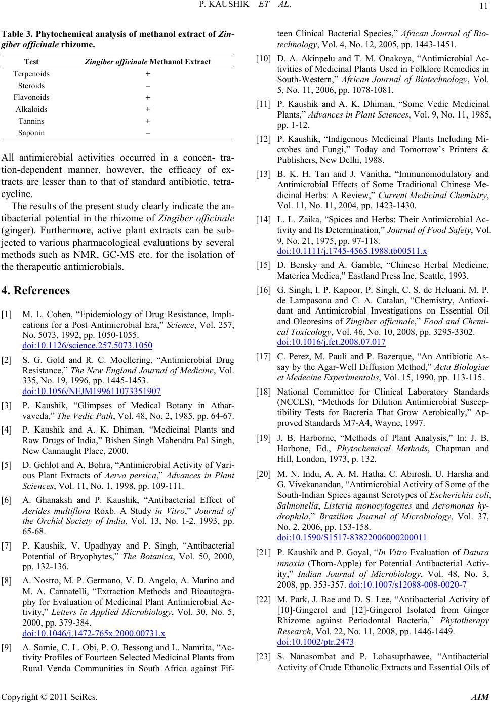
Table 3. Phytochemical analysis of methanol extract of Zin-
giber officinale rh izome.
Test Zingiber offici nale Methanol Extract
Terpenoids +
Steroids –
Flavonoids +
Alkaloids +
Tannins +
Saponin –
All antimicrobial activities occurred in a concen- tra-
tion-dependent manner, however, the efficacy of ex-
tracts are lesser than to that of standard antibiotic, tetra-
cycline.
The results of the present study clearly indicate the an-
tibacterial potential in the rhizome of Zingiber officinale
(ginger). Furthermore, active plant extracts can be sub-
jected to various pharmacological evaluations by several
methods such as NMR, GC-MS etc. for the isolation of
the therapeutic antimicrobials.
4. References
[1] M. L. Cohen, “Epidemiology of Drug Resistance, Impli-
cations for a Post Antimicrobial Era,” Science, Vol. 257,
No. 5073, 1992, pp. 1050-1055.
doi:10.1126/science.257.5073.1050
[2] S. G. Gold and R. C. Moellering, “Antimicrobial Drug
Resistance,” The New England Journal of Medicine, Vol.
335, No. 19, 1996, pp. 1445-1453.
doi:10.1056/NEJM199611073351907
[3] P. Kaushik, “Glimpses of Medical Botany in Athar-
vaveda,” The Vedic Path , Vol. 48, No. 2, 1985, pp. 64-67.
[4] P. Kaushik and A. K. Dhiman, “Medicinal Plants and
Raw Drugs of India,” Bishen Singh Mahendra Pal Singh,
New Cannaught Place, 2000.
[5] D. Gehlot and A. Bohra, “Antimicrobial Activity of Vari-
ous Plant Extracts of Aerva persica,” Advances in Plant
Sciences, Vol. 11, No. 1, 1998, pp. 109-111.
[6] A. Ghanaksh and P. Kaushik, “Antibacterial Effect of
Aerides multiflora Roxb. A Study in Vitro,” Journal of
the Orchid Society of India, Vol. 13, No. 1-2, 1993, pp.
65-68.
[7] P. Kaushik, V. Upadhyay and P. Singh, “Antibacterial
Potential of Bryophytes,” The Botanica, Vol. 50, 2000,
pp. 132-136.
[8] A. Nostro, M. P. Germano, V. D. Angelo, A. Marino and
M. A. Cannatelli, “Extraction Methods and Bioautogra-
phy for Evaluation of Medicinal Plant Antimicrobial Ac-
tivity,” Letters in Applied Microbiology, Vol. 30, No. 5,
2000, pp. 379-384.
doi:10.1046/j.1472-765x.2000.00731.x
[9] A. Samie, C. L. Obi, P. O. Bessong and L. Namrita, “Ac-
tivity Profiles of Fourteen Selected Medicinal Plants from
Rural Venda Communities in South Africa against Fif-
teen Clinical Bacterial Species,” African Journal of Bio-
technology, Vol. 4, No. 12, 2005, pp. 1443-1451.
[10] D. A. Akinpelu and T. M. Onakoya, “Antimicrobial Ac-
tivities of Medicinal Plants Used in Folklore Remedies in
South-Western,” African Journal of Biotechnology, Vol.
5, No. 11, 2006, pp. 1078-1081.
[11] P. Kaushik and A. K. Dhiman, “Some Vedic Medicinal
Plants,” Advances in Plant Sciences, Vol. 9, No. 11, 1985,
pp. 1-12.
[12] P. Kaushik, “Indigenous Medicinal Plants Including Mi-
crobes and Fungi,” Today and Tomorrow’s Printers &
Publishers, New Delhi, 1988.
[13] B. K. H. Tan and J. Vanitha, “Immunomodulatory and
Antimicrobial Effects of Some Traditional Chinese Me-
dicinal Herbs: A Review,” Current Medicinal Chemistry,
Vol. 11, No. 11, 2004, pp. 1423-1430.
[14] L. L. Zaika, “Spices and Herbs: Their Antimicrobial Ac-
tivity and Its Determination,” Journal of Food Safety, Vol.
9, No. 21, 1975, pp. 97-118.
doi:10.1111/j.1745-4565.1988.tb00511.x
[15] D. Bensky and A. Gamble, “Chinese Herbal Medicine,
Materica Medica,” Eastland Press Inc, Seattle, 1993.
[16] G. Singh, I. P. Kapoor, P. Singh, C. S. de Heluani, M. P.
de Lampasona and C. A. Catalan, “Chemistry, Antioxi-
dant and Antimicrobial Investigations on Essential Oil
and Oleoresins of Zingiber officinale,” Food and Chemi-
cal Toxicology, Vol. 46, No. 10, 2008, pp. 3295-3302.
doi:10.1016/j.fct.2008.07.017
[17] C. Perez, M. Pauli and P. Bazerque, “An Antibiotic As-
say by the Agar-Well Diffusion Method,” Acta Biologiae
et Medecine Experimentalis, Vol. 15, 1990, pp. 113-115.
[18] National Committee for Clinical Laboratory Standards
(NCCLS), “Methods for Dilution Antimicrobial Suscep-
tibility Tests for Bacteria That Grow Aerobically,” Ap-
proved Standards M7-A4, Wayne, 1997.
[19] J. B. Harborne, “Methods of Plant Analysis,” In: J. B.
Harbone, Ed., Phytochemical Methods, Chapman and
Hill, London, 1973, p. 132.
[20] M. N. Indu, A. A. M. Hatha, C. Abirosh, U. Harsha and
G. Vivekanandan, “Antimicrobial Activity of Some of the
South-Indian Spices against Serotypes of Escherichia coli,
Salmonella, Listeria monocytogenes and Aeromonas hy-
drophila,” Brazilian Journal of Microbiology, Vol. 37,
No. 2, 2006, pp. 153-158.
doi:10.1590/S1517-83822006000200011
[21] P. Kaushik and P. Goyal, “In Vitro Evaluation of Datura
innoxia (Thorn-Apple) for Potential Antibacterial Activ-
ity,” Indian Journal of Microbiology, Vol. 48, No. 3,
2008, pp. 353-357. doi:10.1007/s12088-008-0020-7
[22] M. Park, J. Bae and D. S. Lee, “Antibacterial Activity of
[10]-Gingerol and [12]-Gingerol Isolated from Ginger
Rhizome against Periodontal Bacteria,” Phytotherapy
Research, Vol. 22, No. 11, 2008, pp. 1446-1449.
doi:10.1002/ptr.2473
[23] S. Nanasombat and P. Lohasupthawee, “Antibacterial
Activity of Crude Ethanolic Extracts and Essential Oils of
Copyright © 2011 SciRes. AIM
P. KAUSHIK ET AL.
12
Spices against Salmonellae and Other Enterobacteria,”
KMITL Science and Technology Journal, Vol. 5, No. 3,
2005, pp. 527-538.
doi:10.1111/j.1472-765X.2004.01605.x
[24] C. Thongson, P. M. Davidson, W. Mahakarnchanakul,
and J. Weiss, “Antimicrobial Activity of Ultrasound-As-
sisted Solvent-Extracted Spices,” Letters in Applied Mi-
crobiology, Vol. 39, No. 5, 2004, pp. 401-406.
[25] H. C. Chen, M. D. Chang and T. J. Chang, “Antibacterial
Properties of Some Spice Plants before and after Heat
Treatment,” Chinese Journal of Microbiology and Im-
munology, Vol. 18, 1985, pp. 190-195.
Copyright © 2011 SciRes. AIM