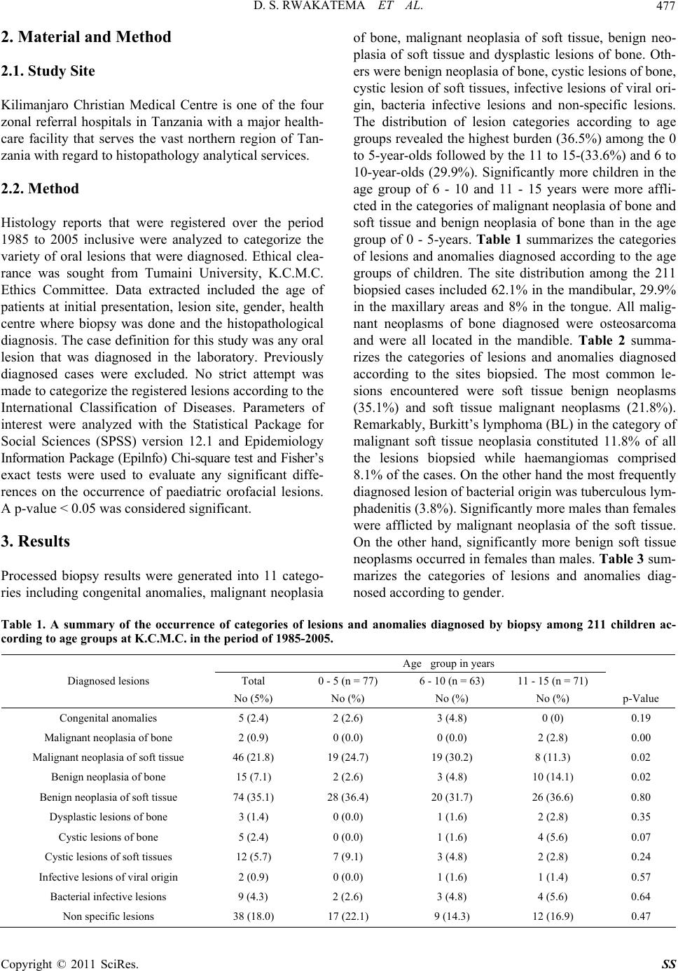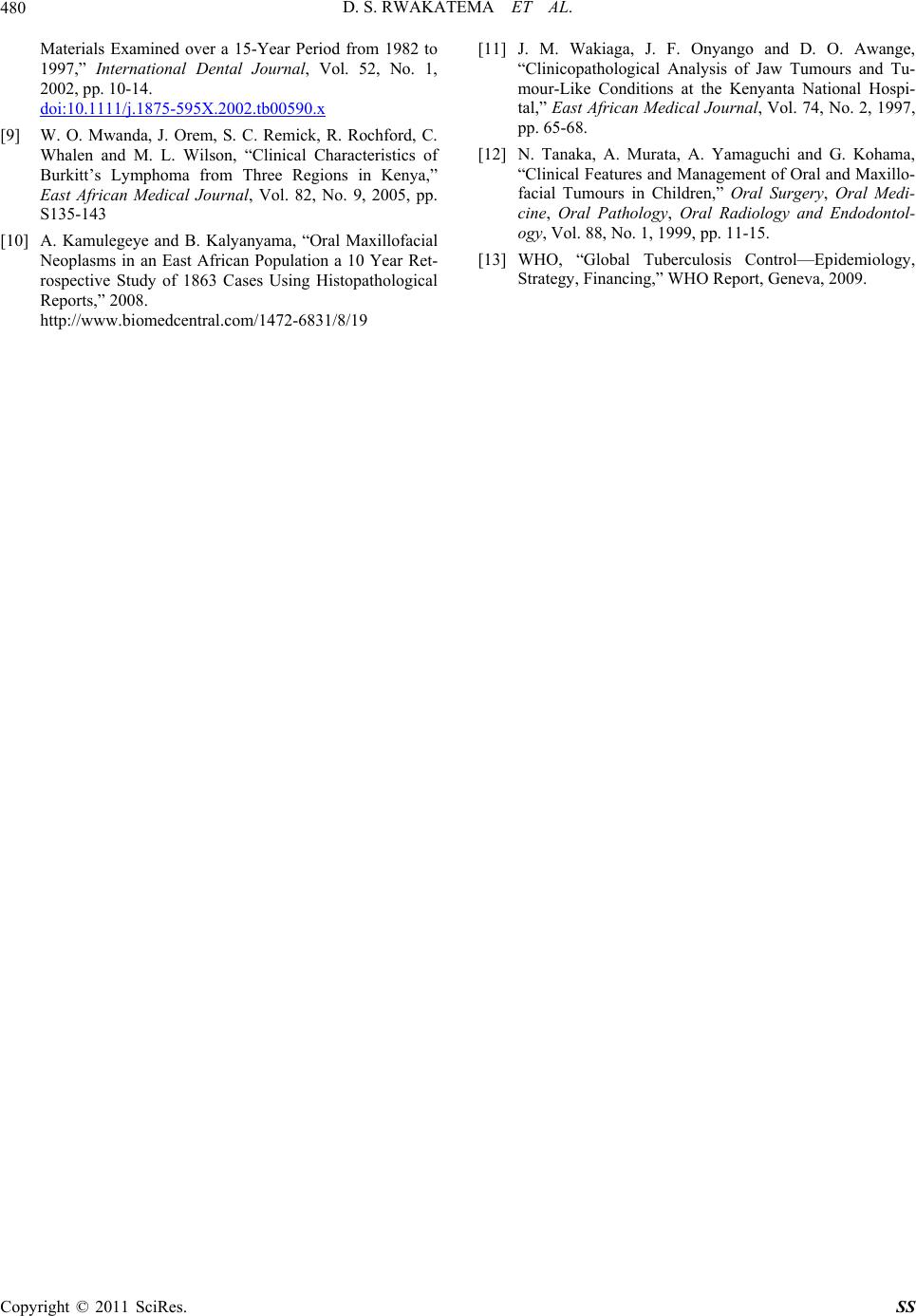
D. S. RWAKATEMA ET AL.479
of the diagnosed lesions were malignant neoplasms of
the soft tissues which was half the number of malignant
neoplasm findings at Muhimbili National Hospital in
Tanzanian children of a similar age group [8]. Since BL
was categorized into malignant neoplasm of the soft tis-
sues its occurrence took a larger portion in this category.
The same trend was also observed in other studies in this
region [6,8,11]. In healthcare, the desirable resources to
optimally manage malignant diseases can be elaborate
and expensive. The present findings should, therefore,
provide useful guidelines for health planners and mana-
gers as to how to logically and proportionately utilize
available resources.
With the present findings and others available else-
where in the region [1,8], sufficient information should
evolve to facilitate the possibilities of mapping out the
pattern and gravity of diseases and conditions that may
be prevalent in children across the East African region.
This may give the region the capacity of comparing in-
formation regarding these lesions with other countries
worldwide. For instance the present investigation has
highlighted the dissimilarities of the occurrence of malig-
nant neoplasms as compared to a similar report in South
American children [3].
In addition to, the essence of highlighting the pattern
and burden of malignant disease in a population cohort
such as the present one, it is also important to note the
significant burden of benign neoplasms. Early in life be-
nign neoplasms if not timely diagnosed and treated, are
often most debilitating and difficult to manage. In the
present investigation, as in several other studies [4,7,12]
the haemangioma was the most commonly diagnosed
neoplasm. This lesion and pleomorphic adenoma de-
pending on their location and extent, pose significant
challenges in terms of definitive management not only in
the professional context but also the man-hours and
school-hours that may be lost while seeking treatment for
these minors. The occurrence of 3.8% of cases of tuber-
culous lymphadenitis in this study was quite alarming
and probably was an indicator of the HIV infection/
AIDS burden in this population [13].
Although they occurred rarely, malignant and benign
neoplasms of bone were significantly higher at the age
group of 11 - 15 years than at the other two age groups.
This is the age of accelerated growth especially in the
bone. Higher chances of bone neoplastic changes are
likely to occur in this period of growth. Overall, the pre-
sent investigation has yielded useful baseline information
that is certainly most pertinent for those in clinical prac-
tice who must maintain logical indices of diagnosing
diverse lesions in the orofacial region. In the absence of
analytical population-based studies, laboratory-based au-
dits shall remain relevant and should be encouraged.
5. Conclusions
Evidently, there is a considerable diversity and prepon-
derance of soft tissue than skeletal oral lesions based on
the present audit. Remarkably, clinicians should maintain
a high index of suspicion regarding the high frequency of
diagnosing BL in such a population.
6. Acknowledgements
The authors are grateful to the authorities of Kilimanjaro
Christian Medical Centre Histopathology Department for
allowing retrieval of biopsy reports of this study. The
authors appreciate the help from Miss Sally Musinde of
the Department of Oral and Maxillofacial Surgery, Sch-
ool of Dental Sciences, University of Nairobi, Kenya for
her involvement in the preparation of the manuscript.
7. References
[1] E. A. O. Dimba, J. Gichana, A. A. K. Limo, K. A. Wakoli,
M. L. Chindia and D. O. Awange, “An Audit of Oral Dis-
ease at a Nairobi Centre, 2000-2004,” International Den-
tal Journal, Vol. 57, No. 6, 2007, pp. 439-444.
[2] P. E. Pertersen, “The World Oral Health Report 2003
Continuous Improvement of Oral Health in the 21st Cen-
tury the Approach of the WHO Oral Health Global Pro-
gramme.” Community Dental Oral Epidemiology, Vol. 31,
No. 1, 2003, pp. 3-24. doi:10.1046/j..2003.com122.x
[3] G. S. Lima, et al, “A Survey of Oral and Maxillofacial
Biopsies in Children: A Single-Centre Retrospective
Study of 20 Years in Pelotas-Brazil,” Journal of Applied
Oral Science, Vol. 16, No. 6, pp. 397-402.
doi:10.1590/S1678-77572008000600008
[4] M. Sato, N. Tanaka, T. Sato and T. Amagasa. “Oral and
Maxillofacial Tumours in Children: A Review,” British
Journal of Oral & Maxillofacial Surgery, Vol. 35, No. 2,
1997, pp. 92-95. doi:10.1016/S0266-4356(97)90682-3
[5] Y. Wang, H. Chang, J. Y. Chang, G. Hung and M. Guo,
“Retrospective Survey of Biopsied Oral Lesions in Pedi-
atric Patients,” Journal of Formos Medical Association,
Vol. 108, No. 11, 2008, pp. 862-871.
doi:10.1016/S0929-6646(09)60418-6
[6] S. B. Aregbesola, V. I. Ugboko, J. A. Akinwande, G. F.
Arole and O. O. Faga, “Orofacial Tumours in Suburban
Nigerian Children and Adolescents,” British Journal of
Oral and Maxillofacial Surgery, Vo. 43, No. 3, 2005, pp.
226-231. doi:10.1016/j.bjoms.2004.11.006
[7] T. A-Khateeb, A. A Hamasha and N. M. Almasri, “Oral
and Maxillofacial tumours in North Jordanian Children
and Adolescents: A Retrospective Analysis over 10
Yeras,” International Journal of Oral and Maxillofacial
Surgery, Vol. 32, No. 1, 2003, pp. 78-83.
doi:10.1054/ijom.2002.0309
[8] B. M. Kalyanyama, M. I. N. Matee and E. Vahahula,
“Oral Tumours in Tanzanian Children Based on Biopsy
Copyright © 2011 SciRes. SS