Surgical Science, 2011, 2, 496-498
doi:10.4236/ss.2011.210109 Published Online December 2011 (http://www.SciRP.org/journal/ss)
Copyright © 2011 SciRes. SS
Purpura Fulminans in Infantile Streptococcal Septicemia
Saurabh Piparsania1, Nitin Rajput1, Kulde ep Singh1, Priti Zade1, Milind Joshi2*
1Department of Paediatrics, Sri Aurobindo Institute of Medical S c iences, Indore, India
2Department of Paediatric surgery, Sri Aurobindo Institute of Medical Sciences, Indore, India
E-mail: *milindj79@yahoo.com
Received June 12, 201 1; revised September 15, 2011; accepted October 14, 2011
Abstract
Purpura fulminans is a hemorrhagic condition associated predominantly with meningococcal and other gram
negative septicemias. It occurs mainly in infants and younger children. Features include tissue necrosis,
small vessel thrombosis, disseminated intravascular coagulation, multi-organ failure and death. Other causes
include clotting factor deficiencies and idiopathic varieties. The condition is uncommon due to gram positive
bacterial sepsis. We report one such case with gram positive bacterial infection.
Keywords: Purpura Fulminans, Gram Positive Sepsis
1. Introduction
Purpura fulminans is a hemorrhagic condition associated
predominantly with meningococcal and other gram nega-
tive septicemias. It occu rs mainly in infants and younger
children. Features include tissue necrosis, small vessel
thrombosis, disse minated intravascular coagu lation, multi-
organ failure and death. Other causes include clotting
factor deficiencies and idiopathic varieties. The condition
is uncommon due to gram positive bacterial sepsis. We
report a case of purpura fulminans caused by alpha-
hemolytic streptococcal sepsis and meningitis in a 4
month old baby girl who was referred to our institute
with digital gangrene and necrotic and hemorrhagic pur-
purae.
2. Case Report
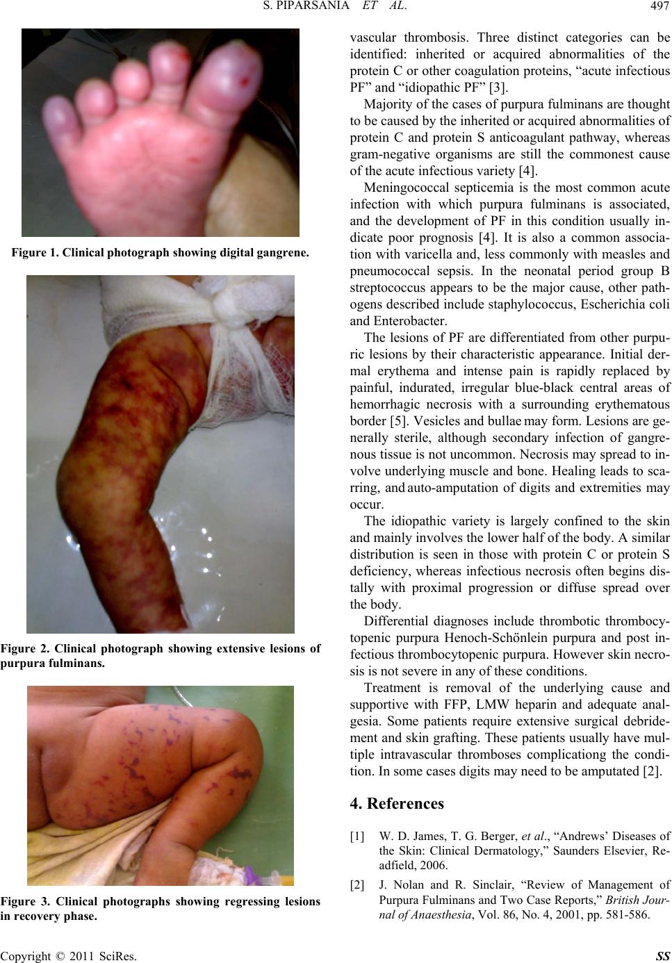
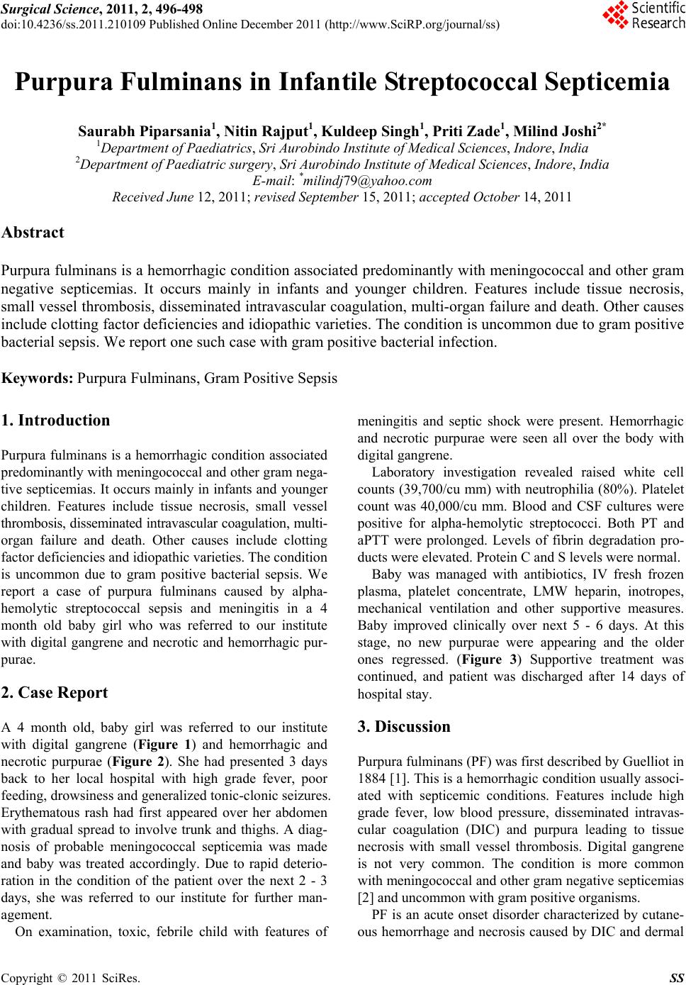
A 4 month old, baby girl was referred to our institute
with digital gangrene (Figure 1) and hemorrhagic and
necrotic purpurae (Figure 2). She had presented 3 days
back to her local hospital with high grade fever, poor
feeding, drowsiness and generalized tonic-clonic seizures.
Erythematous rash had first appeared over her abdomen
with gradual spread to involve trunk and thighs. A diag-
nosis of probable meningococcal septicemia was made
and baby was treated accordingly. Due to rapid deterio-
ration in the condition of the patient over the next 2 - 3
days, she was referred to our institute for further man-
agement.
On examination, toxic, febrile child with features of
meningitis and septic shock were present. Hemorrhagic
and necrotic purpurae were seen all over the body with
digital gangrene.
Laboratory investigation revealed raised white cell
counts (39,700/cu mm) with neutrophilia (80%). Platelet
count was 40,000/cu mm. Blood and CSF cultures were
positive for alpha-hemolytic streptococci. Both PT and
aPTT were prolonged. Levels of fibrin degradation pro-
ducts were elevated. Protein C and S levels were normal.
Baby was managed with antibiotics, IV fresh frozen
plasma, platelet concentrate, LMW heparin, inotropes,
mechanical ventilation and other supportive measures.
Baby improved clinically over next 5 - 6 days. At this
stage, no new purpurae were appearing and the older
ones regressed. (Figure 3) Supportive treatment was
continued, and patient was discharged after 14 days of
hospital stay.
3. Discussion
Purpura fulminan s (PF) was first described by Guelliot in
1884 [1]. This is a hemorrhagic condition usua lly associ-
ated with septicemic conditions. Features include high
grade fever, low blood pressure, disseminated intravas-
cular coagulation (DIC) and purpura leading to tissue
necrosis with small vessel thrombosis. Digital gangrene
is not very common. The condition is more common
with meningococcal and other gram negative septicemias
[2] and uncommon with gram positive organisms.
PF is an acute onset disorder characterized by cutane-
ous hemorrhage and necrosis caused by DIC and dermal