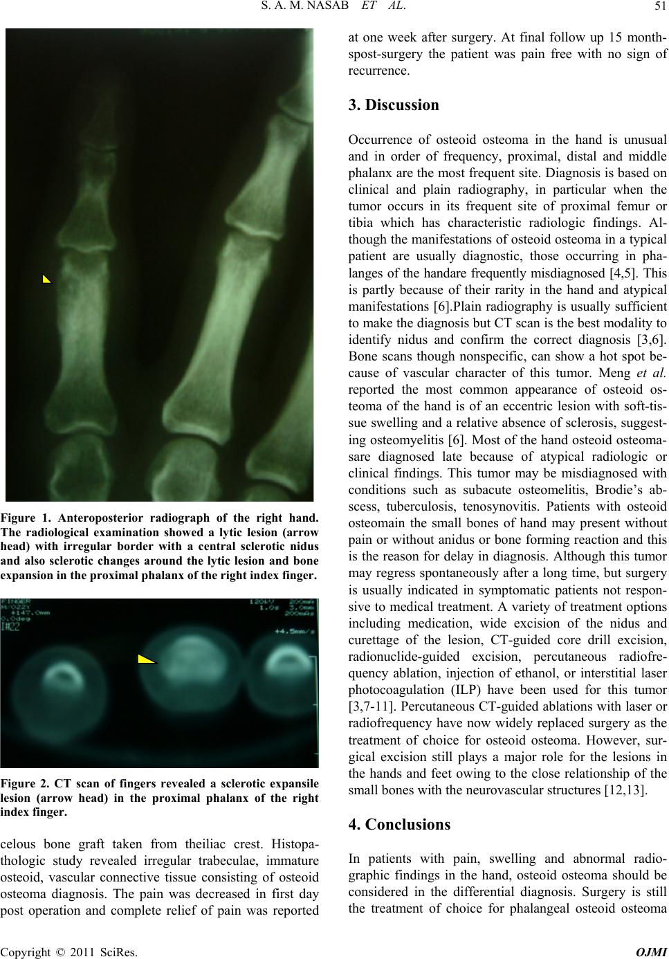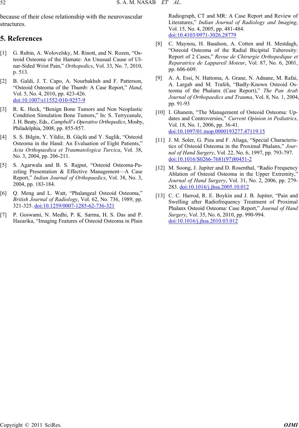
S. A. M. NASAB ET AL.
Copyright © 2011 SciRes. OJMI
52
because of their close relationship with the neurovascular
structures.
5. References
[1] G. Rubin, A. Wolovelsky, M. Rinott, and N. Rozen, “Os-
teoid Osteoma of the Hamate: An Unusual Cause of Ul-
nar-Sided Wrist Pain,” Orthopedics, Vol. 33, No. 7, 2010,
p. 513.
[2] B. Galdi, J. T. Capo, A. Nourbakhsh and F. Patterson,
“Osteoid Osteoma of the Thumb: A Case Report,” Hand,
Vo l. 5, N o. 4 , 2010, pp. 423-426.
doi:10.1007/s11552-010-9257-9
[3] R. K. Heck, “Benign Bone Tumors and Non Neoplastic
Condition Simulation Bone Tumors,” In: S. Terrycanale,
J. H. Beaty, Eds., Campbell’s Operative Orthopedics, Mosby,
Philadelphia, 2008, pp. 855-857.
[4] S. S. Bilgin, Y. Yildiz, B. Güçlü and Y. Saglik, “Osteoid
Osteoma in the Hand: An Evaluation of Eight Patients,”
Acta Orthopaedica et Traumatologica Turcica, Vol. 38,
No . 3, 2004, pp. 20 6-211 .
[5] S. Agarwala and B. S. Rajput, “Osteoid Osteoma-Pu-
zzling Presentation & Effective Management—A Case
Report,” Indian Journal of Orthopaedics, Vol. 38, No. 3,
2004, pp. 183-184.
[6] Q. Meng and L. Watt, “Phalangeal Osteoid Osteoma,”
British Journal of Radiology, Vol. 62, No. 736, 1989, pp.
321-325. doi:10.1259/0007-1285-62-736-321
[7] P. Goswami, N. Medhi, P. K. Sarma, H. S. Das and P.
Hazarika, “Imaging Features of Osteoid Osteoma in Plain
Radiograph, CT and MR: A Case Report and Review of
Literatures,” Indian Journal of Radiology and Imaging,
Vo l. 15, No. 4, 2005, pp. 481-48 4.
doi:10.4103/0971-3026.28779
[8] C. Maynou, H. Baudson, A. Cotten and H. Mestdagh,
“Osteoid Osteoma of the Radial Bicipital Tuberosity:
Report of 2 Cases,” Revue de Chirurgie Orthopedique et
Reparatrice de Lappareil Moteur, Vol. 87, No. 6, 2001,
pp. 606-609.
[9] A. A. Essi, N. Hattoma, A. Grane, N. Adnane, M. Rafai,
A. Largab and M. Trafeh, “Badly-Known Osteoid Os-
teoma of the Phalanx (Case Report),” The Pan Arab
Journal of Orthopaedics and Trauma, Vol. 8, No. 1, 2004,
pp. 91-93
[10] I. Ghanem, “The Management of Osteoid Osteoma: Up-
dates and Controversies,” Current Opinion in Pediatrics,
Vol. 18, No. 1, 2006, pp. 36-41.
doi:10.1097/01.mop.0000193277.47119.15
[11] J. M. Soler, G. Piza and F. Aliaga, “Special Characteris-
tics of Osteoid Osteoma in the Proximal Phalanx,” Jour-
nal of Hand Surgery, Vol. 22, No. 6, 1997, pp. 793-797.
doi:10.1016/S0266-7681(97)80451-2
[12] M. Soong, J. Jupiter and D. Rosenthal, “Radio Frequency
Ablation of Osteoid Osteoma in the Upper Extremity,”
Journal of Hand Surgery, Vol. 31, No. 2, 2006, pp. 279-
283. doi:10.1016/j.jhsa.2005.10.012
[13] C. C. Harrod, R. E. Boykin and J. B. Jupiter, “Pain and
Swelling after Radiofrequency Treatment of Proximal
Phalanx Osteoid Oste oma: Case Report,” Journal of Hand
Surgery, Vol . 35 , No. 6, 20 10, pp. 990-994 .
doi:10.1016/j.jhsa.2010.03.012