Vol.3, No.12, 742-747 (2011)
doi:10.4236/health.2011.312123
C
opyright © 2011 SciRes. Openly accessible at http://www.scirp.org/journal/HEALTH/
Health
Suppressive effects of saliva against enamel
demineralization caused by acid beverages
Shoji Takahashi1*, Shigeru Watanabe1, Takashi Ogihara1, Koji Watanabe1, Kun Xuan2,
Xiaojing Wang2
1Division of Pediatric Dentistry, Department of Human Development & Fostering, School of Dentistry, Meikai University School of
Dentistry, Sakado, Japan; *Corresponding Author: s-takahashi@dent.meikai.ac.jp
2Department of Pediatric Dentistry, College of Stomatology, Fourth Military Medical University, Xi’an, China.
Received 30 September 2011; revised 10 November 2011; accepted 27 November 2011.
ABSTRACT
This study aimed to clarify the ability of the bu-
ffer systems of saliva to inhibit enamel demin-
eralization after intake of an acid beverage. In
the first experiment, titrable acidity tests were
carried out. Ten milliliters of saliva stimulated
by chewing gum base was obtained from 10
healthy adult subjects and the pH of each saliva
sample was measured. The beverages used for
the experiment were a carbonated soft drink (pH
2.2), a sports drink (pH 3.5), and 100% orange
juice (pH 3.8). Distilled water adjusted to the pH
of each saliva sample was used as a control. In
the second experiment, the suppressive ability
of saliva against enamel demineralization was
quantitatively analyzed using quantitative light-
induced fluorescence (QLF). Aliquots of stimu-
lated saliva obtained from a subject were mixed
with 15 ml of 100% orange juice in sal iv a:orang e
juice ratios of 1/30, 1/15, 1/10 and 1/5, and bo-
vine teeth w ere soaked for 24 hours in the solu-
tions. The Q of the Q L F analyses of the e namel
was then measured. The lowest titrant volume
which reduced the pH of the initial saliva (7.7 on
average) to pH 5.4 w as that of the orange juice.
No relationship was found between the buffer
cap aci ty and the pH of th e acid bev erages. From
the QLF measurement, the saliva-orange juice
group showed a significantly decreased amount
of enamel demineralization (p < 0.01 at 20% level)
compared with the distilled water-orange juice
group. In conclusion, saliva acts as a buffer to
suppress enamel demineralization caused by
low-pH beverages.
Keywords: Erosion; Acid Beverage; Saliva;
Buffering Capacity; QLF
1. INTRODUCTION
A large number of the soft drinks we consume regu-
larly, such as sports drinks, carbonated soft drinks and
fruit juices, are acidic (pH 2.2 or more) and have been
shown to cause acid erosion, depending on the amount
and pattern of consumption [1,2]. In the human oral cav-
ity, the salivation rate changes substantially, depending
on the properties of the ingested solutions [3,4], and the
repeated occurrence of intake of a soft drink and saliva-
tion to dilute it maintains the homeostasis of the oral pH
environment [5,6].
With regard to the acid buffering capacity of saliva,
Lilienthal [7] demonstrated a high acid buffering activity
of salivary bicarbonates by titrating hydrochloric acid
solution into saliva. Several studies have investigated the
acid buffering capacity of saliva [8-10]; however, the
acid buffering activity of saliva against low-pH soft
drinks and its effect on dental demineralization have not
yet been fully elucidated.
In this study, with the aim of obtaining evidence for
oral hygiene instruction in dental practice, we performed
titration of different types of soft drinks into stimulated
saliva and the quantitative observation of soft-drink-
induced demineralization of bovine tooth enamel and the
inhibitory effect of saliva on demineralization, using
quantitative light-induced fluorescence (QLF) [11,12].
2. MATERIAL AND METHODS
This study was performed in accordance with the
guidelines of the ethics committee of Meikai University
School of Dentistry (approval number: A0913).
2.1. Experiment 1. Titration of Different
Types of Soft Drinks into Stimulated
Saliva
This experiment involved 10 healthy adult subjects,
S. Takahashi et al. / Health 3 (2011) 742-747
Copyright © 2011 SciRes. Openly accessible at http:// www.scirp.org/journal/HEALTH/
743743
consisting of 4 males aged 25 to 40 years and 6 females
aged 25 to 40 years, with no tooth defects and who were
not receiving any medication. Mechanically-stimulated
saliva samples were collected from each subject by in-
structing them to chew gum base (1.0 g) and the saliva
was immediately subjected to pH measurement. Ten
milliliters each of the collected saliva samples were ti-
trated with Coca Cola (Coca-Cola (Japan) Co., Limited,
Tokyo, Japan; pH 2.2; hereinafter referred to as “the
carbonated soft drink”), Pocari Sweat (Otsuka Pharma-
ceutical, Co., Ltd., Tokyo, Japan; pH 3.5; hereinafter
referred to as “the sports drink”) and 100% orange juice
(Kirin Tropicana, Co., Ltd., Tokyo, Japan; pH 3.8). Dis-
tilled water adjusted with sodium hydroxide to the pH of
each stimulated saliva sample was used as a control. The
volume of each titrant required to lower the pH of saliva
and control solutions to 5.4, the approximate critical pH
level for demineralization, was determined and com-
pared among the three types of beverage and between
saliva and control.
All saliva samples were collected at 3:00 pm and im-
mediately subjected to pH measurement. In addition,
subjects were instructed to refrain from drinking, eating
or smoking for 2 hours before the start of each experi-
ment.
2.2. Experiment 2. Quantitative Observation
of the Demineralization Inhibitory Effect
of Saliva on Bovine Tooth Enamel
Ten bovine maxillary incisor teeth were used in this
experiment. A 1-mm square window was formed over
one-third of the labial and incisal surface of each tooth
with nail varnish. The beverage used in this experiment
was 100% orange juice. A mechanically-stimulated sa-
liva sample was collected from a single subject by in-
structing him or her to chew gum base (1.0 g) and the
saliva was immediately subjected to pH measurement.
The saliva sample was added to 100% orange juice to
prepare solutions containing saliva at ratios of 1:30, 1:15,
1:10 and 1:5. The sample teeth were soaked in 15 ml
each of the saliva-containing orange juice solutions at
room temperature for 24 hours while shaking in a water
bath shaker (Taitec, Aichi, Japan). Distilled water ad-
justed to the pH of the stimulated saliva sample (with
sodium hydroxide) mixed with 100% orange juice was
used as control.
Each soaked tooth was washed, dried and subjected to
evaluation of the degree of demineralization with a QLF
apparatus (Inspektor Research Systems BV, Amsterdam,
Netherlands). More specifically, ultraviolet light at a
wavelength of 370 ± 80 nm was irradiated to the surface
of a tooth with visible light blocked, and the fluores-
cence light reflected from the area near the enamel-den-
tin junction was allowed to pass through a 520-nm filter
and captured by a CCD camera as image data [11]. QLF
results were evaluated using ΔQ (% mm2), a parameter
representing the mean amount of demineralization.
For statistical analysis of the results of each experi-
ment, the Student t-test was used for two-group com-
parison and Scheffe’s multiple comparison test was used
for multigroup comparison.
3. RESULTS
3.1. Experiment 1
The mean pH of mechanically-stimulated saliva sam-
ples collected from 10 subjects instructed to chew gum
base was 7.70 ± 0.19. Ten milliliters of each of the saliva
samples was titrated with the carbonated soft drink,
sports drink and 100% orange juice. The results of the
titration experiment are shown in Figures 1-3. The
volumes of the carbonated soft drink and sports drink
required to lower the pH of the saliva samples to the
critical pH level for demineralization were about 7 and 4
times higher than those required to lower the pH of dis-
tilled water adjusted to the same pH levels, with signifi-
cant differences between saliva and control (p < 0.01)
(Figure 4). The volume of 100% orange juice required
Figure 1. Titration curve with the carbonated soft drink (pH 2.2).
S. Takahashi et al. / Health 3 (2011) 742-747
Copyright © 2011 SciRes. Openly accessible at http:// www.scirp.org/journal/HEALTH/
744
Figure 2. Titration curve with the sports drink (pH 3.5).
Figure 3. Titration curve with the 100% orange juice (pH 3.8).
p < 0.01
p < 0.05
Figure 4. The comparison of volume required to reduce the pH of saliva to the
critical pH level for demineralization.
to reduce the pH of saliva to the critical pH level for
demineralization was significantly lower than those of
the sports drink and carbonated soft drink.
3.2. Experiment 2
The results of a comparison of the degrees of demin-
eralization of sample teeth soaked in 100% orange juice
(ΔQ), orange juice containing saliva at different ratios
(ΔQs) and control (ΔQw) are shown in Figure 5. Com-
pared with the orange juice containing no saliva, ΔQs
values significantly increased and the amount of demin-
eralization significantly decreased with increasing mix-
ture ratio. Although a slight increase in ΔQw was ob-
served, the degree of decrease in demineralization with
increasing mixture ratio was lower in saliva-containing
distilled water than in saliva-containing orange juice.
A comparison between ΔQs and ΔQw showed that the
amount of demineralization in saliva-containing orange
juice was lower than that in saliva-containing distilled
ater at all mixture ratios. A significant difference was w
S. Takahashi et al. / Health 3 (2011) 742-747
Copyright © 2011 SciRes. Openly accessible at http:// www.scirp.org/journal/HEALTH/
745745
p < 0.05p < 0.01
Figure 5. The comparison of the degrees of demineralization of sample teeth
soaked in 100% orange juice (ratio 0 as control), orange juice containing sa-
liva at different ratios.
found between the two solutions at a mixture ratio of 1:5
(p < 0.01).
The experiment also revealed that demineralization of
bovine tooth enamel began after 3 hours of soaking in
100% orange juice (Table 1).
4. DISCUSSION
4.1. Acid Buffering Effect of Saliva and
Titratable Acidity of Soft Drinks
In experiment 1, stimulated saliva samples collected
from subjects were titrated with three types of soft
drinks and changes in the pH of the saliva samples were
measured. The volume of each beverage required to re-
duce the pH of distilled water to the critical level of 5.4
was significantly lower than that required to lower the
pH of the saliva samples, clearly demonstrating an acid
buffering effect of saliva. The magnitude of the acid
buffering capacity of saliva is in proportion to the con-
tent of bicarbonate in saliva, which is known to increase
with increasing rate of salivation in response to stimuli
[9,13]. Thus, the mechanically-stimulated saliva samples
collected from the subjects instructed to chew gum base
are considered to have a higher acid buffering capacity
Ta b l e 1 . The progression of Q of the teeth soaking in 100%
orange juice.
Soaking time (hour) Q (% mm2)
0 0
1 0
3 –0.24 ± 0.13
4.5 –0.39 ± 0.19
5 –1.54 ± 0.45
6 –3.70 ± 1.16
12 –6.47 ± 2.06
24 –16.61 ± 4.29
than unstimulated saliva. Of the three types of soft
drinks, 100% orange juice, which had the highest pH,
lowered the pH of the saliva samples at the earliest time
point. The major acid components of each beverage are
as follows: phosphoric acid for carbonated soft drinks,
citric acid and fruit-derived organic acid for sports
drinks, and several types of fruit-derived organic acid for
100% orange juice drinks. The titratable acidities (buff-
ering capacity) of different fruit juice drinks, including
orange juice drinks, which show relatively higher pH
levels than other soft drinks, have been measured by
titrating each beverage with a sodium hydroxide solution
[14-18]. The results of these studies suggest that the
magnitude of the acid buffering capacity of a solution
does not necessarily match its pH levels. The results of
the present experiments were also consistent with previ-
ous findings. The reason for this has been known, but
Larsen and Nyvad suggested that fruit-derived organic
acid binds to calcium contained in a solution to form an
organic-calcium complex, which exerts a strong buffer-
ing effect [16].
4.2. Demineralization Inhibitory Effect of a
Saliva-Containing Soft Drink
The present study used the ΔQ value for evaluation of
demineralization. The ΔQ value has been shown to be as
reliable as the ΔZ value, which reflects mineral loss in a
caries lesion and is used in microradiography [19]. Sev-
eral studies have employed the ΔQ value as a measure of
the degree of demineralization [11,19].
The risk for tooth demineralization caused by soft
drinks has traditionally been evaluated based only on
their pH levels. The results of the present study suggest
the need for reconsidering this risk.
In experiment 2, the amount of demineralization de-
creased with increasing mixture ratio at a greater degree
in saliva-containing soft drinks than in saliva-containing
S. Takahashi et al. / Health 3 (2011) 742-747
Copyright © 2011 SciRes. Openly accessible at http:// www.scirp.org/journal/HEALTH/
746
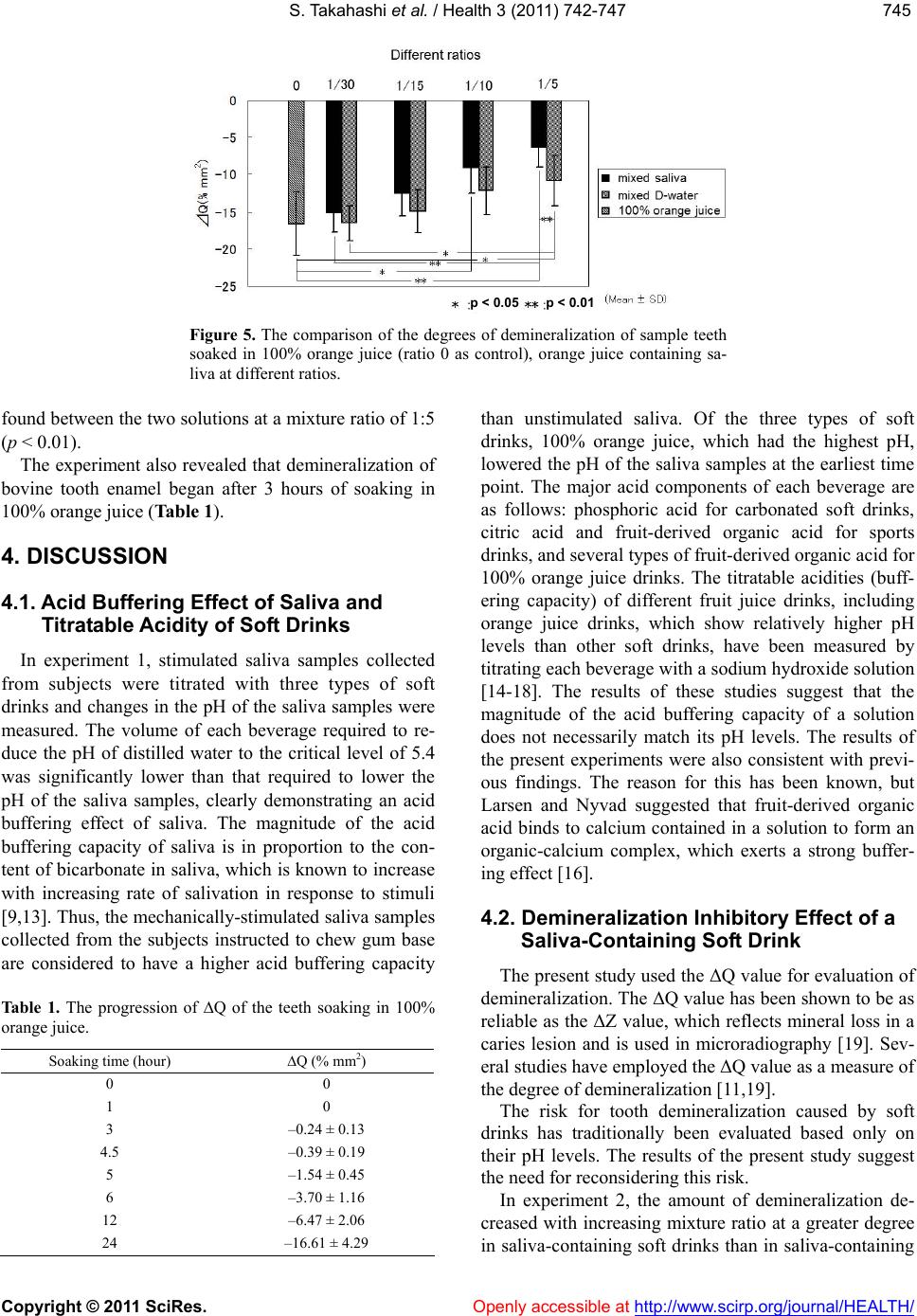
distilled water. This result appeared to depend greatly on
the acid buffering capacity of saliva. The mixture of
stimulated saliva with 100% orange juice resulted in a
greater degree of increase in pH with increasing mixture
ratio than when saliva was mixed with distilled water.
The increase in pH was about 0.1 at a saliva mixture
ratio of 1:5. Larsen et al. [20] showed that the amount of
apatite crystal that can be dissolved in 1.5 L of distilled
water was 0.5 g at pH 5, 5 g at pH 4 and 85 g at pH 3,
suggesting that at a pH level of 4 or less, a slight change
in pH can significantly affect the amount of enamel
demineralization. The results of the present experiments
thus suggest that the acid buffering effect of saliva is a
significant factor that inhibits tooth demineralization.
The pH levels of solutions after 24 hours of soaking of
bovine teeth were higher by about 2.0 than those imme-
diately after soaking. This may be explained by the pre-
vious finding that carbonates are released from the
enamel during the process of demineralization and con-
verted into carbonic acid, which increases pH [20].
The inhibition of demineralization of bovine teeth by
the addition of saliva appears to be mediated not only by
the aforementioned acid buffering effect of saliva but
also by remineralization via the supply of saliva-derived
minerals to the demineralized portion. It is known that
saliva is supersaturated with respect to minerals, such as
hydroxyapatite or tooth enamel and thus the calcium and
phosphate ions in saliva serve as remineralization pro-
moting factors. The concentrations of these ions, as well
as bicarbonates, are higher in stimulated saliva than in
unstimulated saliva. It is thus possible that in the present
experiments, minerals contained in the stimulated saliva
samples were supplied to the demineralized portion of
the bovine tooth enamel, which might have resulted in
the observed inhibition of demineralization. Another
possible factor for demineralization inhibition is the
protective effect of a pellicle formed on the surface of
the enamel. The time required for pellicle formation
varies substantially depending on the experimental en-
vironment. Meurman et al. [21] reported that it takes 7
days until a pellicle is formed in vitro while Hannig et al.
[22] reported that it only takes 3 minutes before a pelli-
cle is formed and exerts its protective effect in vivo. In
the oral cavity with normal salivation, the tooth enamel
is always protected by a pellicle and exposed to saliva
supersaturated with respect to tooth minerals. It is thus
likely that the demineralization inhibitory effect of saliva
is constantly exerted in the oral cavity.
In conclusion, saliva acts as a buffer to suppress
enamel demineralization caused by low-pH beverages.
5. ACKNOWLEDGEMENTS
The author thanks the subjects for their cooperation and prof. C.
Dawes, Department of Oral Biology, University of Manitoba, Winni-
peg, Canada, for helpful suggestions and comments.
REFERENCES
[1] ten Cate, J.M. and Imfeld, T. (1996) Dental erosion,
summary. European Journal of Oral Sciences, 104,
241-244. doi:10.1111/j.1600-0722.1996.tb00073.x
[2] Vissink, A., Burlage, F.R., Spijkervet, F.K., Jansma, J.
and Coppes, R.P. (2003) Prevention and treatment of the
consequences of head and neck radiotherapy. Critical
Reviews in Oral Biology & Medicine, 14, 213-225.
doi:10.1177/154411130301400306
[3] Watanabe, S. and Dawes, C. (1988) The effects of dif-
ferent foods and concentrations of citric acid on the flow
rate of whole saliva in man. Archives of Oral Biology, 33,
1-5. doi:10.1016/0003-9969(88)90089-1
[4] Watanabe, S. and Dawes, C. (1988) A comparison of the
effects of tasting and chewing foods on the flow rate of
whole saliva in man. Archives of Oral Biology, 33,
761-764. doi:10.1016/0003-9969(88)90010-6
[5] Dawes, C. (1983) A mathematical model of salivary
clearance of sugar from the oral cavity. Caries Research,
17, 321-334. doi:10.1159/000260684
[6] Dawes, C., Watanabe, S., Biglow-Lecomte, P. and Dibdin,
G.H. (1989) Estimation of the velocity of the salivary
film at some different locations in the mouth. Journal of
Dental Research, 68, 1479-1482.
doi:10.1177/00220345890680110201
[7] Lilienthal, B. (1955) An analysis of the buffer systems in
saliva. Journal of Dental Research, 34, 516-530.
doi:10.1177/00220345550340040701
[8] Larsen, M.J., Jensen, A.F., Madsen, D.M. and Pearce,
E.I.F. (1999) Individual variations of pH, buffer capacity,
and concentrations of calcium and phosphate in un-
stimulated whole saliva. Archives of Oral Biology, 44,
111-117. doi:10.1016/S0003-9969(98)00108-3
[9] Bardow, A., Moe, D., Nyvad, B. and Nauntofte, B. (2000)
The buffer capacity and buffer systems of human whole
saliva measured without loss of CO2. Archives of Oral
Biology, 45, 1-12. doi:10.1016/S0003-9969(99)00119-3
[10] Moritsuka, M., Kitasako, Y., Burrow, M.F., Ikeda, M. and
Tagami, J. (2006) The pH change after HCl titration into
resting and stimulated saliva for a buffering capacity test.
Australian Dental Journal, 51, 170-174.
doi:10.1111/j.1834-7819.2006.tb00422.x
[11] Ando, M., Hall, A.F., Eckert, G.J., Schemehorn, B.R.,
Analoui, M. and Stookey, G.K. (1997) Relative ability of
laser fluorescence techniques to quantitate early mineral
loss in vitro. Caries Research, 31, 125-131.
doi:10.1159/000262387
[12] de Josselin de Jong, E., Sundstrom, F., Westerling, H.,
Tranaeus, S., ten Bosch, J.J. and Angmar-Mansson, B.
(1995) A new method for in vivo quantification of
changes in initial enamel caries with laser fluorescence.
Caries Research, 29, 2-7. doi:10.1159/000262032
[13] Edgar, W.M. (1992) Saliva: Its secretion, composition
and functions. Brazilian Dental Journal, 172, 305-312.
doi:10.1038/sj.bdj.4807861
[14] Owens, B.M. (2007) The potential effects of pH and
buffering capacity on dental erosion. General Dentistry,
S. Takahashi et al. / Health 3 (2011) 742-747
Copyright © 2011 SciRes. http://www.scirp.org/journal/HEALTH/Openly accessible at
747747
55, 527-531.
[15] Edwards, M., Creanor, S.L., Foye, R.H. and Gilmour,
W.H. (1999) Buffering capacities of soft drinks: the po-
tential influence on dental erosion. Journal of Oral Re-
habilitation, 26, 923-927.
doi:10.1046/j.1365-2842.1999.00494.x
[16] Larsen, M.J. and Nyvad, B. (1999) Enamel erosion by
some soft drinks and orange juice relative to their pH,
buffering effect and contents of calcium phosphate. Car-
ies Research, 33, 81-87. doi:10.1159/000016499
[17] Zero, D.T. and Lussi, A. (2005) Erosion-chemical and
biological factors of importance to the dental practitioner.
International Dental Journal, 55, 285-290.
[18] Jensdottir, T., Holbrook, P., Nauntofte, B., Buchwald, C.
and Bardow, A. (2006) Immediate erosive potential of
cola drinks and orange juice. Journal of Dental Research,
85, 226-230. doi:10.1177/154405910608500304
[19] Pretty, I.A., Edgar, W.M. and Higham, S.M. (2002) De-
tection of in vitro demineralization of primary teeth using
quantitative light-induced fluorescence(QLF). Interna-
tional Journal of Paediatric Dentistry, 12, 158-167.
doi:10.1046/j.1365-263X.2002.00357.x
[20] Larsen, M.J., Pearce, E.I.F. and Jensen, S.J. (1993) Notes
on the dissolution of human dental enamel in dilute acid
solutions at high solid/solution ratio. Caries Research, 27,
87-95. doi:10.1159/000261523
[21] Meurman, J.H. and Frank, R.M. (1991) Scanning elec-
tron microscopic study of the effect of salivary pellicle
on enamel erosion. Caries Research, 25, 1-6.
doi:10.1159/000261335
[22] Hannig, M., Fiebiger, M., Guntzer, M., Dobert, A.,
Zimehl, R. and Nekrashevych, Y. (2004) Protective effect
of the in situ formed short-term salivary pellicle. Ar-
chives of Oral Biology, 49, 903-910.
doi:10.1016/j.archoralbio.2004.05.008