Open Journal of Stomatology, 2011, 1, 158-164
doi:10.4236/ojst.2011.14023 Published Online December 2011 (http://www.SciRP.org/journal/ojst/ OJST
).
Published Online December 2011 in SciRes. http://www.scirp.org/journal/OJST
Comparison of the arch forms and dimensions in various
malocclusions of the Turkish population
Sultan Olmez, Servet Dogan
Ege University, Izmir, Turkey.
Email: sultanolmez@gmail.com
Received 17 July 2011; revised 27 August 2011; accepted 9 September 2011.
ABSTRACT
Objectives: The aim of this study is to determine the
distribution of morphological differences in the cli-
nical mandibular arch forms seen in Angle Class I, II
and III malocclusions in Turkish population and to
examine the effect of gender on arch dimension pa-
rameters. Material and methods: This study ha s been
conducted on pretreatment mandibular study models
of 600 individuals (362 girls, 238 boys) aged between
14 and 19. On the photocopies derived from these
models, 4 linear and 2 proportional measurements
have been made. The samples have been evaluated as
square, ovoid and tapered (OrthoForm-3M Unitek)
arch for m templates. Results: The most frequent arch
form encountered among all the groups was the ta-
pered one (62.5%) followed by the ovoid (27.3%) and
the square one (10.2%). Gender difference influences
on morphological structure was apparent. Generally,
compared with girls, arch width and depth were
found to be more in boys. Conclu sions: The mos t fre-
quent arch form seen in Angle malocclusion groups
was the tapered one, followed by the less frequent
ovoid and squ are ones.
Keywords: Arch Form; Arch Dimension; Sex Differ-
ences
1. INTRODUCTION
While a parameter curve displays a perfect conformity
with the arches, in 40% - 50% of the patients th is accor-
dance shows a decrease [1]. It can be accepted that in at
least half of the patients the preformed arch wires don’t
seem to be functional [2]. Because of these reasons, the
routinely used superelastic preformed arch wires have to
be in various forms with individual malocclusion adap-
tations.
Most of the studies conducted on arch form are fo-
cused on findin g a single shape in perf ect conformity f or
the dental arch of a specific sample [3]. Despite individ-
ual differences, when the ethnical variations are taken
into consideration; the application of a single ideal arch
form for every individual could effect the post treatment
functional, esthetic and stable arch form outcomes [4].
In 1932 Chuck [5] classified the arch forms as tapered,
ovoid and square for the first time. These arch forms can
also be expressed as narrow, normal and wide. Especial-
ly in determining the arch wire forms utilized at the ini-
tial phase of the treatment, he advocates that making a
choice between these three forms would be better than
using a single arch form [5]. For this reason, in leveling
and arrangement phases, the most convenient arch form
type according to the ethnical origin and malocclusion of
the patients should be chosen from the preformed su-
perelastic arch wires [4,6].
There are some studies aimed at determining the arch
forms specific to various ethnical groups [4,6-8]. In a
study where Ko ok, Y. A. et al. [4] determined the ethni-
cal differences between Korean and North Caucasian
groups, arch forms passing through clinical brackets
which is appraised as a valuable approach in modern
orthodontic technics were used.
Although there have been studies one on the evalua-
tion of arch forms in various groups, no such research
has been performed on the Turkish population; this fact
has urged us to carry out this study. The aim of this study
is to determine the differences of clinical mandibular
arch forms in Angle Class I, II, and III malocclusions in
the Turkish population by identifying its morphological
variations and to evaluate gender differences with re-
spect to arch dimension parameters.
2. MATERIAL AND METHODS
This study consisted of 600 subjects’ (362 female, 238
male) pretreatment mandibular dental casts between the
ages of 14 and 19 years, among whom 200 were Angle Cl-
ass I, 200 were Class II and 200 were Class III (Table 1).
The following inclusion criterias were used before as-
sessing the patients:
S. Olmez et al. / Open Journal of Stomatology 1 (2011) 158-164 159
Table 1. Distribution of sex, age and arch forms accordig to the
Angle classifications.
Samples Number BoysGirls
Mean
Age
(years)
Standard
Deviation
T apered A rch F orm
Class I
Class II
Class III
135
130
110
49
52
42
86
78
68
15.63
15.36
16.17
1.72
1.67
1.79
Ovoid Arch Form
Class I
Class II
Class III
50
57
57
14
28
21
36
29
36
15.16
15.56
16.35
1.72
1.69
1.77
Square Arch Form
Class I
Class II
Class III
15
13
33
8
3
21
7
10
12
15.47
16.08
15.61
1.36
1.60
1.64
Total 600 238 362 15.63 1.71
1) Angle Class I, II, and III malocc lus io n s.
2) Permanent dentition without atrision or fractures
in the incisal edges or cusp tip s in permanent den-
tition.
3) No restorations extending to the approximal faces,
cusp tips or incisal edge s.
4) Arch discrepancies (crowding or diastemas) less
than 3 mm.
The photocopies (Rex-Rotary DSm635 AficioTM, 2005,
Japan) of the occlusal surfaces of the mandibular models
and the arrangement of the datas according to the XY
coordinate was done as Nojima, K. et al. [6] suggested.
On the photocopied images the most facial portions of
13 proximal contact areas were marked to determine the
clinical bracket po ints (Figure 1).
Figure 1. Black dots; points digitized on occlusal
photocopy which represent the most facial portions of
13 proximal contact areas, red dots; clinical bracket
points that place in the middle of the proximal points
of each teeth.
Figure 2 shows the arch shape differences between
the three arch forms; tapered, ovoid and square (Ortho-
form, 3M Unitek, Calif) when superimposed as describ-
ed by R. P. McLaughlin, J. C. Bennett and H. J. Trevisi
[9]. One of the 3 arch forms that best fits with the sam-
ple’s arch that consists 8 teeth’s clinical bracket points
between 1st premolars were selected.
The following 4 linear and 2 proportional measure-
ments of arch dimensions were taken (Figure 3):
Inter-canine width: the distance between the ca-
nine clinical bracket points.
Inter-molar width: the distance between the first
molar clinical bracket points.
Canine depth: the shortest distance from a line
connecting the canine clinical bracket points to
the origin between the central inciso rs.
Figure 2. The superimposition of
mandibular arch forms.
Figure 3. Determination of the clinical bracket points and 4
linear and 2 proportional measurements of arch dimensions. 1)
intercanine width; 2) intermolar width; 3) intercanine depth; 4)
intermolar depth; 5) canine W/D ratio; 6) molar W/D ratio.
C
opyright © 2011 SciRes. OJST
S. Olmez et al. / Open Journal of Stomatology 1 (2011) 158-164
Copyright © 2011 SciRes.
160
Table 2. The Dahlberg’s Error of the Method, lower and upper
limit values.
Molar depth: the shortest distance from a line
connecting the first molar clinical bracket points
to the origin between the central incisors. Measurements Error of the Standard
Method (Sm) Lower
Limit Upper
Limit
3-3 width 0.12 0.09 0.17
6-6 width 0.12 0.98 0.76
3-3 depth 0.16 0.12 0.23
6-6 depth 0.14 0.11 0.20
3-3 W/D 0.17 0.13 0.24
6-6 W/D 0.01 0.01 0.02
Canine W/D ratio: the ratio of the inter-canine
width and the canine depth.
Molar W/D ratio: the ratio of the inter-molar
width and the molar depth.
3. STATISTICAL ANALYSIS
The dental data from Angle classification and arch form
groups were statistically assessed by using SPSS 15.00
and MedCalc v.11.2 statistical software programs in Ege
University, Faculty of Medicine, Department of Biosta-
tistics and Medical Informatics. All analyses were tested
at the significance level of 0.05. Dahlberg’s [10] error of
method formula was used to calculate the error of meas-
ured data. According to this method, 50 mandibular den-
tal cast photocopies were randomly selected and all mea-
surements were repeated on these models. The differ-
ences between the two measurements were determined
and used in the following formula to find out the stan-
dard error of the method with lower and upper borders
(Table 2).
4. RESULTS
Mandibular arch forms of 600 patients were evaluated
according to the Angle classification. In Angle Class I
samples, the tapered arch form showed the highest fre-
quency as 67.5% whereas ovoid and square arch forms
were less frequent ( Figure 4). The arch form distribution
in Angle Class II malocclusion samples were 65% ta-
pered, 28.5% ovoid, and 6.5% square arch forms. In An-
gle Class III samples, the tapered arch form showed the
highest frequency as 55%, following ovoid (28.5%) and
square (16.5%) arch forms. The arch form distribution
between Angle Class I and II was statistically insignifi-
cant (p = 0.71). The difference within three classification
was due to Angle Class III malocclusion.
2
d
2
Sm nx
(Erro r of the Standa r d Met h od )
According to the formula, the error of the method is
closer to 0 (zero) means the measurement accuracy, as it
gets closer to 1 (one) the incidence of the method error
rises. Our findings were between 0.01 and 0.17 which
indicates accuracy of the method.
Even though the arch form distribution in Angl e Class
III group was similar to the other two groups, the square
arch form showed higher frequency among the groups.
This result was statistically significant. After evaluation
of the best fitted arch form for each dental cast, a second
evaluation was done by the same author (SO) to define
the intra-rater agreement and the Kappa value was 0.83
indicating “very good” agreement.
As the arch form classificatio n show s up ordered data,
Weighted Kappa statistics were used to evaluate the in-
tra-rater agreement. The effect of sex on arch form dis-
tribution were evaluated by using chi-square test. Arch
dimension measurements in Angle classes and arch form
classes were evaluated by using Oneway-ANOVA and
Bonferroni Tests which is one of the Post Hoc tests. The
effect of sex on arch dimension measurements were eva-
luated by using independent samples tests.
Evaluation of arch dimensions for Angle classifica-
tions are shown in Table 3 with no difference in terms of
inter-canine width (p = 0.59). Molar width in Class III
was higher than Class I and II (p < 0.001 and p < 0.05,
Figure 4. The distribution of tapered, ovoid and square arch forms according
to the Angle classification.
OJST
S. Olmez et al. / Open Journal of Stomatology 1 (2011) 158-164 161
respectively). Canine depth showed the least value in
Class III assigning flatter anterior portion of the arch
where as there was no statistically significant difference
between Class I and III regarding canine depth. Molar
depth measurements from highest to least were Class II,
I and III which was also statistically significant. The
canine and molar W/D ratio s were increased in Class III
as an expected outcome.
Table 4 shows the arch dimension measurements of
the regrouped dental cast photocopies according to the
square, ovoid and tapered arch forms. Mandibular arch
forms showed increasing inter-canine width, inter-molar
width, canine W/D ratio, and molar W/D ratio and de-
creasing canine depth, and molar depth as the mandibu-
lar arches changed in form from tapered to ovoid and
ovoid to square.
The effect of sex on arch form distribution was evalu-
ated by using chi-square test. Although there was no
differences between the groups, square arch form was
more frequent in boys. The effect of sex on arch dimen-
sion measurements were evaluated by using independent
samples tests. Measurements concerning these p values
are presented in Table 5. Inter-canine width and inter-
molar width measurements were higher in boys than in
girls (0.76 mm and 1.34 mm, respectively). Similar rela-
tionship occurred in arch depth measurements with ca-
nine depth being 0.22 mm (p < 0.05) and molar depth
being 0.99 mm (p < 0.001) mo r e in boys. The can ine and
molar W/D ratios were not effected by sex. Both values
were less in boys which was not statistically significant.
5. DISCUSSION
In the present stud y the age distribu tion was limited with
14 - 19 in order to eliminate the variations in arch di-
mensions related with age. After examining the differ-
ences in arch width in relationship with age, Bishara et
al. [11] stated that although they had observed a reduc-
tion in canine width b etween 13 - 26 and 26 - 45 in men
and women, only the reduction detected in women be-
tween 26 - 45 was statistically important. Even though
there is an increase in mandibular canine width until 13
years; this increase is found to be statistically important
in boys until 8 and in g irls until 13 years of age. After 13
years of age, the canine width shows a reduction in 25
and 45 years. In Bishara’s study the inter-molar width
didn’t show a significant change between 13 - 26 and 26 -
45 years.
In most of the conventional studies, the tubercule tips
and incisal edges are taken as landmarks in determining
the arch form. In our study, the clinical bracket points
which correspond to bracket slots were used as land-
marks for the identification of the mandibular arch forms.
Table 3. Comparison of arch parameters by Angle classifications.
1. Angle Class I 2 . Angle Class II 3. Angle Class III
Variable Mean SD Mean SD Mean SD
Significant differences
between groups
Intercanine width (mm) 27.89 1.69 28.05 1.88 27.89 1.86 NS
Intermolar width (mm) 48.00 2.52 48.16 2.48 48.88 2.87 2 - 3*; 1 - 3**
Canine depth (mm) 5.36 1.10 5.77 1.05 5.15 1.08 1 - 2***; 2 - 3***
Molar depth (mm) 26.79 2.09 27.32 2.24 26.21 2.15 1 - 2*; 2 - 3***; 1 - 3*
Canine W/D ratio 5.47 1.43 5.03 0.99 5.72 1.67 1 - 2**; 2 - 3***
Molar W/D ra tio 1.80 0.17 1.77 0.17 1.88 0.19 2 - 3***; 1 - 3***
NS, Not Significant (p > 0.05); *p < 0.05; **0.01 < p < 0.05; ***p < 0.001.
Table 4. Comparison of arch parameters by Arch forms.
1. Tapered Ar ch Form 2. Ovoid Arch Form 3. Square Arch Form
Variable Mean SD Mean SD Mean SD
Significant differences
between groups
Intercanine width (mm) 27.52 1.68 28.58 1.87 28.84 1.71 1 - 2***; 1 - 3***
Intermolar width (mm) 47.26 2.30 49.67 2.04 51.47 1.96 1 - 2***;2 - 3***;1 - 3***
Canine depth (mm) 5.72 1.07 5.12 0.95 4.42 0.91 1 - 2***;2 - 3***;1 - 3***
Molar depth (mm) 27.17 2.13 26.48 2.03 25.11 2.20 1 - 2**;2 - 3***;1 - 3***
Canine W/D ratio 4.99 1.18 5.80 1.34 6.84 1.76 1 - 2***;2 - 3***;1 - 3***
Molar W/D ratio 1.75 0.14 1.89 0.14 2.07 0.19 1 - 2***;2 - 3***;1 - 3***
* p < 0.05; ** 0.01 < p < 0.05; ***p < 0.001.
C
opyright © 2011 SciRes. OJST
S. Olmez et al. / Open Journal of Stomatology 1 (2011) 158-164
162
Table 5. Arch parameters in boys and girls.
N Mean SD Min Max p
Intercanine width
Girls
Boys
362
238
27.64
28.40
1.74
1.82
22.50
23.00
33.00
34.00
***
Intermolar width
Girls
Boys
362
238
47.81
49.15
2.59
2.55
40.00
43.00
56.00
56.00
***
Canine depth
Girls
Boys
362
238
5.34
5.56
1.04
1.19
2.00
2.00
9.00
9.00
*
Molar depth
Girls
Boys
362
238
26.38
27.37
2.03
2.33
20.00
22.00
33.00
34.00
***
Canine W/D ratio
Girls
Boys
362
238
5.42
5.38
1.41
1.44
3.22
3.13
14.50
15.50
NS
Molar W/D ratio
Girls
Boys
362
238
1.82
1.81
0.17
0.19
1.45
1.39
2.52
2.55
NS
NS, Not Signif i cant (p > 0.05); *,p < 0.05; ***, p < 0.001.
The aim in specification of the “bracket” arch form was
to evaluate the final arch form which will be obtain ed by
the use of fix orthodontic appliances in patients who
have referred to our clinic due to orthodontic disorders.
In recent studies this arch form which is thought to be
more realistic is preferred in determining the individual
arch form [4,6-9].
Arch form templates used in the evaluation of photo-
copies of mandibular models are the 3 type of (narrow,
normal and wide) arch forms specified by Bennett, Mc-
Laughlin and Trevisi [9] and used by Chuck [5] for the
first time in 1932. The transversal difference produced
between the three arch forms by superimposition is spe-
cified as 6 mm [9].
According to our study, there was no significant dif-
ference with respect to arch form variance between Class
I and Class II arches. Tapered arch form was seen in
high frequency in both groups whereas the sequence of
ovoid arch form was less. Similar results have been ob-
tained in studies performed by Felton et al. [12].
In studies aiming at determination and difference of
arch forms between races [4,6-8], square arch form is the
most frequent one in Class III malocclusion individuals.
Howeve r, in our study when Class I and II ar e co mp a r ed ,
although the frequency of square arch form was more in
Class III arches, the sequence of tapered arch form was
higher.
Upon examination of the arch dimension differences
between Angle classes, while our study didn’t reveal a
statistically significant difference between classes in
terms of canine width; molar width in Class III arches
were found to be 0.88 mm more with respect to Class I
and 0.72 mm more with respect to Class II. The molar
width increase in Class III arches can be explained by
lingual tipping of the anterior teeth in Class III devel-
opment and flattening of the anterior area besides the
lateral growth of the tongue due to the decrease of the
molar depth [4,13]. The findings regarding a difference
of 1 mm on average were statistically significant and
also assumed to be clinically significant since arch form
tends to return toward the origin al or even narrower pre-
treatment form after retention period. Therefore minimal
treatment changes would be in great significance to pre-
vent post treatment relapse tendency [3]. In the report
published by Braun et al. [13] on arch dimension differ-
ences between Angle classifications, it is also similarly
stated that; starting from the premolar area mandibular
arches with Class III malocclusion are averagely 2.1 mm
wider with respect to Class I mandibular arches. Basaran
et al. [14] compared the dental arch widths in Class I,
Class II div 1 and Class III groups and found no differ-
ence with regard to mandibular canine width in the three
groups; which was similar with our results. When man-
dibular molar width was considered, while no difference
could be detected between Class I and Class III, upon
comparison of these groups with Class II div 1; statistic-
cally significant differences were observed.
In our study, while canine width was more in Class II
arches with respect to Class I and III, Class III arches
were less than Class I and Class II in terms of molar
depth. Our results were similar with the results of Kook
et al. [4] and Bayome et al. [7]. Accordingly, Class II
canine width is more than the Class I samples. Braun et
al. [15] stated that when compared with Class I arches,
C
opyright © 2011 SciRes. OJST
S. Olmez et al. / Open Journal of Stomatology 1 (2011) 158-164 163
Class II mandibular arches generally show decreased
arch width and depth. At the same time, it was shown
that the Class III mandibular arches have averagely 3.3
mm less depth with regard to Class I. In their compari-
son of Caucasian and Japanese mandibular arch forms
Nojima et al. [6] found that the Class I arches are deeper
for both of the ethnical groups with regard to Class II
arches and this is not consistent with our results. In the
same study it was concluded that Class III arches are the
shallowest and widest of all.
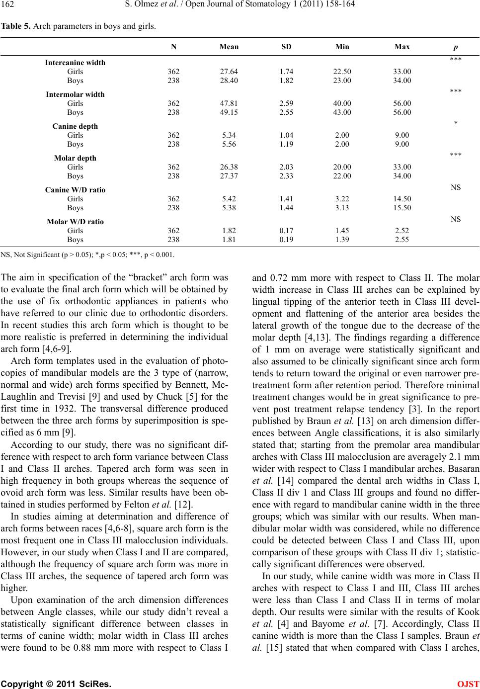
Apart from this, it was shown that the Class II arches
possess the least canine W/D ratio followed by Class I
and Class III. Class III arches have the highest molar
W/D ratio followed by Class I and Class II arches. In the
study of Kook et al. [4] both the canine and the molar
W/D ratio is the least in Class II arches followed by
Class I and Class III. Similar results have been found in
studies report ed by Nojima et al. [6] and Gafni et al. [8].
By examining the arch dimensions with regard to
gender, it was found that the arch dimension is remarka-
bly higher in boys than girls in the permanent dentition.
These findings are in accordance with Bishara [13]. In a
study where especially arch width, depth and chord
measurements were evaluated, Cassidy et al. [16] found
that these values are 3% - 5% higher in boys. In Carter
and McNamara’s study [17] it was stated that the arch
depth decreases in canine, first and second premolar and
first molar teeth area in both genders. In Ward et al.’s
[18] study the resu lts showed no differences in boys and
girls.
In most of the studies, although the values are less in
girls, there is a relationship with the gender and arch
dimension of the samples. In the study done by Raberin
et al. [19] there were significant differences related with
gender only in the transversal dimensions. In present
study even though there are significant differences with
respect to gender and canine/molar width, both of the
measurements are found to be higher in boys. Although
boys possess a wider arch form than girls, there is an
overall agreement that there is no gender variance with
respect to arch form [20,21]. As it can be derived from
our results, no statistically significant variances were
found between gender and arch form.
6. CONCLUSIONS
Due to the lack of studies aimed at dental arch form
variances in Turkey, in our study:
1) It was determined that the most frequently seen
arch form in the Angle malocclusion groups was the
tapered, the least frequent one was the ovoid and the
square one, respectively.
2) Arch widths and depths were found to be more
in boys when compared with girls.
3) No significant differences were found between
gender and arch form variances.
4) In the evaluation of arch dimension measure-
ments with regard to Angle malocclusion groups, An-
gle Class III had the highest values in molar width
and the least values in canine and molar depth meas-
urements.
With this study, it is foreseen that the arch form
should be determined in relation with each patients’ pre-
treatment mandibular dental model and especially in
relation with each patients’ ethnic group in order to
achieve an esthetic, functional and stable arch form out-
come.
REFERENCES
[1] White, L.W. (1978) Individualized ideal arches. Journal
of Clinical Orthodontics, 12, 779-787.
[2] Engel, G.A. (1979) Preformed arch wires: Reliability of
fit. American Journal of Orthodontics, 76, 497-504.
doi:10.1016/0002-9416(79)90254-9
[3] De La Cruz, A.R., Sampson, P., Little, R.M., Årtun, J.
and Shapiro, P.A. (1995) Long-term change in arch form
after orthodontic treatment and retention. American Jour-
nal of Orthodontics and Dentofacial Orthopedics, 107,
518-530. doi:10.1016/S0889-5406(95)70119-2
[4] Kook, Y.A., Nojima, K., Moon, H.B., McLaughlin, R.P.
and Sinclair, P.M. (2004) Comparison of arch forms be-
tween Korean and North American white populations.
American Journal of Orthodontics and Dentofacial Or-
thopedics, 126, 68 0- 68 6.
doi:10.1016/j.ajodo.2003.10.038
[5] Chuck, G.C. (1932) Ideal arch form. Angle Orthodontist,
4, 312-327.
[6] Nojima, K., McLaughlin, R.P., Isshiki, Y. and Sinclair,
P.M. (2001) A comparative study of Caucasion and Japa-
nese mandibular clinical arch forms. Angle Orthodontist,
71, 195-200.
[7] Bayome, M., Sameshima, G.T., Nojima, K., Baek, S.H.
and Kook, Y.A. (2011) Comparison of arch forms be-
tween Egyptian and North American white populations.
American Journal of Orthodontics and Dentofacial Or-
thopedics, 139, e245-e252.
doi:10.1016/j.ajodo.2009.11.012
[8] Gafni, Y., Tzur-Gadassi, L., Nojima, K., McLaughlin,
R.P., Abed, Y. and Redlich, M. (2011) Comparison of
arch forms between Israeli and North American white
populations. American Journal of Orthodontics and
Dentofacial Orthopedics, 139, 339-344.
doi:10.1016/j.ajodo.2009.03.047
[9] McLaughlin, R.P., Bennett, J.C. and Trevisi, H.J. (2001)
Systemized orthodontic treatment mechanics. Mosby,
Edinburgh.
[10] Dahlberg, G. (1949) Statitistic methods for medical and
biological students. Interscience Publication, New York.
[11] Bishara, S.E., Jakobsen, J.R., Treder, J. and Nowak, A.
(1997) Arch width changes from 6 weeks to 45 years of
age. American Journal of Orthodontics and Dentofacial
Orthopedics, 111, 401-409.
C
opyright © 2011 SciRes. OJST
S. Olmez et al. / Open Journal of Stomatology 1 (2011) 158-164
Copyright © 2011 SciRes.
164
OJST
doi:10.1016/S0889-5406(97)80022-4
[12] Felton, J.M., Sinclair, P.M., Jones, D.L. and Alexander,
R.G. (1987) A computerized analysis of the shape and
stability of mandibular arch form. American Journal of
Orthodontics and Dentofacial Orthopedics, 92, 478-483.
doi:10.1016/0889-5406(87)90229-0
[13] Braun, S., Hnat, W.P., Leschinsky, R. and Legan, H.L.
(1999) An evaluation of the shape of some popular nickel
titanium alloy preformed arch wires. American Journal
of Orthodontics and Dentofacial Orthopedics, 11, 6-12.
[14] Basaran, G., Hamamci, N. and Hamamci, O. (2008) Com -
parison of dental arch widths in different types of maloc-
clusions. Journal of Orthodontics, 9, 20-28.
[15] Braun, S., Hnat, W.P., Fender, D.E. and Legan, H.L.
(1998) The form of the human dental arch. Angle Ortho-
dontist, 68, 29-36.
[16] Cassidy, K.M., Harris, E.F., Tolley, E.A. and Keim, R.G.
(1998) Genetic influence on dental arch form in ortho-
dontic patients. Angle Orthodontist, 68, 445-454.
[17] Carter, G.A. and McNamara, J.A. (1998) Longitudinal
dental arch changes in adults. American Journal of Or-
thodontics and Dentofacial Orthopedics, 114, 88-99.
doi:10.1016/S0889-5406(98)70243-4
[18] Ward, D.E., Workman, J., Brown, R. and Richmond, S.
(2006) A 20-year longitudinal study of orthodontic treat-
ment. Angle Orthodontist, 76, 6-13.
[19] Raberin, M., Laumon, B., Martin, J. and Brunner, F.
(1993) Dimensions and form of dental arches in subjects
with normal occlusions. American Journal of Orthodon-
tics and Dentofacial Orthopedics, 104, 67-72.
doi:10.1016/0889-5406(93)70029-N
[20] Collins, B.P. and Harris, E.F. (1998) Arch form in Ameri-
can blacks and whites with malocclusions. Journal of the
Tennessee Dental Association, 78, 15-18.
[21] Ferrario, V.F., Sforza, C., Miani, A.Jr. and Taraglia, G.
(1994) Mathematical definition of the shape of dental
arches in human permanent healthy dentitions. European
Journal of Orthodontics, 16, 287-294.