Open Journal of Stomatology, 2011, 1, 172-178
doi:10.4236/ojst.2011.14026 Published Online December 2011 (http://www.SciRP.org/journal/ojst/ OJST
).
Published Online December 2011 in SciRes. http://www.scirp.org/journal/OJST
Clinical and radiographic evaluation of surgical treatment of
zygomatic fractures using 1.5 mm miniplates system
Paulo Norberto Hasse, Walter Cristiano Gealh*, Cassiano Costa Silva Pereira,
Luiz Francisco Coradazzi, Osvaldo Magro Filho, Idelmo Rangel Garcia Junior
Maxillofacial Surgery Department, Araçatuba Dentistry School, Universidade Estadual Paulista, São Paulo, Brazil.
Email: *waltergealh@gmail.com
Received 10 July 2011; revised 25 August 2011; accepted 5 September 2011.
ABSTRACT
Fractures of the zygomaticomaxillary complex are am-
ong the most common face traumas. Based upon the
complexity and great variety of reported diagnoses
and treatments, the proposal of this study wa s to eva-
luate, clinically and radiographically, unilateral zy-
gomatic fractures treated through internal rigid fixa-
tion with miniplates and screws of 1.5 mm. Material
and Method: 15 patients with unilateral fractures of
the zygomaticoma xillary complex were analyzed, and
compared with 15 patients without fractures so that a
comparative analysis of the area and the perimeter of
the orbital cavities could be made, as well as the dis-
tance from the nasal point to the zygomatic promi-
nence between both groups. Results: In the radiogra-
phic analysis, the both groups presented similarity in
the perimeter and in the area of the orbital cavities.
Concerning the distance from the nasal point to the
zygomatic prominence, only the operated group show-
ed a significant difference between the sides, even
though clinically the observation of the asymmetry
had been absent or discreet. Conclusions: The treat-
ment of unilateral fractures of the zygomaticomaxil-
lary complex with the use of plates and screws of the
1.5 mm system proved to be effective, showing good
esthetic results and low complication rates.
Keywords: Zygomatic Fractures; Internal Fracture Fixa-
tion; Facial Injuries
1. INTRODUCTION
The treatment of the fractures of the zygomaticomaxil-
lary complex (ZMC) is amply discussed in the literature,
being that three great conflicts still persist in relation to
the applied therapy [1]. The first concerns the best way
for surgical reduction of the fractures. The second is
related to the necessity to fix them or not after the reduc-
tion, and the third concerns the number of fixation points
necessary so that the fractures of the ZMC are stabilized
[2].
Treating with mi nipl at es and screws provi ded faci lity of
use, good stability, bio-compatibility, as well as the pos-
sibility of being used in areas where the bone is not so
thick, with little soft tissue overlap, minimizing the sym-
ptoms of local esthetic discomfort [3].
Technological development together with surgical ne-
cessity stimulated the deve lopment of extremely delicate
systems of plates and screws with guarantees of pro-
moting adequate resistance to bending and three-dimen-
sional mechanical stability assuring predictability in the
treatment of facial fractures [4-6].
Considering the high rate of fractures in the ZMC and
the disagreements about treatment we proposed to carry
out the present study to evaluating the zygomatic fra-
ctures treated with a 1.5 mm fixation system.
2. MATERIAL AND METHODS
Thirty patients were analyzed clinically and radiogra-
phically. Fifteen had been submitted to surgical treat-
ment of a non-comminuted unilateral fracture of the zygo-
maticomaxillary complex, and made up the Treated Group
(TG) and fifteen peoples without fractures of the ZMC
were called the Con trol Group (CG).
On the fifteen patients of the TG access to the fronto-
zygomatic region was carried out through a supraciliary
incision. The second region to be accessed varied ac-
cording to the case, using the infra-orbital region and/or
zygomaticomaxillary pillar. The surgical reduction was
carried out through the use of the Caroll-Girard screw
and the internal fix ation was done in all the patients with
titanium miniplates and screws of the system of 1.5 mm.
Posteroanterior X-rays for the cheeks of the face (Wa-
ter’s X-ray) were carried out in the thirty patients, fifteen
in the TG and fifteen in the CG. After obtaining all radio-
graphs, fifteen being from the operated patients and fif-
teen from the salutary patients without fractures, the scan-
ning of the same were done on a “ScanJet 4c/T” scanner
P. N. Hasse et al. / Open Journal of Stomatology 1 (2011) 172-178 173
(Hewlett-Packard). For all of the radiographs a size of
standard image was established, so that there would not
be differences among them. After obtaining the images,
the analysis was carried out by way of the program Im-
ageLab 2000/2.4—a program of analysis and digital pro-
cessing/computerized image (Figure 1).
Two radiographic points were established in order to
evaluate the symmetry of the zygomatic bone from both
sides. The first point was defined by the intersection of the
nose bones (nasal pyramid) and the nasal septum, being
called the nasal point (NP). The second point, called the
zygomatic point (ZP), was established from the most
sideward point and outside the zygomatic arch.
In this way, the operated side (OpS) was compared to
the opposite side (OS) in the Treated Group, and in the
Control Group, th e right side (RS) and the left side (LS)
was compared, in order to obtain fundamental values for
the statistical study.
The clinical evaluation of the TG was carried out with
the intention of determining clinically the possible con-
sequences resulting from the surgical procedures, as well
as the persistent symptomatology originating from the
trauma. Pointing out that in order to do so, only one ob-
server was used, and he considered the following pa-
rameters for evaluation: ocular movement, dystopia, dip-
lopia, paresthesia, limitation of mouth opening, maloc-
clusion, discomfort, ectropion, entropion, apparent sclera,
enophthalmos, exophthalmos, facial symmetry and the
presence of post operative infection. The fifteen patients
from the TG were observed in frontal norm, mento-oc-
cipital, profile, and supero-inferior.
The data obtained was submitted to the test of vari-
ance for analysis of normality by the Kolmogorov-Smir-
nov test and presented normal distribution, which al-
lowed for the carrying out of the parametric t-student
test for matched data (p < 0.05).
3. RESULTS
In the analysis of the Treated Group (TG), the patients’
ages varied from 21 to 64 years (mean = 33 years). Of
these, 87% were men and 13% were women.
In relation to the side of the face that had a higher in-
cidence of fractures of the zygomatic bone, 67% were on
the left side and 33% on the right side. The postoperative
Figure 1. Standard scanning posteroanterior X-rays on the program ImageLab 2000 / 2.4.
C
opyright © 2011 SciRes. OJST
P. N. Hasse et al. / Open Journal of Stomatology 1 (2011) 172-178
174
periods were 6 months at the minimum and 16 months at
the maximum. Among the etiological factors, automobile
accidents prevailed (33%), followed by motorcycle acci-
dents (26.6%) and physical aggression (20%). Automo-
bile accidents with pedestrians, falls and work accidents
were responsible for one of each being responsible for
6.6% in each group.
In order to analyze and compare the Treated Group
with the Control Group we selected individuals of the
same sex and age as the patients with the ZMC fracture.
In the analysis of the Control Group the ages varied from
16 to 62 (mean = 37.3 years), also with 80% of the pa-
tients in this group being men and the women being only
20%.
The surgical procedures of fixation with mini-plates
and screws of 1.5 mm system in the patients that made
up the Treated Group included different pillars of sup-
port. The zygomaticomaxillary pillar was the main choi-
ce, used in 14 patients (93.3%), followed by the frontal-
zygomatic region in 13 patients (86.6%) and the infra-
orbital border involved in 5 (33.3%) operated patients.
The internal fixations, depending on the sustaining pil-
lars, presented combinations among themselves. The to-
tal of 11 patients (73.3%) received fixation in two osse-
ous pillars, 3 patients (20%) in three pillars and only 1
patient (6.6%) in one sustaining pillar.
The numbers obtained from the perimeter, area and
distance from the NP to the ZP in the Treated Group and
Control Gro up are c ont ai ned in Table 1.
Through the Image lab 2000 Program, it was possible
to enhance the osseous outlines in order to facilitate the
demarcations of the NP and the ZP (Figures 2 and 3).
4. STATISTICAL ANALYSIS
4.1. Control Group
The analysis of the matched data of the perimeter be-
tween the right side and the left side did not present any
significant difference (p = 0.782), with measurements of
62.32 mm for the right side and 62.43 mm for the left
side.
For the area, the average of the right side was 263.32
mm and 264.38 for the lef t side, there not being any sig-
nificant statistical difference (p = 0.378).
In measuring the distance from the NP to the ZP, the
average values 42.45 mm for the right side and 42.57
mm for the left side, were also not statistically signify-
cant (p > 0.243).
4.2. Treated Gr oup
The analysis of the matched data of the perimeter be-
tween the operated side and the opposite side did not
present any significant difference (p = 0.249), with val-
ues of 62.90 mm for the operated side and 62.15 mm for
the opposite side.
For the area, the average of the operated side was
263.86 mm and 269.19 mm for the opposite side, not
having any statistical difference between them.
From the measuring of the distance from the NP to the
ZP, the average values of 44.50 mm for the right side
and 45.16 mm for the left side presented a statistically
significant difference (p = 0.003).
4.3. Treated Group × the Control Group
Mean and standard deviation from the perimeter, area
and distance from the NP to the ZP between the Control
Group and Treated Group is on Table 2.
Table 1. Perimeter, area and distance from the nasal point (NP) to the zygomatic point (ZP) from the orbital cavities on the operated
side (OpS) and the opposite side (OS) in the Treated Group (TG) and Control Group (CG).
Patient Perimeter OpS (mm)
TG/CG Area OpS (mm)
TG/CG Distance OpS (mm)
TG/CG Perim eter OS (mm)
TG/CG Area OS (mm)
TG/CG Distance OS (mm)
TG/CG
1 63.9/56.3 279.0/219.5 40.26/39.66 61.8/60.6 275.2/217.4 39.64/40.05
2 62.4/63.8 267.0/263.4 43.95/40.84 58.2/62.5 269.2/261.1 43.58/40.74
3 66.4/64.2 304.3/287.5 47.18/42.53 65.0/65.3 301.2/285.4 48.88/42.85
4 73.4/62.3 325.2/267.5 47.98/41.21 70.8/62.4 322.9/266.4 48.15/41.52
5 62.2/59.7 295.0/221.9 44.28/46.44 62.3/59.1 290.6/220.9 46.13/46.97
6 58.6/55.8 230.4/227.0 45.46/40.82 60.3/54.5 234.9/224.2 46.12/40.00
7 63.7/66.5 275.6/297.7 41.35/46.15 63.4/66.8 274.5/304.4 42.49/46.27
8 58.3/64.2 221.3/280.4 43.11/41.79 58.9/65.9 228.9/281.3 44.48/41.89
9 61.5/61.7 235.7/252.2 44.03/42.30 62.1/61.5 246.1/251.7 44.86/42.03
10 73.8/69.8 317.2/321.5 46.17/41.57 72.4/68.5 310.5/322.5 46.82/42.07
11 62.3/61.6 254.6/258.1 45.19/38.82 58.1/60.9 250.2/258.7 45.79/38.91
12 56.0/64.8 205.9/288.0 45.58/41.33 61.2/64.5 210.8/288.4 46.66/41.94
13 62.3/58.5 269.7/237.5 44.84/45.49 61.9/57.6 359.9/252.5 44.97/45.08
14 59.8/61.7 244.4/256.5 42.89/43.05 59.8/62.5 239.0/257.9 43.29/43.12
15 58.9/63.9 232.6/271.1 45.20/44.71 56.1/63.8 224.0/272.9 45.51/45.12
Mean TG/CG 62.4/61.7 263.4/262.9 44.07/41.93 61.7/62.0 268.7/263.8 44.60/42.13
C
opyright © 2011 SciRes. OJST
P. N. Hasse et al. / Open Journal of Stomatology 1 (2011) 172-178 175
Table 2. Mean and standard deviation from the perimeter, area and distance from the nasal point (NP) to the zygomatic point (ZP)
between the Control Group and Treated Group (significant difference for p < 0.05).
Variable Mean standard deviation t p
Perimeter
Control Group 62.37 3.62 0.142 0.888
Treated Group 62.53 4.71
Area
Control Group 263.85 29.02 0.305 0.762
Treated Group 266.53 38.41
Distance NP t o ZP
Control Group 42.51 2.30 4.024 <0.001
Treated Group 44.83 2.16
Figure 2. Enhancing the osseous outlines in order to facilitate the demarcations of the NP, ZP and to calculate the perimeter and area
of the orbitas.
present any significant difference (p = 0.888), with val-
ues of 62.37 mm fo r the control group and 62.5 3 for the
treated group. The analysis of the matched data for the
area between the control group and the treated group did
not present any significant difference (p = 0.762), with
values of 263.8 5 for the con trol grou p and 266.53 for the
treated group.
In measuring the distance from the NP to the ZP the
mean values of 42.51 mm for the control group and
44.83 mm for the treated group presented a significant
statistical difference (p < 0.001).
In the clinical analysis among the patients of the Treat-
ed Group no cases of ectropion, entropion, enophthalmos,
exophthalmos, diplopia, sclera apparent, malocclusion or
C
opyright © 2011 SciRes. OJST
P. N. Hasse et al. / Open Journal of Stomatology 1 (2011) 172-178
176
Figure 3. Line of the distance from the NP to the ZP.
mouth opening limitation were found. There was only
one case of asymmetry that was classified as “discreet”,
with an appearance of distopia, but without any com-
plaints by the patient. Only one patient reported a dis-
creet paresthesia in the region of the alveolar mucosa
innerved by the superior anterior alveolar nerve associ-
ated with a slight discomfort after nine postoperative
months.
5. DISCUSSION
From the analysis of the collected data, we observed that
traffic accidents (with auto mobiles and motorcyc les) were
responsible for 59.9% of the fractures, and in second
place, patients who were victims of physical aggression
made up 20% of the cases. This corroborated the rates
from Covington et al. [7], who pointed out as main etio-
logical factors, accidents caused by motor vehicles, with
78.6% of the cases, being 68.8% automobiles and 9.8%,
motorcycles.
One of the most controversial top ics in the literature is
about the number of fixation points that are necessary to
avoid the post-surgical dislocation of the fractured zygo-
matic maxillary complex [8-10]. The great majority of
the authors are divided about the need for two or three
places, with a variation of the complexity of the trauma
and the degree of dislocation of the fractured segment [7,
11].
Whenever the primary stabilization of the ZMC frac-
ture was necessary fixation in the region of the zygoma-
tic maxillary pillar (ZM) was chosen, totaling 14 patients
(93.3% of the cases). The frontozygomatic region (FZ)
was fixed in 13 patients (86% of the cases), and the in-
fraorbital rim (IO) in 5 patients (33.3% of the cases).
The combination of the fixation sites followed the fol-
lowing statistics, 9 patients (60%—FZ and ZM), 3 pa-
tients (20%—FZ, ZM and IO), and 1 patient (6.6%) for
each group left over (FZ and IO; IO and ZM; only ZM).
Although there are reports of stability of fractures fixed
only in one point [7,12,13] chose, whenever possible to
put fixation in the two pillars of the ZMC.
The internal fixations carried ou t in the patients in this
study, according to the integrity of the osseous pillars,
presented combinations among them, being that in 11
patients (73.3%) two points of fixation were used, in 3
C
opyright © 2011 SciRes. OJST
P. N. Hasse et al. / Open Journal of Stomatology 1 (2011) 172-178 177
cases (20%), there was the need for osteossynthesis in
three pillars and in only 1 patient (6.6%) one fixation
point was used.
Davidson et al. [14] analyzed, in an in vitro study,
several forms of fixation, combining steel wires, plates
and screws, obtaining a total of 25 different combina-
tions. By means of traction that simulated the action of
the masseter muscles they calculated the rotation and the
dislocation after the application of the forces on the dif-
ferent planes. The means of fixation that received the
best results were: fixation with steel wires in three points
and fixation with plates and screws in three points.
However, the stability reached by fixation with plates
and screws in two points was similar to the previous
fixations, highlighting the zygomatic pillar as the strate-
gic point for the opposition of the forces of th e masseter
muscle.
In relation to the combinations and the choice of os-
seous pillars, the combination between the frontozygo-
matic region and the zygomatic maxillary making up a
total of nine (60%) of the fifteen patients treated.
The zygomatic maxillary pillar should be the region of
choice for unstable fractures of the zygomatic bone, for
it acts as a direct antagonist to the action of traction
provoked by the masseter muscle [15].
Strong and Sykes [16] propose a combination of sys-
tems of mini-plates and micro-plates in the different pil-
lars of support of the ZMC. They recommend the use of
microplates of 1.0 or 1.2 mm in the infra-orbital border,
1.5 or 1.7 mm in the frontozygomatic region and 2.0 mm
in the zygomatic maxillary pillar region.
The data obtained from this study are in agreement
with the results obtained in other studies [17]. The diffe-
rences obtained, for the perimeter as well and for the
area of the operated side and the opposite side of the
Treated Group were not statistically significant.
The good stability obtained from the fixation in two
points gains force when compared with the sides obtain-
ed from the Control Group, in other words, the patients
who did not have fractures of the ZMC. In those we
verified that the differences in the values obtained be-
tween the right side and the left side, in referring to the
perimeter and, as well as the area, were also not statistic-
cally significant.
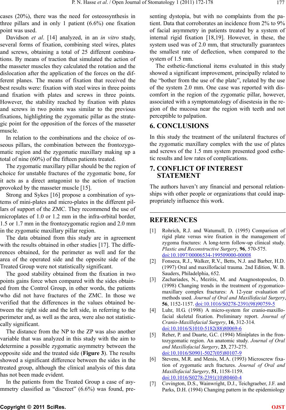
The distance from the NP to the ZP was also another
variable that was analyzed in this study with the aim to
determine a possible zygomatic asymmetry between the
opposite side and the treated side (Figure 3). The results
showed a significant difference between the sides in the
treated group, although the clinical analysis of this data
has not been made evident.
In the patients from the Treated Group a case of asy-
mmetry classified as “discreet” (6.6%) was found, pre-
senting dystopia, but with no complaints from the pa-
tient. Data that corroborates an incidence from 2% to 9%
of facial asymmetry in patients treated by a system of
internal rigid fixation [18,19]. However, in these, the
system used was of 2.0 mm, that structurally guarantees
the smallest rate of deflection, when compared to the
system of 1.5 mm.
The esthetic-functional items evaluated in this study
showed a significant improvement, principally related to
the “bother from the use of the plate”, related by the use
of the system 2.0 mm. One case was reported with dis-
comfort in the region of the zygomatic pillar, however,
associated with a symptomatology of disestesia in the re-
gion of the mucosa near the region with teeth and not
perceptible to palpation.
6. CONCLUSIONS
In this study the treatment of the unilateral fractures of
the zygomatic maxillary complex with the use of plates
and screws of the 1.5 mm system presented good esthe-
tic results and low rates of complications.
7. CONFLICT OF INTEREST
STATEMENT
The authors haven’t any financial and personal relation-
ships with other people or organizations that could inap-
propriately influence this work.
REFERENCES
[1] Rohrich, R.J. and Watumull, D. (1995) Comparison of
rigid plate versus wire fixation in the management of
zygoma fractures: A long-term follow-up clinical study.
Plastic and Reconstructive Surgery, 96, 570-575.
doi:10.1097/00006534-199509000-00008
[2] Fonseca, R.J., Walker, R.V., Betts, N.J. and Barber, H.D.
(1997) Oral and maxillofacial trauma. 2nd Edition, W. B.
Sauders, Philadelphia, 652.
[3] Zachariades, N., Mezitis, M. and Anagnostopoulos, D.
(1998) Changing trends in the treatment of zygomatico-
maxillary complex fractures: A 12-year evaluation of
methods used. Journal of Oral and Maxillofacial Surgery,
56, 1152-1157. doi:10.1016/S0278-2391(98)90759-5
[4] Luhr, H.G. (1998) A micro-system for cranio-maxillo-
facial skeletal fixation. Preliminary report. Journal of
Cranio-Maxillofacial Surgery, 16, 312-314.
doi:10.1016/S1010-5182(88)80069-6
[5] Reher, P. and Duarte, G.C. (1994) Miniplates in the fron-
tozygomatic region. An anatomic study. Journal of Oral
and Maxillofacial Surgery, 23, 273-275.
doi:10.1016/S0901-5027(05)80107-9
[6] Stevens, M.R. and Menis, M.A. (1993) Microscrew fixa-
tion of zygomatic arch fractures. Journal of Oral and
Maxillofacial Surgery, 51, 1158-1159.
doi:10.1016/S0278-2391(10)80460-4
[7] Covington, D.S., Wainwright, D.J., Teichgraeber, J.F. and
Parks, D.H. (1994) Changing pattern in the epidemiology
C
opyright © 2011 SciRes. OJST
P. N. Hasse et al. / Open Journal of Stomatology 1 (2011) 172-178
Copyright © 2011 SciRes.
178
OJST
and treatment of zygoma fractures: 10 year review. Jour-
nal of Trauma, 37, 243-248.
doi:10.1097/00005373-199408000-00016
[8] Dal, S.F, Ellis, E.III and Throckmorton, G.S. (1992) The
effects of zygomatic complex fracture on masseteric
muscle force. Journal of Oral and Maxillofacial Surgery,
50, 791-799. doi:10.1016/0278-2391(92)90267-4
[9] Ellis, E.III and Kittidumkerng, W. (1996) Analysis of
treatment for isolated zygomatico-maxillary complex fra-
ctures. Journal of Oral and Maxillofacial Surgery, 54,
386-400. doi:10.1016/S0278-2391(96)90107-X
[10] Rinehart, G.C., Marsh, J.L., Hemmer, K.M. and Bresina,
S. (1989) Internal fixation of malar fractures: An experi-
mental biophysical study. Plastic and Reconstructive
Surgery, 84, 21-28.
doi:10.1097/00006534-198907000-00003
[11] Karlan, M.S. and Cassisi, N.J. (1979) Fractures of the
zygoma. A geometric, biomechanical, and surgical analy-
sis. Archives of Otolaryngology, 105, 320-327.
doi:10.1001/archotol.1979.00790180018004
[12] Eisele, D.W. and Duckert, L.G. (1987) Single-point stabi-
lization of zygomatic fractures with the minicompression
plate. Archives of Otolaryngology—Head & Neck Sur-
gery, 113, 267-270.
doi:10.1001/archotol.1987.01860030043005
[13] Tarabichi, M. (1994) Transsinus reduction and one-point
fixation of malar fractures. Archives of Otolaryngology—
Head & Neck Surgery, 120, 620-625.
doi:10.1001/archotol.1994.01880300036005
[14] Davidson, J., Nickerson, D. and Nickerson, B. (1991)
Zygomatic fractures: Comparison of methods of internal
fixation. Plastic and Reconstructive Surgery, 87, 585-
587.
[15] Gruss, J.S. and Mackinnon, S.E. (1986) Complex maxil-
lary fractures: Role of buttress reconstruction and imme-
diate bone grafts. Plastic and Reconstructive Surgery, 78,
9-22. doi:10.1097/00006534-198607000-00002
[16] Strong, E.B. and Sykes, J.M. (1990) Zygoma complex
fractures. Facial Plastic Surgery, 14, 105-115.
doi:10.1055/s-0028-1085306
[17] Ellis, E.III, El-Attar, A. and Moos, K.F. (1985) An analy-
sis of 2067 cases of zygomatico-orbital fracture. Journal
of Oral and Maxillofacial Surgery, 43, 417-428.
doi:10.1016/S0278-2391(85)80049-5
[18] Sands, T., Symington, O., Katsikeris, N. and Brown, A.
(1993) Fractures of the zygomatic complex: A case report
and review. Journal of the Canadian Dental Association,
59, 749-757.
[19] Schortinghuis, J., Bos, R.R. and Vissink, A. (1999) Com-
plications of internal fixation of maxillofacial fractures
with microplates. Journal of Oral and Maxillofacial Surgery,
57, 130-135. doi:10.1016/S0278-2391(99)90224-0