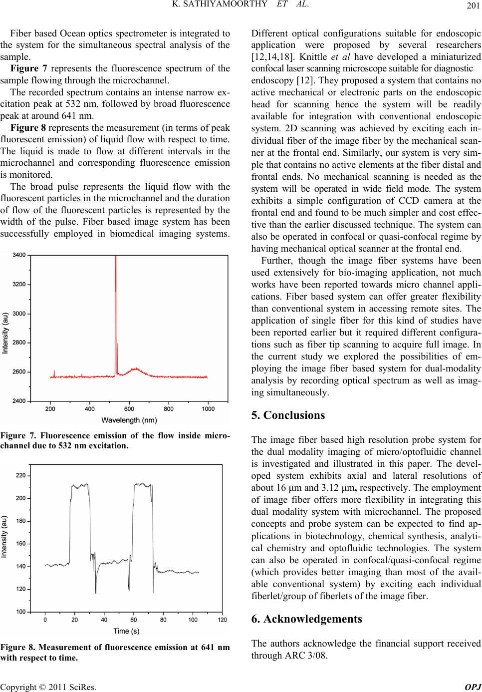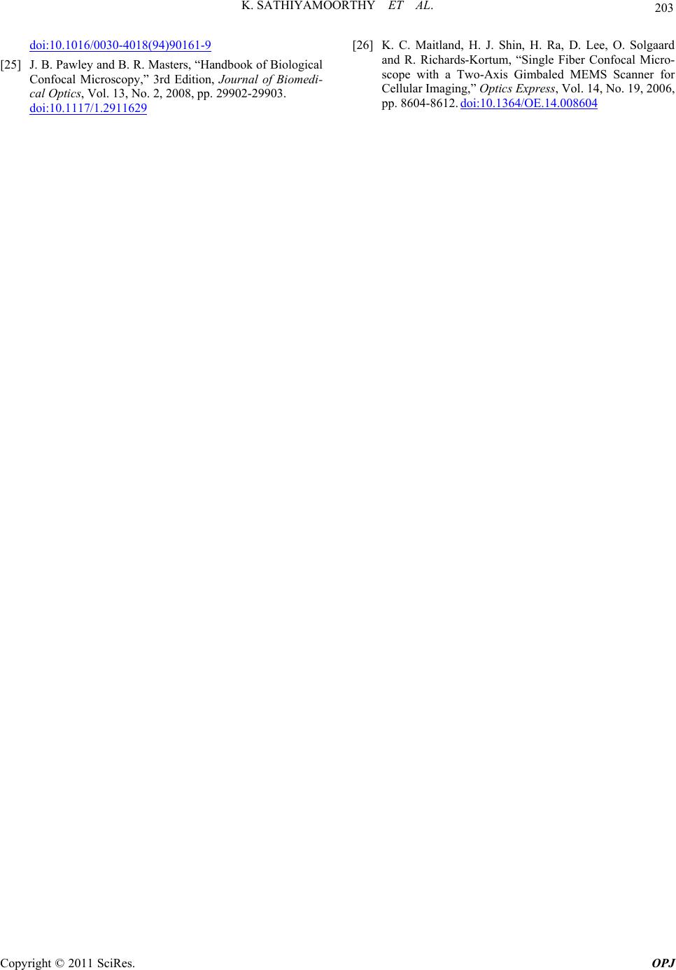
K. SATHIYAMOORTHY ET AL.
202
7. References
[1] A. Ashkin, “Optical Trapping and Manipulation of Neu-
tral Particles Using Lasers,” Proceedings of the National
Academy of Sciences of the United States of America, Vol.
94, No. 10, 1997, pp. 4853-4860.
doi:10.1073/pnas.94.10.4853
[2] P. Domachuk, M. Cronin-Golomb, B. Eggleton, S. Mut-
zenich, G. Rosengarten and A. Mitchell, “Application of
optical trapping to beam manipulation in optofluidics,”
Optics Express, Vol. 13, No. 19, 2005, pp. 7265-7275.
doi:10.1364/OPEX.13.007265
[3] D. Hoffmann, J. O'Brien, D. Brennan and M. Loughran,
“Optically Encoded Silicon Microbeads: Detection and
Characterisation in a Microfluidic System,” Sensors and
Actuators B: Chemical, Vol. 122, No. 2, 2007, pp. 653-
658.
[4] H. Kinoshita, M. Oshima, S. Kaneda and T. Fujii, “Vali-
dation of Confocal Micro-PIV Technique by Poiseuille
Flow Measurement,” International Conference on Micro-
technologies in Medicine and Biology, 9-12 May 2006,
pp. 78-80.
[5] S. Kuhn, B. S. Phillips, E. J. Lunt, A. R. Hawkins and H.
Schmidt, “Ultralow Power Trapping and Fluorescence
Detection of Single Particles on an Optofluidic Chip,”
Lab on a Chip, Vol. 10, No. 2, 2010, pp. 189-194.
doi:10.1039/b915750f
[6] S.-K. Lee, S.-H. Kim, J.-H. Kang, S.-G. Park, W.-J. Jung,
S.-H. Kim, G.-R. Yi and S.-M. Yang, “Optofluidics
Technology Based on Colloids and Their Assemblies,”
Microfluidics and Nanofluidics, Vol. 4, No. 1, 2008, pp.
129-144. doi:10.1007/s10404-007-0218-8
[7] R. Lima, S. Wada, S. Tanaka, M. Takeda, T. Ishikawa,
K.-I. Tsubota, Y. Imai and T. Yamaguchi, “In Vitro
Blood Flow in a Rectangular PDMS Microchannel: Ex-
perimental Observations Using a Confocal Micro-PIV
system,” Biomedical Microdevices, Vol. 10, No. 2, 2008,
pp. 153-167. doi:10.1007/s10544-007-9121-z
[8] C. Monat, P. Domachuk and B. J. Eggleton, “Integrated
Optofluidics: A New River of Light,” Nature Photonics,
Vol. 1, No. 2, 2007, pp. 106-114.
doi:10.1038/nphoton.2006.96
[9] J.-C. Roulet, R. Völkel, H. P. Herzig, E. Verpoorte, N. F.
de Rooij and R. Dändliker, “Performance of an Integrated
Microoptical System for Fluorescence Detection in Mi-
crofluidic Systems,” Analytical Chemistry, Vol. 74, No.
14, 2002, pp. 3400-3407. doi:10.1021/ac0112465
[10] K. Carlson, M. Chidley, K.-B. Sung, M. Descour, A.
Gillenwater, M. Follen and R. Richards-Kortum, “In Vivo
Fiber-Optic Confocal Reflectance Microscope with an
Injection-Molded Plastic Miniature Objective Lens,” Ap-
plied Optics, Vol. 44, No. 10, 2005, pp. 1792-1797.
doi:10.1364/AO.44.001792
[11] T. Dabbs and M. Glass, “Fiber-Optic Confocal Micro-
scope: FOCON,” Applied Optics, Vol. 31, No. 16, 1992,
pp. 3030-3035. doi:10.1364/AO.31.003030
[12] J. Knittel, L. Schnieder, G. Buess, B. Messerschmidt and
T. Possner, “Endoscope-Compatible Confocal Micro-
scope Using a Gradient Index-Lens System,” Optics
Communications, Vol. 188, No. 5-6, 2001, pp. 267-273.
doi:10.1016/S0030-4018(00)01164-0
[13] A. Osdoit, M. Genet, A. Perchant, S. Loiseau, B. Abrat
and F. Lacombe, “In Vivo Fibered Confocal Reflectance
Imaging: Totally Non-Invasive Morphological Cellular
Imaging Brought to the Endoscopist,” In: G. J. Tearney
and T. D. Wang, Eds., SPIE, San Jose, 2006, pp. 608208-
608210. doi:10.1117/12.646659
[14] K.B. Sung, C. Liang, M. Descour, T. Collier, M. Follen,
A. Malpica and R. Richards-Kortum, “Near Real Time in
Vivo Fibre Optic Confocal Microscopy: Sub-Cellular
Structure Resolved,” Journal of Microscopy, Vol. 207,
No. 2, 2002, pp. 137-145.
doi:10.1046/j.1365-2818.2002.01049.x
[15] B. A. Flusberg, E. D. Cocker, W. Piyawattanametha, J. C.
Jung, E. L. M. Cheung and M. J. Schnitzer, “Fiber-Optic
Fluorescence Imaging,” Nature Methods, Vol. 2, No. 12,
2005, pp. 941-950. doi:10.1038/nmeth820
[16] V. M. Murukeshan and N. Sujatha, “All Fiber Based
Multispeckle Modality Endoscopic System for Imaging
Medical Cavities,” Review of Scientific Instruments, Vol.
78, No. 5, 2007, pp. 53106-53108.
doi:10.1063/1.2737772
[17] “Bibliography on Confocal Microscopy and Its Applica-
tions,” Journal of Scanning Microscopies: Scanning, Vol.
16, No. 1, 1994, pp. 33-56.
[18] T. Wilson, R. Juskaitis, N. P. Rea and D. K. Hamilton,
“Fibre Optic Interference and Confocal Microscopy,” Op-
tics Communications, Vol. 110, No. 1-2, 1994, pp. 1-6.
doi:10.1016/0030-4018(94)90161-9
[19] M. Gu and D. Bird, “Fibre-Optic Double-Pass Confocal
Microscopy,” Optics & Laser Technology, Vol. 30, No. 2,
1998, pp. 91-93. doi:10.1016/S0030-3992(98)00014-0
[20] V. M. Murukeshan, N. Sujatha, L. S. Ong, A. Singh and
L. K. Seah, “Effect of Image Fiber on the Speckle Fringe
Pattern in Image Fiber-Guided DSPI Endoscopy,” Optics
& Laser Technology, Vol. 39, No. 3, 2007, pp. 527-531.
doi:10.1016/j.optlastec.2005.11.001
[21] N. Sujatha, V.M. Murukeshan, L. S. Ong and L. K. Seah,
“An All Fiber Optic System Modeling for the Gastroin-
testinal Endoscopy: Design Concepts and Fluorescent
Analysis,” Optics Communications, Vol. 219, No. 1-6,
2003, pp. 71-79.
[22] K. Sathiyamoorthy, V. K. Mohankumar and M. V.
Matham, “Variable Focal Lengths Image Fiber Based
Microscope System for Ciomedical Application,” Photo-
nics Global Conference, Singapore, 14-16 Decenmber
2010, pp. 1-3.
[23] Y. Li, T. Wang, H. Kosaka, S. Kawai and K. Kasahara,
“Fiber-Image-Guide-Based Bit-Parallel Optical Inter-
connects,” Applied Optics, Vol. 35, No. 35, 1996, pp.
6920-6933. doi:10.1364/AO.35.006920
[24] T. Wilson, R. Juškaitis, N. P. Rea and D. K. Hamilton,
“Fibre Optic Interference and Confocal Microscopy,”
Optics Communications, Vol. 110, No. 1-2, 1994, pp. 1-6.
Copyright © 2011 SciRes. OPJ