Vol.2, No.4, 406-412 (2011)
doi:10.4236/as.2011.24052
C
opyright © 2011 SciRes. Openly accessible at http://www.scirp.org/journal/AS/
Agricultural Scienc es
Molecular cloning of a phosphotriesterase-related
protein gene of silkworm and its expression analysis
in the silkworm infected with Bombyx mori cytoplasmic
polyhedrosis virus
Xiu Wang1, Kun Gao2,3, Ping Wu2,3, Guangxing Qin2,3, Ting Liu2,3, Xijie Guo2,3*
1College of Biotechnology and Chemical Engineering, Jiangsu University of Science and Technology, Zhenjiang, China;
2Sericultural Research Institute, Jiangsu University of Science and Technology, Zhenjiang, China;
3Sericultural Research Institute, Chinese Academy of Agricultural Sciences, Zhenjiang, China; *Corresponding Author: guoxijie@126.com
Received 9 September 2011; revised 20 October 2011; accepted 30 October 2011.
ABSTRACT
Bombyx mori cytoplasmic polyhedrosis virus is
one of the major viral p athogens for the silkworm.
The immune response of silkworm to the virus
infection is obscure. A phosphotriesterase-re-
lated protein gene of silkworm, Bombyx mori
(BmPTERP) was found in our previous mi-
croarry analysis of the midgut infected with the
virus. In the present study, we cloned and ana-
lyzed the full-length cDNA of BmPTERP gene by
means of rapid amplification of complementary
DNA ends (RACE) and bioinformatic analy sis for
exploring its functions in interaction between
the silkworm and the virus. The nucleotide se-
quence of the gene is 1349-bp and contains a
131 bp 5’UTR and a 165 bp 3’UTR. The 1053 bp
open reading frame encodes a 350 amino acid
protein. The deduced protein contains specific
hits of phosphotriesterase-related proteins and
belongs to the amidohydrolase superfamily. RT-
PCR analysis revealed that BmPTERP gene was
expressed in all the tissues tested, including
midgut, hemocyte, gonad, fat body and silk gland.
Real-time quantitative polymerase chain reac-
tion analysis indicated that the relative tran-
script of BmPTERP gene in the infected midgut
was 19.32 fold lower than that in normal midgut
at 72 hours post inoculation.
Keywords: Silkworm; Cytoplasmic Polyhedrosis
Virus; Phosphotriesterase-Related Protein; Gene
1. INTRODUCTION
Phosphotriesterase-related protein (also called phos-
photriesterase homology protein, PHP) is a member of
amidohydrolase superfamily and exhibits higher sequence
identity, and high sequence similarity to phosphotries-
terase (PTE) [1]. PTE is a group of bacterial enzyme that
catalyzes the hydrolysis of a wide range of organophos-
phate triesters including organophosphate insecticides
and chemical nerve agents [2]. PTE exists as a homodimer
with one active site per monomer. The active site is lo-
cated next to a binuclear metal center, at the C-terminal
end of a TIM alpha-beta barrel motif and contains tow
zinc ions in native enzyme. However, these ions can be
replaced with other metals such as cobalt, cadmium,
nickel or manganese and the enzyme still remains active.
The hydrolysis reaction of PTE is sufficient to utilize the
nucleophilicity of the bridging hydroxide according to
theoretical study of the phosphotriesterase reaction mecha-
nism [2].
The PTE has attracted much interest in recent years
because of their potential ability in the decontamination
of hazardous organophosphate compounds [3]. The most
efficient PTEs have been identified from several micro-
bial species, such as Pseudomonas diminuta, Sulfolobus
solfataricus and Flavobacterium [4]. The directed evolu-
tion research of PTE resulted in improvement in func-
tional expression and enzymatic activity [5,6]. Data col-
lected showed that PTE was evolved from the family of
phosphotriesterase-related proteins [7-9]. The research
on its structure and expression would elucidate the evo-
lutionary story of PTEs. To date, the phosphotriesterase-
related proteins have been isolated from E. coil and
some other organisms and grouped into a single family
[7,9]. Although considerably homologous in sequence
and in the structure of the bimetal catalytic site, phos-
photriesterase-related proteins differs from PTE and ex-
hibits no phosphotriesterase activity [1,7]. But, subse-
quent studies reported a weak esterase activity and PTE
activity in an E. coli PHP mutant [1].
X. Wang et al. / Agricultural Sciences 2 (2011) 406-412
Copyright © 2011 SciRes. Openly accessible at http://www.scirp.org/journal/AS/
407407
Bombyx mori cytoplasmic polyhedrosis virus (Bm-
CPV), which belongs to the genus Cypovirus in the fam-
ily Reoviridae, is a significant virus to the silkworm,
often causing severe economic damages to the sericul-
tural industry [10,11]. Unfortunately, to date, the im-
mune mechanism of silkworm to CPV infection remains
obscure. In our previous studies, a differentially expressed
gene similar to the genes of phosphotriesterase-related
proteins was identified in the CPV-infected silkworm by
using microarry analysis [12]. In the present study, we
first cloned the gene of phosphotriesterase-related pro-
tein from silkworm, Bombyx mori (BmPTERP) by means
of RACE method. Furthermore, the expression patterns
of BmPTERP in midgut and some other tissues after
infection with BmCPV were analyzed by quantitative
real-time PCR. The results provided useful information
for further study of the immune mechanism of silkworm
to virus infection.
2. MATERIALS AND METHODS
2.1. Silkworm Strain
The silkworm strain p50 was provided by the National
Silkworm Genetic Resource Preservation Center of
Chinese Academy of Agricultural Science. The larvae of
silkworm were reared with mulberry leaves at standard
temperature of 25˚C and under a photoperiod of 12 h
light and 12 h dark up to the fourth molting for virus
inoculation.
2.2. Virus Inoculation
BmCPV was suspended in disinfected distilled water
to a concentration of 108 polyhedra per mL. One mL
viral suspension was spread totally on 10 pieces of mul-
berry leaves which were nearly 15 cm2 each. 25 newly
molted fifth instar larvae were fed on it. The control lar-
vae were treated with the same amount of mulberry
leaves spread with distilled water. The infection does
was calculated as 4 × 10⁵ polyhedra per larva.
2.3. Collection of Midgut and Other Tissues
Midgut of BmCPV-infected and control larvae were
collected at 24, 48 and 72 h post-inoculation by dissect-
ing the larvae on ice. The midgut were quickly washed
in diethylpyrocarbonate (DEPC)-treated water to remove
the attached leaf pieces and then immediately frozen in
liquid nitrogen. At the same time, other tissues namely
gonad, silk gland, hemocyte and fat body were also col-
lected at 48 h post inoculation and frozen in liquid ni-
trogen before being stored at –80˚C. The same tissues of
five larvae for midgut and of ten larvae for gonad, silk
gland, hemocyte and fat body at each time point were
mixed for RNA extraction and following experiments.
2.4. Isolation of Total RNA
Total RNA was extracted respectively from midgut,
gonad, silk gland, hemocyte and fat body collected at
different time point post inoculation by using Trizol re-
agent (Invitrogen, Carlsbad, CA, US) and subjected to
DNase I treatment according to the manufacturer’s pro-
tocol. The concentration of total RNA was determined
by using a Biophotometer (Eppendorf, Hamburg, Ger-
many) to measure the absorbance at 260 nm and 280 nm.
RNAs with the A260:A280 value of 1.9 to 2.0 were
stored at –80˚C and used for further study.
2.5. Rapid Amplification of cDNA Ends
(RACE)
Full-length cDNA of BmPTERP gene was synthesized
using 2 μg total RNA of midgut as a template with
SMARTTM RACE cDNA Amplification Kit (Clontech).
Specific primers for 5’RACE and 3’RACE were de-
signed based on BmPTERP gene cDNA fragment re-
vealed in our previous study. The primer for 5’RACE
was 5’-TTACAACTGCCTCCTTCGCGGCCTGA-3’ and
that for 3’RACE was 5’-CGGCTGCGGTGTCAGTTT-
CCATCCTC-3’. 5’-RACE was performed in a reaction
system of 25 μL containing 17.25 μL PCR-Grade water,
2.5 μL 10 × Advantage 2 PCR buffer, 0.5 μL dNTP Mix
(10 μM), 0.5 μL 50 × Advantage 2 Polymerase Mix, 1.25
μL 5’-RACE-Ready cDNA, 2.5 μL UPM (10×), 0.5 μL
5’-specific primer (10 μM) and as the following proce-
dures: 32 cycles of 95˚C for 30 s, 68˚C for 3 min. 3’
RACE was performed in a reaction system of 25 μL
containing 17.25 μL PCR-Grade water, 2.5 μL 10 × Ad-
vantage 2 PCR buffer, 0.5 μL dNTP Mix (10 μM), 0.5
μL 50 × Advantage 2 Polymerase Mix, 1.25 μL 5’-
RACE-Ready cDNA, 2.5 μL UPM (10×), 0.5 μL 3’-
specific primer (10 μM) with the same procedures. The
PCR products were examined by electrophoresis in 1%
agarose gel and the fragment sizes were determined rela-
tive to marker DNA. The appropriate band was purified,
cloned into pGEM-T Easy vector and sequenced by
Sangom Biotech Co. Ltd. (Shanghai).
2.6. Sequence Anal ysis, Multiple Sequence
Alignment and Phylogenetic Analysis
The sequences were searched in GenBank with
BLASTx for comparative analysis and assembled with
the obtained fragments. The sequences were analyzed
using the BLAST algorithm at NCBI (http://www.ncbi.
nlm.gov/blastn). The gene structure was predicted with
Gene Structure Display Server (GSDS, http://gsds.cbi.
pku.edu.cn). The deduced amino acid sequence of Bm-
X. Wang et al. / Agricultural Sciences 2 (2011) 406-412
Copyright © 2011 SciRes. Openly accessible at http://www.scirp.org/journal/AS/
408
PTERP was analyzed with Protparam software (www.
expasy.ch/tools/protparam.html) and ProtScale software
(http://www.expasy.org/tools/protscale.html). The signal
peptide was predicted with the SignalP 3.0 (http://www.
cbs.dtu.dk/services/SignalP/). Multiple protein sequences
were aligned using the MegAlign program by CLUSTAL
W method in DNASTAR software package. A phyloge-
netic tree was constructed by MEGA 4.0 software.
2.7. RT-PCR
Total RNA extracted as described above from differ-
ent tissues of fifth instar silkworm, including hemocyte,
silk gland, fat body, gonad and midgut were used as
template to synthesize cDNA using the Prime ScriptTM
RT Reagent Kit (TaKaRa). The PCR reaction was per-
formed as the procedures with denaturation for 2 min at
94˚C, 30 cycles of 30 s at 94˚C, 30 s at 54˚C and 10 min
at 72˚C.
2.8. Quantitative Real-Time PCR of
BmPTERP Gene
Total RNAs extracted respectively from the midgut of
BmCPV-infected and control larvae at 24, 48 and 72 h
were adjusted with diethylpyrocarbonate (DEPC) H2O to
a concentration of 500 ng/μL. 1000 ng of each of the
total RNAs was reverse transcribed in 20 μL of reaction
system containing 4 μL 5 × PrimeScript buffer (for Real
time), 1 μL PrimerScript RT Enzyme Mix I, 1 μL Oligo
dT Primer (50 μM), 1 μL Random 6 mers (100 μM), 2
μL Total RNA, 11 μL RNase Free dH2O using the Prime
ScriptTM RT Reagent Kit (TaKaRa). Quantitative real-
time PCR was performed using 1 μL of diluted first-
strand cDNA (1/10) in each 25 μL reaction volume ac-
cording to the manufacturer’s instructions for the SYBR
Premix Ex TaqTM (TaKaRa). Specific primers for Bm-
PTERP gene and β-actin were designed by Primer Pre-
mier 5.0 software (Primer Premier, Palo Alto, CA, US).
For BmPTERP gene, the forward primer was 5’-ATTTA-
GACCGAACCCTACTTG-3’ and the reverse primer was
5’-TCGTGGGACATTAACACTTT-3’ and for β-actin,
they were 5’-AATGGCTCCGGTATGTGC-3’ and 5’-
TTGCTCTGTGCCTCGTCT-3’ respectively. Reactions
were run in triplicate for the same pooled samples on an
Opticon lightcycler (BioRad, Hercules, CA, US) using
the following thermal cycling parameters: 95˚C for 10 s,
40 cycles of 95˚C for 5 s, 60˚C for 20 s, 72˚C for 5 s.
Following amplification, melting curves were constructed.
Data were analyzed and normalized relative to β-actin
transcript levels by Opticon Monitor Analysis software
(MJ Research, Waltham, MA, US). The relative quanti-
tative method was used to evaluate the differential ex-
pression of BmPTERP gene [13]. Ct for amplified target
product of BmPTERP gene and internal control β-actin
was determined for each sample to normalize the differ-
ences in the amount of template and the efficiency of
RT-PCR (ΔCt = Cttarget Ctβ-actin). The RNA of the normal
silkworm larvae was used as calibrator and the ΔCt for
each sample was subtracted from the ΔCt of the calibra-
tor to calculate the difference ΔΔCt. 2−ΔΔCt was used to
calculate the relative expression level of BmPTERP gene.
Relative expression level is expressed as average ± SE.
For other tissues, the quantitative real-time PCR of
BmPTERP gene were performed with the same proce-
dure described above but using the total RNA from a
specific tissue as template.
3. RESULTS
3.1. Occurrence of Infection
Inoculation of BmCPV with the concentration of 4 ×
105 polyhedra per larva to the fifth instar silkworm
caused infection and disease to all the larvae. It was con-
firmed by the appearance of white wrinkles on the mid-
gut as typical symptoms at approximately 72 h post-
inoculation and the observation of polyhedra under a
microscope.
3.2. cDNA Cloning and Sequence Analysis
of BmPTERP Gene
The full length cDNA of the phosphotriesterase-re-
lated protein gene of the silkworm, Bombyx mori (Bm-
PTERP gene) was cloned by RACE method and then
sequenced. The obtained full length cDNA of BmPTERP
gene is 1349-bp, containing a 131-bp 5’untranslated
region (UTR), a 165-bp 3’untranslated region and a
1053-bp open reading frame (ORF). The nucleotide se-
quence of the cloned gene has been deposited in Gen-
Bank with the accession number HQ391899. The ORF
which locates in the region from 132-bp to 1184-bp en-
codes a putative protein of 350 amino acids. A putative
polyadenylation signal AATAAA was detected in the
3’UTR 14-bp upstream from the poly(A) tail.
For analysis of the gene structure, the cloned sequence
of BmPTERP gene was aligned with the genome se-
quence of Bombyx mori by Blastn. It was found that the
full cDNA was completely contained in the genome se-
quence of Bombyx mori (GenBank accession number:
BABH01012392.1) . The result by GSDS (Genes struc-
ture display server) showed that the gene sequence of
BmPTERP gene contains six exons and five introns
(Figure 1). Each exons-intron boundary comforms to the
“GT-AG” rule.
3.3. Characteristic and Phylogenetic
Analysis of BmPTERP
Analysis by using Protparam software revealed that
X. Wang et al. / Agricultural Sciences 2 (2011) 406-412
Copyright © 2011 SciRes. http://www.scirp.org/journal/AS/
409409
3.5. Expression Analysis of BmPTERP Gene
in Normal and CPV-Infected Midgut of
Silkworm
the molecular weight of the putative BmPTERP was
39.03-KDa and isoelectric point 5.72. Analysis of the
amino acid sequence with ProtScale software indicated
that BmPTERP has stronger hydrophilicity. No typical
signal peptide was predicted by SignalP 3.0, which sug-
gested that BmPTERP may be a soluble protein. Ho-
mology analysis of the protein indicated that BmPTERP
shares 69% identity to the phosphotriesterase-related
protein of Helicoverpa zea (ADK73626.1) and 54%
identity to the phosphotriesterase-related protein of Apis
mellifera (XP_395159.2). A phylogenetic tree was con-
structed (Figure 2), showing that BmPTERP is clustered
well with phosphotriesterase-related protein from Heli-
coverpa zea.
BmCPV infects the midgut and multiplies mainly in
the columnar cells of the midgut of silkworm larvae.
Therefore, the differential expression of BmPTERP gene
in midgut of both CPV-infected and normal silkworm
larvae at different time points 24, 48 and 72 h post in-
oculation was analyzed in details. Representative ampli-
fication plots of real-time PCR were used to differentiate
transcript level in normal and CPV-infected midget of
silkworm. The results showed that the transcript level of
BmPTERP gene has significant difference between the
midgut of CPV-infected and normal larva at 72 h point,
while at 24, 48 h, its transcript level has no significant
difference. The Ct values and the standard deviations of
A (β-action transcript level in BmCPV infected midgets
at 72 h point), B (β-action transcript level in normal
midgut at 72 h point), C (PTERPBm transcript level in
normal midgut at 72 h point) and D (PTERPBm tran-
script level in BmCPV infected midgut at 72 h point)
were 19.16 ± 0.11, 20.3 ± 0.13, 24.3 ± 0.31, 27.49 ± 0.23,
respectively. The qPCR distinguished that the expression
of BmPTERP gene was obviously down-regulated in the
BmCPV-infected silkworm at 72 h post inoculation. Its
relative expression in the infected midgut was calculated
to be approximately 19.32 fold lower than that in normal
midgut at 72 hours post inoculation (Figure 5).
3.4. Expression Analysis of BmPTERP Gene
in Different Tissues
RT-PCR analysis of the RNA from different tissues of
fifth instar larvae of silkworm revealed that the gene of
BmPTERP was expressed in all the five tested tissues,
namely midgut, fat body, silk gland, hemocyte and go-
nad (Figure 3). The highest transcript level was found in
fat body and the lowest transcript level in silk gland as
compared to other tissues. Furthermore, the gene showed
differential expression in the tissues of silkworm in-
fected with BmCPV as compared to the normal ones. At
48 hours post inoculation, significant difference in the
gene expression was found in gonads, while almost no
changes in the hemocyte (Figure 4).
Figure 1. Genomic structure of BmPTERP gene. The gene contains six exons and five introns. The se-
quences of BABH01012392.1 were downloaded according to the result of alignment. On the basis of
cDNA sequence of BmPTERP gene and downloaded sequence of BABH01012392.1, the gene structure
of BmPTERP gene was analyzed by gene structure display server, GSDS (http://gsds.cbi.pku.edu.cn/).
Exon1, 145bp(4149-4293); exon2, 252bp(4515-4766); exon3, 258bp(5684-5941); exon4, 165bp(6988-
7152); exon5, 218bp(8045-8262); exon6, 279bp(8658-8936).
HQ 391899 B ombyx mori
A DK 73626. 1 Heli coverpa z ea
X P 395159. 2 Api s mellifera
X P 003488340. 1 Bom bus i m pat i ens
X P 001953466. 1 Dros ophi l a ananas sae
X P 002096927.1 Dros ophi l a yakuba
NP 731339.1 Drosophi l a m el anogaster
100
99
90
100
0.05
Figure 2. Phylogenetic tree of BmPTERP gene of silkworm and homologous sequences from other in-
sects. This was constructed by MEGA 4.0.
Openly accessible at
X. Wang et al. / Agricultural Sciences 2 (2011) 406-412
Copyright © 2011 SciRes. Openly accessible at http://www.scirp.org/journal/AS/
410
Figure 3. Expression analysis of BmPTERP gene in five dif-
ferent tissues. The BmPTERP gene was expressed in all the
five tested tissues. β-actin in corresponding tissue were dis-
played in the bottom panel as internal control.
Figure 4. Relative expression analysis of BmPTERP gene in
different tissues between the BmCPV-infected and normal
silkworm at 48 hours post inoculation. The blank and filled
columns indicate normal and CPV-infected silkworm respect-
tively. Error bars represent standard deviation of triplicate ex-
periments for the same pooled sample.
4. DISCUSSION
Bombyx mori cytoplasmic polyhedrosis virus (Bm-
CPV) belongs to the genus Cypovirus in the family Reo-
viridae. BmCPV infects the midgut epithelium and mul-
tiplies in the cytoplasm of columnar cells and forms in-
clusion bodies which occlude virus particles. The virus
contains segmented, double-stranded RNA as the ge-
nome [14,15]. However, the molecular mechanism of
CPV infection in the silkworm is poorly understood. The
purpose of this study is to provide significant informa-
tion for exploring the molecular mechanism of CPV in-
fection to the silkworm.
In the present study, the gene of a putative phosphot-
riesterase-related protein was cloned by means of RACE
techniques for the first time from the silkworm, Bombyx
mori and analyzed by bioinformatic method. Sequence
analysis convinced that the gene we cloned encodes for a
putative protein showing sequence identity with the
phosphotriesterase-related protein family and belongs to
the amidohydrolase superfamily. We therefore propose
that the gene be called BmPTERP gene. Homologous
analysis showed that the BmPTERP from silkworm
shared high homologies with other known phosphotri-
esterase-related proteins, especially the highest with that
of Helicoverpa zea. Phylogenetic analysis showed that
the BmPTERP was clustered well with phosphotries-
terase-related protein from Helicoverpa zea, indicating
that these two genes are homologues and might share
similar functions. Quantitative real-time RT-PCR showed
that the relative transcript level of BmPTERP gene in the
infected midgut was 19.32 fold lower than that in normal
midgut at 72 hr post inoculation. The same tendency was
also observed by Wu et al. [12] in microarray analy- sis
of BmCPV-infected midgut of silkowrm. It was clear
that after BmCPV invasion, the expression level of Bm-
PTERP gene in midgut changed correspondingly, sug-
gesting that BmPTERP gene of silkworm might be cor-
related to the interaction between the silkworm host and
BmCPV infection.
Figure 5. Relative expression of BmPTERP gene in BmCPV-
infected and normal midgut of silkworm. The relative expres-
sion of BmPTERP gene in the infected midgut was approxi-
mately 19.32 fold lower than that in normal midgut at 72 hours
post inoculation. Error bars represent standard deviation of
triplicate experiments for the same pooled sample.
X. Wang et al. / Agricultural Sciences 2 (2011) 406-412
Copyright © 2011 SciRes. Openly accessible at http://www.scirp.org/journal/AS/
411411
Regarding the function of the phosphotriesterase-re-
lated protein, only few reports were available at present.
In mice, phosphotriesterase has hypothetical function
that catalyzes small cytoplasmic molecules which would
prove toxic and protects mice against organophosphate
neurotoxins [16,17]. Attempts to control agricultural and
forest insects and spread of insect-borne diseases, such
as malaria, using organophosphate insecticides are being
frustrated by the development of resistant strains of in-
sects. Some of these achieve resistance by modification
of acetylcholinesterase, others show a phosphotriesterase
activity apparently different from that of bacterial phos-
photriesterases [18]. However, no activity of the phos-
photriesterase-related protein was commonly detected,
except for a weak esterase activity and PTE activity in
an E. coli PHP mutant [1]. Recently, a PHP gene from
the thermophilic bacterium Geobacillus caldoxylosilyti-
cus TK4 was cloned and overexpressed in E. coli [19].
The recombinant protein showed activities with p-ni-
trophenyl acetate and p-nitrophenyl butyrate. This is the
first reported PHP having an extremely pH- and thermo-
stable esterase activity.
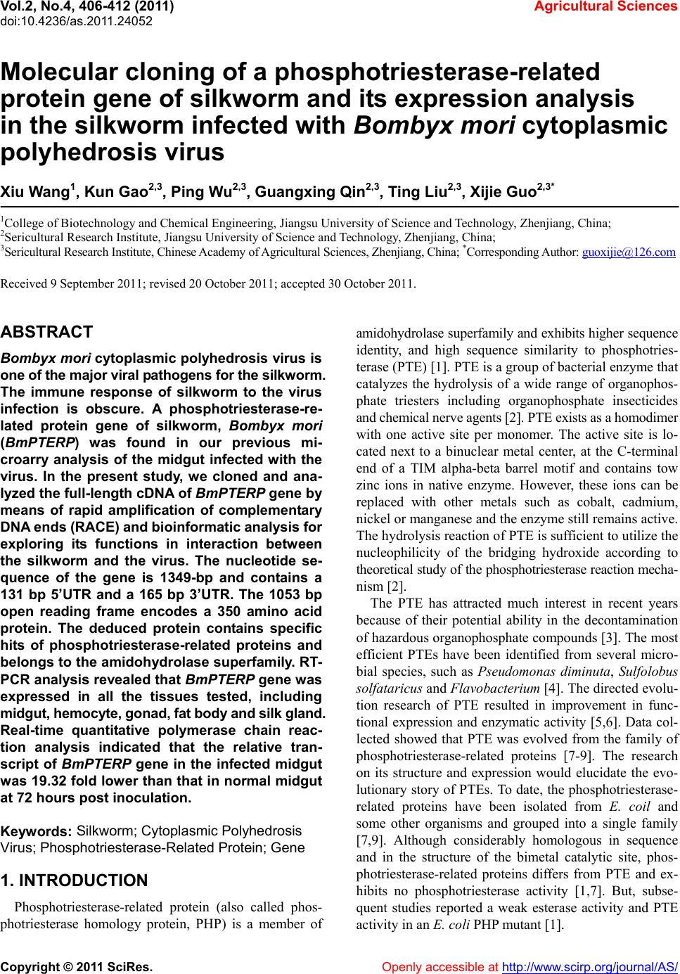
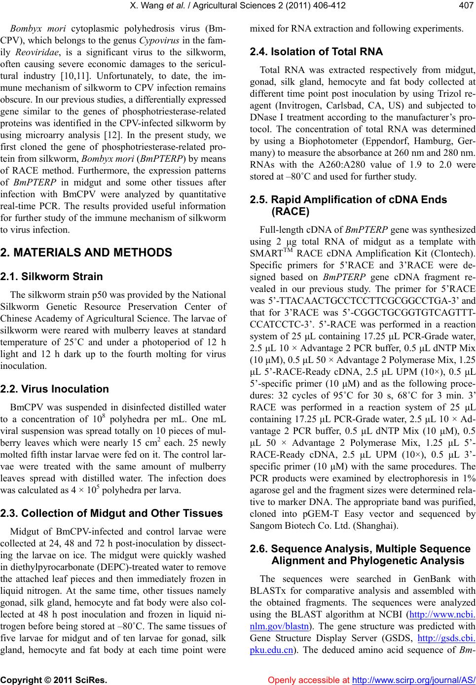
Our result in the present study is the first report that a
phosphotriesterase-related protein gene was cloned from
the silkworm. While, its in vivo function still remains
unclear. At present, the only clue comes from its strong
homology to PHPs of bacteria and other organisms, but
there is not yet any experimental proof that BmPETRP is
itself a phosphotriesterase-related protein. As the disease
of silkworm caused by the infection with BmCPV ad-
vanced, the expression of the BmPETRP gene was ob-
viously down-regulated. This might be attributed to the
facts that a series of physiological and pathological
changes takes place due to the infection. Further func-
tional experimental research should be addressed in our
future work. If BmPETRP does turn out to have the ac-
tivities of PHP or phosphotriesterase predicted from its
homology, it would be very interesting to establish
whether insects have homologous genes, whether these
genes might confer resistance to organophosphate pesti-
cides and most importantly whether these genes might
be involved in the interaction between the silkworm and
the BmCPV infecton.
5. ACKNOWLEDGEMENTS
This research was supported by the National Natural Science Foun-
dation of China (Grant No.30972143) and Natural Science Foundation
of Jiangsu Province (Grant No. BK2010353).
REFERENCES
[1] Roodveldt, C. and Tawfik, D.S. (2005) Shared promis-
cuous activities and evolutionary features in various
members of the amidohydrolase superfamily. Biochemi s-
try, 44, 12728-12736. doi:10.1021/bi051021e
[2] Chen, S.L., Fang, W.H. and Himo, F. (2007) Theoretical
study of the phosphotriesterase reaction mechanism. The
Journal of Physical Chemistry, 111, 1253-1255.
doi:10.1021/jp068500n
[3] Porzio, E., Merone, L., Mandrich, L., Rossi, M. and
Manco, G. (2007) A new phosphotriesterase from Sul-
folobus acidocaldarius and its comparison with the
homologue from Sulfolobus solfata. Biochimie, 89, 625-
636. doi:10.1016/j.biochi.2007.01.007
[4] Merone, L., Mandrich, L. and Rossi, M. (2005) A ther-
mostable phosphotriesterase from the archaeon Sul-
folobus solfataricus: Cloning, overexpression and prop-
erties. Extremophile, 9, 297-305.
doi:10.1007/s00792-005-0445-4
[5] Griffiths, A.D. and Tawfik, D.S. (2003) Directed evolu-
tion of an extremely fast phosphotriesterase by in vitro
compartmentalization. The EMBO Journal, 22, 24-35.
doi:10.1093/emboj/cdg014
[6] Roodveldt, C. and Tawfik, D.S. (2005b) Directed evolu-
tion of phosphotriesterase from Pseudomonas diminuta
for heterologous expression in Escherichia coli results in
stabilization of the metal-free state. Protein Engineering,
Design & Selection, 18, 51-58.
doi:10.1093/protein/gzi005
[7] Buchbinder, J.L., Stephenson, R.C., Dresser, M.J., Pitera,
J.W., Scanlan, T. S. and Fletterick, R.J. (1998) Bio-
chemical characterization and crystallographic structure
of an Escherichia coli protein from the phosphotri-
esterase gene family. Biochemistry, 37, 5096-5106.
doi:10.1021/bi971707+
[8] Scanlan, T.S. and Reid, R.C. (1995) Evolution in action.
Chemistry & Biology, 2, 71-75.
doi:10.1016/1074-5521(95)90278-3
[9] Hou, X.Y., Maser, R.L., Magenheimer, B.S. and Calvet,
J.P. (1996) A mouse kidney- and liver-expressed cDNA
having homology with a prokaryotic parathion hydrolase
(phosphotriesterase)-encoding gene: Abnormal expres-
sion in injured and polycystic kidneys. Gene, 168, 157-
163. doi:10.1016/0378-1119(95)00746-6
[10] Ikeda, K., Nagaoka, S., Winkler, S., Kotani, K., Yagi, H.,
Nakanishi, K., Miyajima, S., Kobayashi, J. and Mori, H.
(2001) Molecular characterization of Bombyx mori cyto-
plasmic polyhedrosis virus genome segment 4. Journal
of Virology, 75, 988-995.
doi:10.1128/JVI.75.2.988-995.2001
[11] Qanungo, K.R., Kundu, S.C., Mullins, J.I. and Ghosh,
A.K. (2002) Molecular cloning and characterization of
Antheraea mylitta cytoplasmic polyhedrosis virus ge-
nome segment 9. Journal of General Virology, 83, 1483-
1491.
[12] Wu, P., Wang, X., Qin, G.X., Liu, T., Jiang, Y.F., Li, M.W.
and Guo, X.J. (2011) Microarray analysis of gene ex-
pression profile in the midgut of silkworm infected with
cytoplasmic polyhedrosis virus. Molecular Biology Re-
ports, 38, 333-341.
doi:10.1007/s11033-010-0112-4
[13] Livak, K.J. and Schmittgen, T.D. (2001) Analysis of
relative gene expression data using real-time quantitative
PCR and the 2−ΔΔCT Method. Methods, 25, 402-408.
doi:10.1006/meth.2001.1262
X. Wang et al. / Agricultural Sciences 2 (2011) 406-412
Copyright © 2011 SciRes. http://www.scirp.org/journal/AS/Openly accessible at
412
[14] Watanabe, H. (2002) Genetic resistance of the silkworm,
Bombyx mori to viral diseases. Current Science, 83,
439-446.
[15] Sun, Y., Wu, A., Dai, R. and Shen, X. (1982) Synthesis of
structural proteins in a cell free system directed by silk-
worm cytoplasmic polyhedrosis virus mRNA synthesized
in vitro. Scientia Sinica, 24, 685-690.
[16] Tuovinen, K., Kaliste-Korhonen, E., Raushel, F.M. and
Hanninen, O. (1996) Protection of organophosphate-in-
activated esterases with phosphotriesterase. Fundamental
and Applied Toxicology, 31, 210-217.
doi:10.1006/faat.1996.0093
[17] Davies, J.A., Buchman, V.L., Krylova, O. and Ninkina,
N.N. (1997) Molecular cloning and expression pattern of
rpr-1, a resiniferatoxin-binding, phosphotriesterase-re-
lated protein, expressed in rat kidney tubules. FEBS Let-
ters, 410, 378-382.
doi:10.1016/S0014-5793(97)00614-5
[18] Vaughan, A., Rodriguez, M. and Hemingway, J. (1995)
The independent gene amplification of electrophoretically
indistinguishable B esterases from the insecticide-resis-
tant mosquito Culex quinquefasciatus. Biochemical Jour-
nal, 305, 651-658.
[19] Yildirim, M., Colak, A., Col, M. and Canakci, S. (2009)
A new recombinant phosphotriesterase homology protein
from Geobacillus caldoxylosilyticus TK4: An extremely
thermo- and pH-stable esterase. Process Biochemistry, 44,
1366-1373. doi:10.1016/j.procbio.2009.07.014