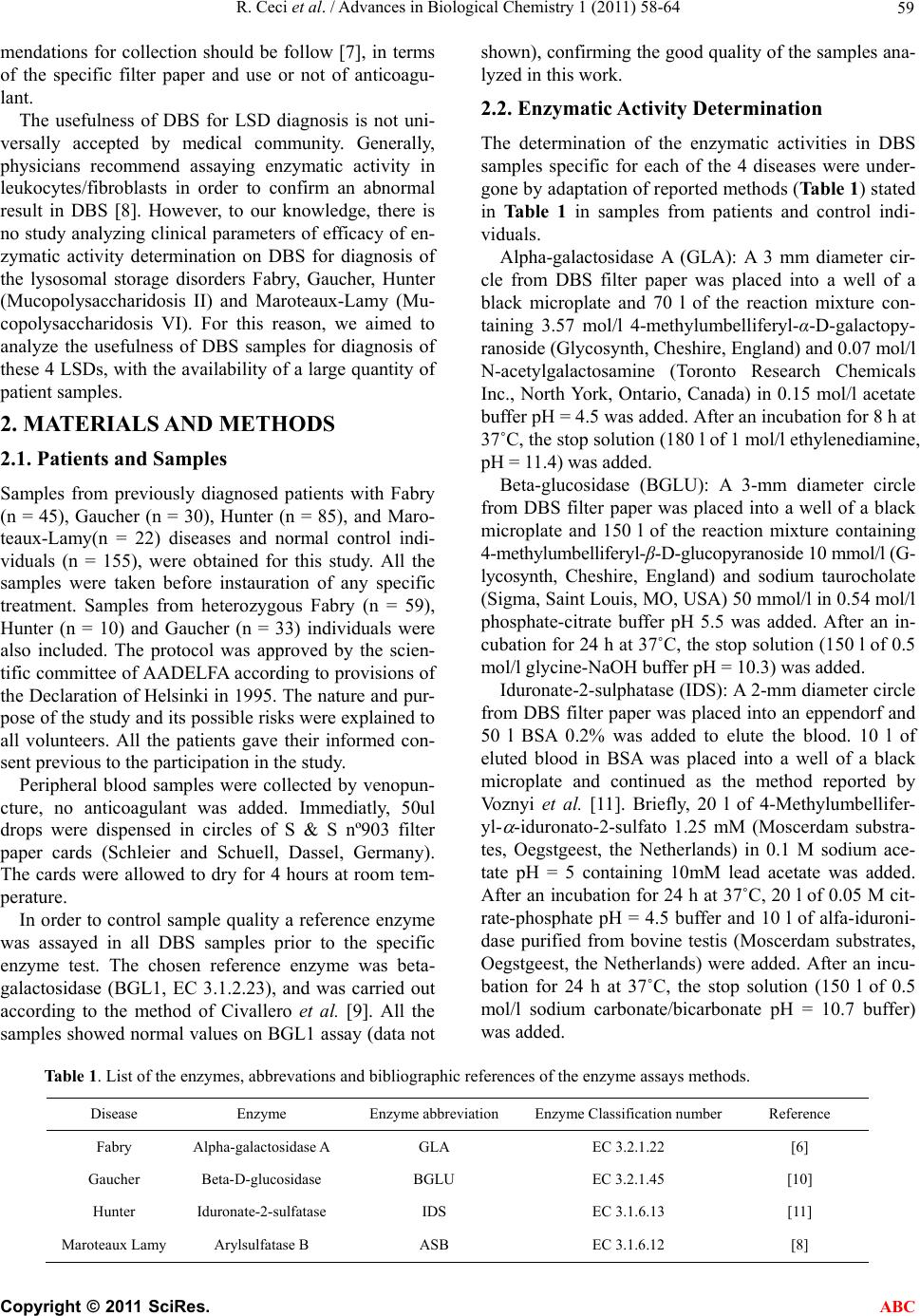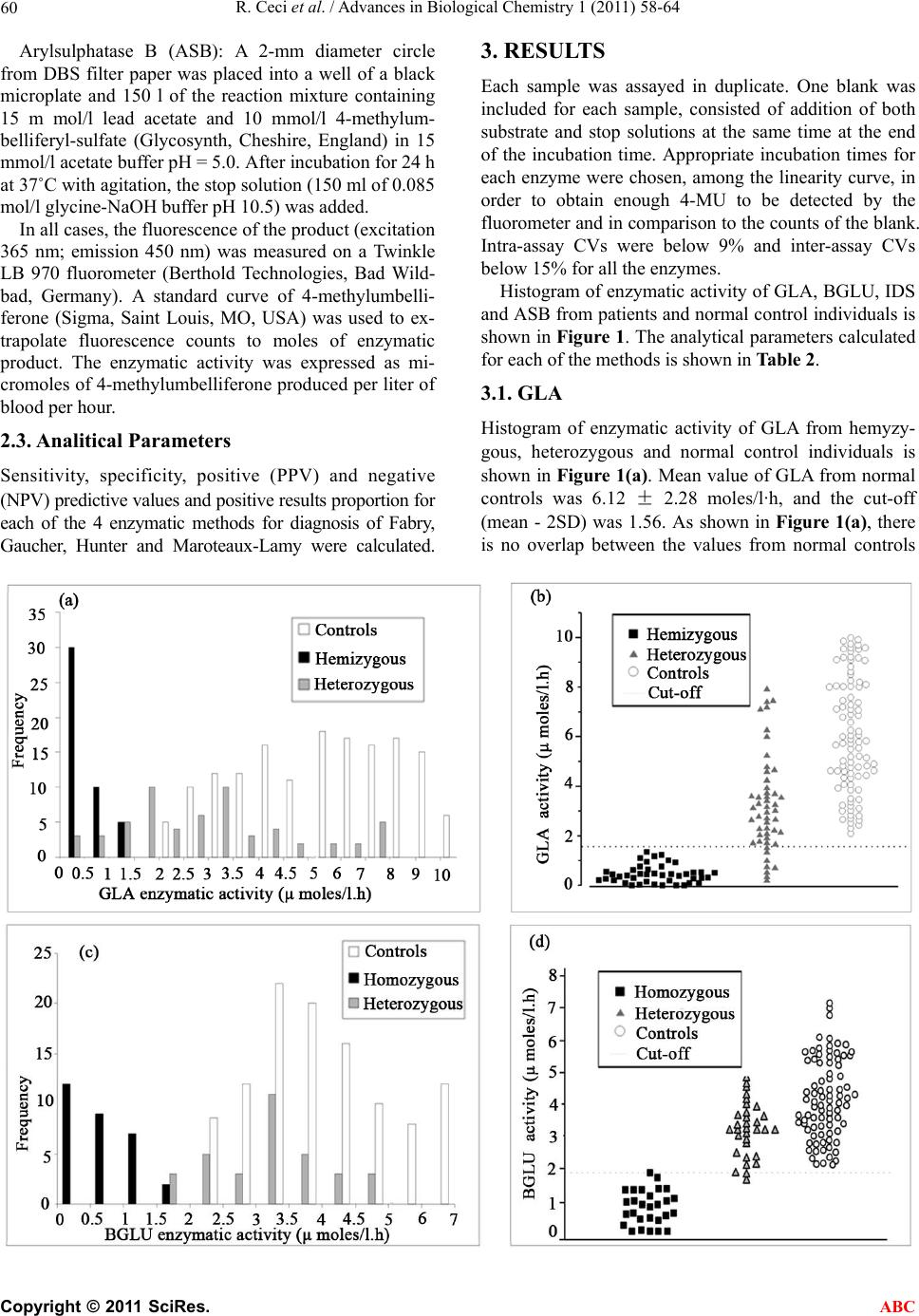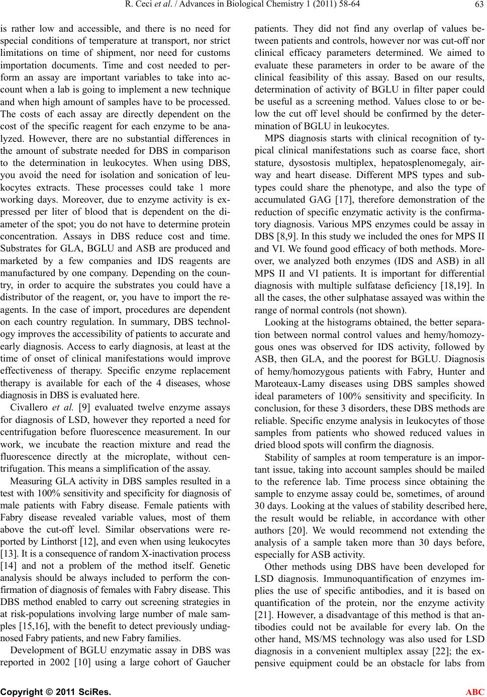
R. Ceci et al. / Advances in Biological Chemistry 1 (2011) 58-64
64
developing countries. Quality control materials to be
used for every method are being prepared [23], which
will be used to the development of a quality control sys-
tem to standardize the assays around the different refer-
ence laboratories.
In conclusion, in this work we analyzed the usefulness
of DBS samples for diagnosis of 4 LSDs. DBS methods
for IDS, ASB and GLA activities shown to be reliable,
however, BGLU DBS assay would need a posterior con-
firmatory step. We recommend evaluating available me-
thods for other lysosomal diseases diagnosis in DBS, in
order to evaluate its usefulness as screening and/or con-
firmatory methods.
5. CONCLUSIONS
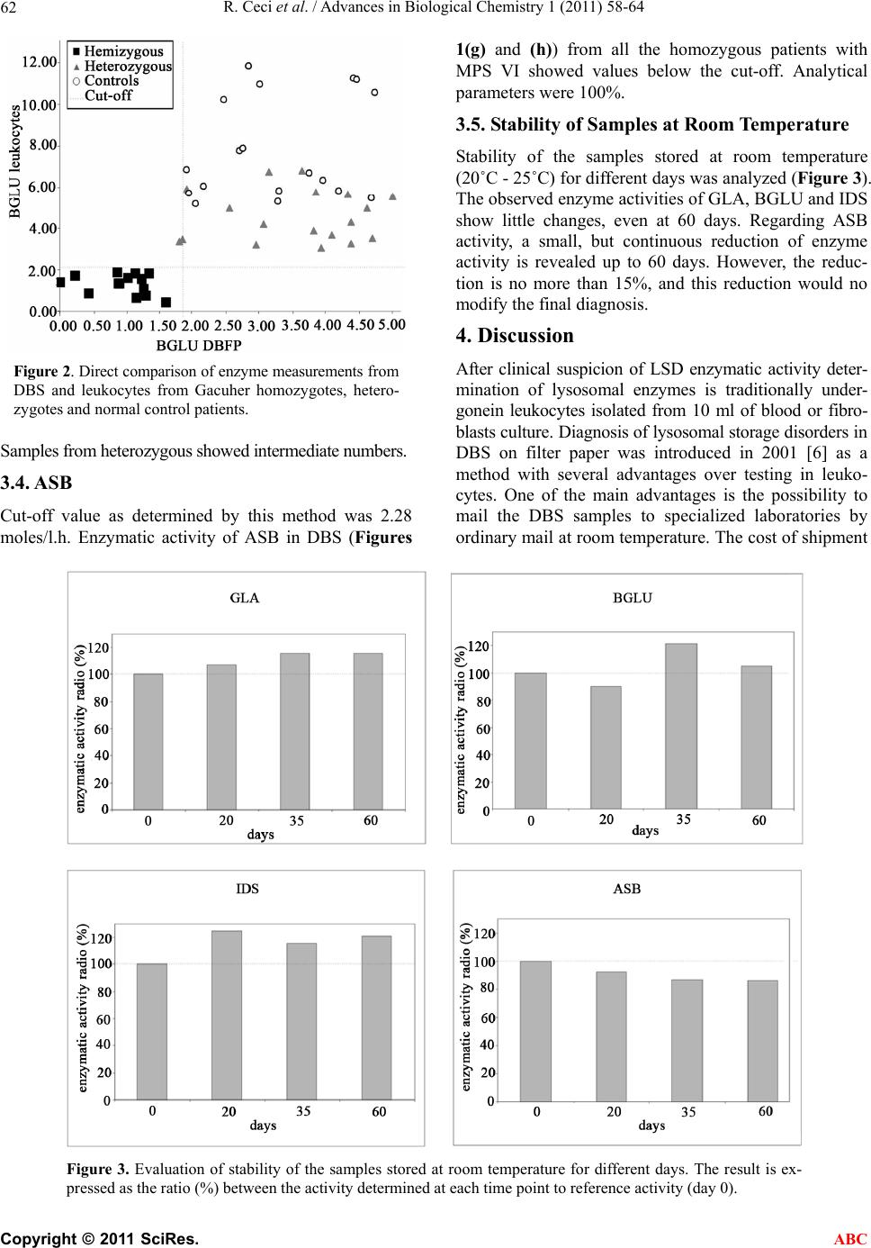
Leukocyte measurement is the only reliable way to di-
agnose Gaucher disease. For Hunter, Fabry and Maro-
teaux-Lamy disorders discrimination between patients
and controls seems adequate by DBS.
6. ACKNOWLEDGEMENTS
This study was supported by grants from SHIRE HGT and Agencia
Nacional de Promoción Científicay Tecnológica.
REFERENCES
[1] Scriver, C.R., Beaudet, A.L., Sly, W. S. and Valle, D.
(2001) Metabolic and molecular basis of inherited dis-
eases. 8th Edition, McGraw Hill, New York.
[2] Beck, M. (2010) Therapy for lysosomal storage disorders.
IUBMB Life, 62, 33-40.
[3] Wenger, D.A., Coppola, S. and Liu, S.L. (2003) Insights
into the diagnosis and treatment of lysosomal storage
diseases. Arch Neurol, 60, 322-328.
doi:10.1001/archneur.60.3.322
[4] Kolodny, E.H. and Mumford, R.A. (1976) Human leu-
kocyte acid hydrolases: Characterization of eleven ly-
sosomal enzymes and study of reaction conditions for
their automated analysis. Clinica Chimica Acta, 70, 247-
257. doi:10.1016/0009-8981(76)90426-5
[5] Coelho, J. and Giugliani, R. (2000) Fibroblasts of skin
fragments as a tool for the investigation of genetic dis-
eases: Technical recommendations. Genetics and Mo-
lecular Biology, 23, 269-271.
doi:10.1590/S1415-47572000000200004
[6] Chamoles, N.A., Blanco, M. and Gaggioli, D. (2001).
Fab- ry disease: Enzymatic diagnosis in dried blood spots
on filter paper. Clinica Chimica Acta, 308, 195-196.
doi:10.1016/S0009-8981(01)00478-8
[7] Olivova, P., van der Veen, K., Cullen, E., et al. (2009)
Effect of sample collection on alpha-galactosidase a en-
zyme activity measurements in dried blood spots on filter
paper. Clinica Chimica Acta, 403, 159-162.
doi:10.1016/j.cca.2009.02.008
[8] Chamoles, N.A., Blanco, M.B., Gaggioli, D., et al. (2001)
Hurler-like phenotype: Enzymatic diagnosis in dried
blood spots on filter paper. Clinica Chimica Acta, 47,
2098-2102.
[9] Civallero, G., Michelin, K., de Mari, J., et al. (2006)
Twelve different enzyme assays on dried-blood filter pa-
per samples for detection of patients with selected inher-
ited lysosomal storage diseases. Clinica Chimica Acta,
372, 98-102. doi:10.1016/j.cca.2006.03.029
[10] Chamoles, N.A., Blanco, M., Gaggioli, et al. (2002)
Gaucher and niemann-pick diseases enzymatic diagnosis
in dried blood spots on filter paper: retrospective diagno-
sis in newborn-screening cards. Clinica Chimica Acta,
317, 191-197. doi:10.1016/S0009-8981(01)00798-7
[11] Voznyi, Y.V., Keulemans, J.L.M., Beyer, E.M., et al.
(2001) A fluorogenic assay for the diagnosis of Hunter
disease (MPS II). Journal of Inherited Metabolic Disease,
24, 675-680.
[12] Linthorst, G., Vedder, A., Aerts, J., et al. (2005) Screen-
ing for fabry disease using whole blood spots fails to
identify one-third of female carriers. Clinica Chimica
Acta, 353, 201-203. doi:10.1016/j.cccn.2004.10.019
[13] Lukacs, Z., Hartung, R., Beck, M., et al. (2007) Direct
comparison of enzyme measurements from dried blood
and leukocytes from male and female fabry disease pa-
tients. Journal of Inherited Metabolic Disease, 30, 614.
[14] Lyon, M.F. (1961) Gene action in the X-chromosome of
the mouse (Mus musculus L.). Nature, 190, 372-373.
doi:10.1038/190372a0
[15] Porsch, D.B., Nunes, A.C., Milani, V., et al. (2008) Fabry
disease in hemodialysis patients in southern Brazil:
prevalence study and clinical report. Renal Failure, 30,
825-830. doi:10.1080/08860220802353777
[16] Gaspar, P., Herrera, J., Rodrigues, D., et al. (2010) Fre-
quency of fabry disease in male and female haemodialy-
sis patients in Spain. BMC Medical Genetics, 11, 19.
doi:10.1186/1471-2350-11-19
[17] Neufeld, E.F. and Muenzer, J. (1995) The mucopolysac-
charidoses. In: Scriver, C.R., Beaudet, A.L., Sly, W.S.,
Valle, D., Eds., The Metabolic and Molecular Bases of
Inherited Disease, McGraw-Hill, New York, 2465.
[18] Cosma, M.P., Pepe, S., Annunziata, I., et al. (2003) The
multiple sulfatase deficiency gene encodes an essential
and limiting factor for the activity of sulfatases. Cell, 11 3,
445-456. doi:10.1016/S0092-8674(03)00348-9
[19] Dierks, T., Schmidt, B., Borissenko, L.V., et al. (2003)
Multiple sulfatase deficiency is caused by mutations in
the gene encoding the human C(alpha)-formylglycine
generating enzyme. Cell, 113, 435-444.
doi:10.1016/S0092-8674(03)00347-7
[20] Poeppl, A.G., Murray, G.J. and Medin, J.A. (2005) En-
hanced filter paper enzyme assay for high-throughput
population screening for fabry disease. Analysis Bio-
chemical, 337, 161-163. doi:10.1016/j.ab.2004.10.007
[21] Meikle, P.J., Grasby, D.J., Dean, C.J., et al. (2006) New-
born screening for lysosomal storage disorders. Mole-
cular Genetics and Metablism, 88, 307-314.
doi:10.1016/j.ymgme.2006.02.013
[22] Li, Y., Scott, C.R., Chamoles, N.A., et al. (2004) Direct
multiplex assay of lysosomal enzymes in dried blood
spots for newborn screening. Clinica Chimica Acta, 50,
1785-1796. doi:10.1373/clinchem.2004.035907
[23] De Jesus, V.R., Zhang, X.K., Keutzer, J, et al. (2009)
Development and evaluation of quality control dried
blood spot materials in newborn screening for lysosomal
storage disorders. Clinica Chimica Acta, 55, 158-164.
doi:10.1373/clinchem.2008.111864
C
opyright © 2011 SciRes. ABC