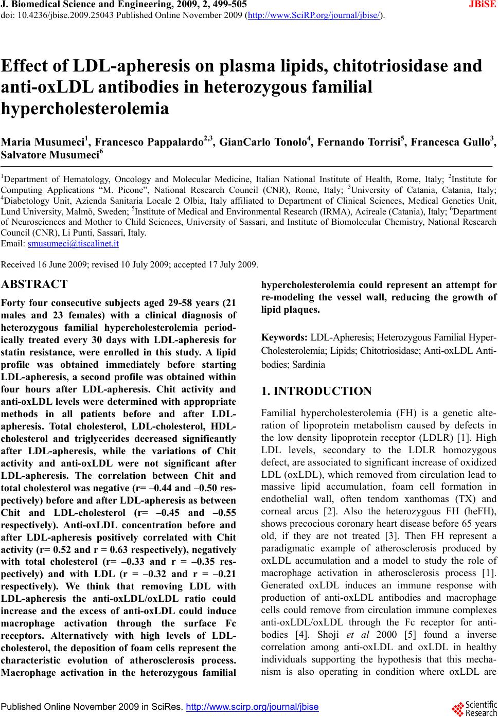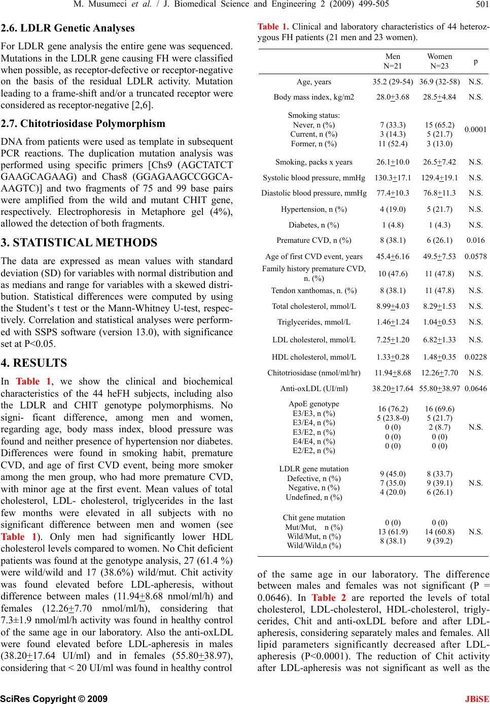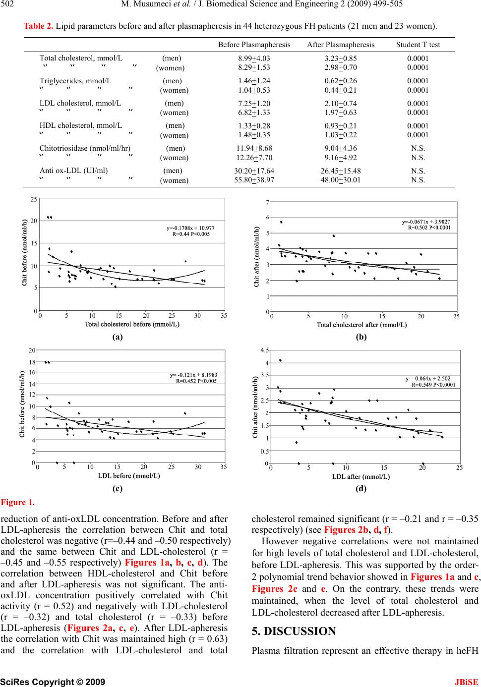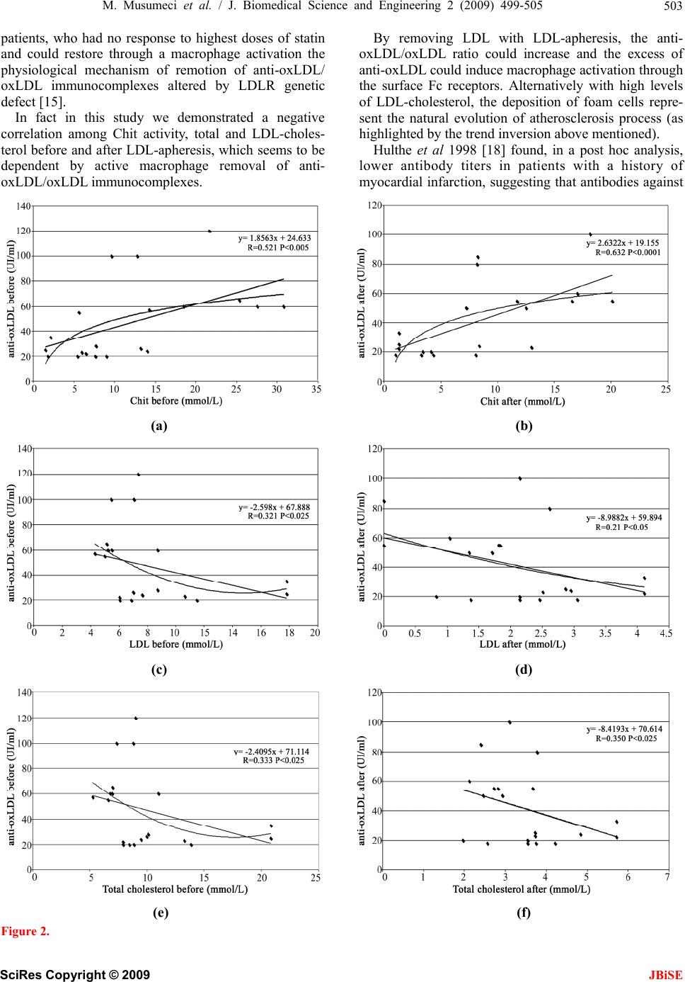
M. Musumeci et al. / J. Biomedical Science and Engineering 2 (2009) 499-505
SciRes Copyright © 2009
505
JBiSE
disease, N. Engl. J. Med., 340, 115−126.
[3] Civeira, F., (2004) Guidelines for the diagnosis and
management of heterozygous familial hypercholestero-
lemia, Atherosclerosis, 173, 55−68.
[4] Kern, F., (1990) Cholesterol metabolism, LDL, and the
LDL receptor, Edited by Myant NB, Academic press,
NewYork, 465.
[5] Shoji, T., Nishizawa, Y., Fukumoto, M., Shimamura, K.,
Kimura J., Kanda, H., et al., (2000) Inverse relationship
between circulating oxidized low density lipoprotein
(oxLDL) and anti-oxLDL antibody levels in healthy sub-
jects. Atherosclerosis; 148, 171−7.
[6] Bertolini, S., Cantafora, A., Averna, M., Cortese, C.,
Motti, C., Martini, S., et al., (2000) Clinical expression
of familial hypercholesterolemia in clusters of mutations
of the LDL receptor gene that cause a receptor-defective
or receptor-negative phenotype, Arterioscler Thromb
Vasc Biol, 20, 41−52.
[7] Hollak, C. E., van Weely, S., van Oers, M. H., and Aerts,
J. M., (1994) Marked elevation of plasma chitotriosidase
activity, A novel hallmark of Gaucher disease, J. Clin.
Invest., 93, 1288−1292.
[8] Boot, R. G., van Achterberg, T. A., van Aken, B. E.,
Renkema, G. H., Jacobs, M. J., Aerts, J. M., et al., (1999)
Strong induction of members of the chitinase family of
proteins in atherosclerosis: Chitotriosidase and human
cartilage gp-39 expressed in lesion macrophages, Arterio-
scler Thromb Vasc Biol, 19, 687−694.
[9] Malaguarnera, L., Di Rosa, M., Zambito, A. M., dell'Ombra,
N., Nicoletti, F., and Malaguarnera, M., (2006) Chitotrio-
sidase gene expression in Kupffer cells from patients with
non- alcoholic fatty liver disease, Gut, 55, 1313−1320.
[10] Artieda, M., Cenarro, A., Gañán, A., Jericó, I., Gonzalvo,
C., Casado, J. M., et al., (2003) Serum chitotriosidase
activity is increased in subjects with atherosclerosis
disease, Arterioscler Thromb Vasc Biol., 23, 1645−1652.
[11] Artieda, M., Cenarro, A., Gañán, A., Lukic, A., Moreno,
E., Puzo, J., et al., (2007) Serum chitotriosidase activity,
a marker of activated macrophages, predicts new
cardiovascular events independently of C-Reactive
Protein, Cardiology, 108, 297−306.
[12] Hansson, G. K., (2005) Inflammation, atherosclerosis,
and coronary artery disease, N. Engl. J. Med., 352,
1685−1695.
[13] van Eijk, M., van Roomen, C. P., Renkema, G. H.,
Bussink, A. P., Andrews, L., Blommaart, E. F., et al.,
(2005) Characterization of human phagocyte-derived
chitotriosidase, a component of innate immunity, Int.
Immunol., 17, 1505−12.
[14] Shaw, P. X., Hörkkö, S., Tsimikas, S., Chang, M. K.,
Palinski, W., Silverman, G. J., et al., (2001) Human-derived
anti-oxidized LDL autoantibody blocks uptake of oxi-
dized LDL by macrophages and localizes to atheroscle-
rotic lesions in vivo, Arterioscler Thromb Vasc Biol, 21,
1333−9.
[15] Bláha, M., Cermanová, M., Bláha, V., Blazek, M., Malý,
J., Siroký, O., et al., (2007) Safety and tolerability of
long lasting LDL-apheresis in familial hyperlipopro-
teinemia. Ther Apher Dial, 11, 9−15.
[16] Hixson, J. E. and Vernier, D. T., (1990) Restriction isotyping
of human apolipoprotein E by gene amplification and
cleavage with HhaI, J. Lipid. Res., 31, 545–8.
[17] Tsukamoto, K., Watanabe, T., Matsushima, T., Kinoshita,
M., Kato, H., Hashimoto, Y., Kurokawa, K., and
Teramoto, T., (1993) Determination by PCR-RFLP of
apo E genotype in a Japanese population, J. Lab. Clin.
Med., 121, 598−602.
[18] Hulthe, J., Wikstrand, J., Lidell, A., Wendelhag, I.,
Hansson, G. K., and Wiklund, O., (1998) Antibody titers
against oxidized LDL are not elevated in patients with
familial hypercholesterolemia, Arterioscler Thromb Vasc
Biol, 18, 1203−1211.
[19] Tinahones, F. J., Gomez-Zumaquero, J. M., Rojo-
Martinez, G
., Cardona, F., Esteva de Antonio, I. E., Ruiz
de Adana, M. S., et al., (2002) Increased levels of
anti-oxidized low-density lipoprotein antibodies are as-
sociated with reduced levels of cholesterol in the general
population, Metabolism, 51, 429−31.
[20] Tinahones, F. J., Gomez-Zumaquero, J. M., Garrido-
Sanchez, L., Garcia-Fuentes, E., Rojo-Martinez, G.,
Esteva, I., et al., (2005) Influence of age and sex on lev-
els of anti-oxidized LDL antibodies and anti-LDL im-
mune complexes in the general population, J. Lipid. Res.,
46, 452−7.
[21] Canudas, J., Cenarro, A., Civeira, F., García-Otín, A. L.,
Arístegui, R., Díaz, C., et al., (2001) Chitotriosidase
genotype and serum activity in subjects with combined
hyperlipidemia: Effect of the lipid-lowering agents,
atorvastatin and bezafibrate, Metabolism, 50, 447−450.
[22] Brizzi, P., Tonolo, G., Bertrand, G., Carusillo, F., Severino,
C., Maioli, M., et al., (2004) Autoantibodies against oxi-
dized low-density lipoprotein (oxLDL) and LDL oxida-
tion status, Clin. Chem. Lab. Med., 42, 164−70.
[23] Frostegård, J., Tao, W., Georgiades, A., Råstam, L.,
Lindblad, U., and Lindeberg, S., (2007) Atheroprotective
natural anti-phosphorylcholine antibodies of IgM sub-
class are decreased in Swedish controls as compared to
non-westernized individuals from New Guinea, Nutr
Metab (Lond), 20, 7.
[24] Binder, C. J., Chang, M. K., Shaw, P. X., Miller, Y. I.,
Hartvigsen, K., Dewan, A., et al., (2002) Innate and acquired
immunity in atherogenesis, Nat. Med., 8, 1218–26.
[25] Palinski, W., Miller, E., Witztum, J. L., (1995) Immunization
of low density lipoprotein (LDL) receptor-deficient rabbits
with homologous malondialdehyde-modified LDL reduces
atherogenesis, Proc. Natl. Acad. Sci., USA, 92, 821–5.
[26] Binder, C. J., Hörkkö, S., Dewan, A., Chang, M. K., Kieu,
E. P., Goodyear, C. S., et al., (2003) Pneumococcal vac-
cination decreases atherosclerotic lesion formation: mo-
lecular mimicry between Streptococcus pneumoniae and
oxidized LDL, Nat. Med., 9, 736–43.
[27] Ameli, S., Hultgardh-Nilsson, A., Regnstrom, J., Calara,
F., Yano, J., Cercek, B., et al., (1996) Effect of immu-
nization with homologous LDL and oxidized LDL on
early atherosclerosis in hypercholesterolemic rabbits,
Arteri-oscler Thromb Vasc Biol, 16, 1074–9.
[28] Pappalardo, F., Musumeci, S., and Motta, S., (2008)
Modeling immune system control of atherogenesis, Bio-
informatics.
[29] Orem, C., Orem, A., Uydu, H. A., Celik, S., Erdol, C., and
Kural, B. V., (2002) The effects of lipid-lowering therapy
on low-density lipoprotein auto-antibodies: Relationship
with low-density lipoprotein oxidation and plasma total
antioxidant status, Coron Artery Dis, 13, 65–71.
[30] Brizzi, P., Isaja, T., D'Agata, A., Malaguarnera, L.,
Malaguarnera, M., and Musumeci, S., (2002) Oxidized
LDL antibodies (OLAB) in patients with beta thala-
ssemia major, J. Atheroscler Thromb, 9, 139−44.