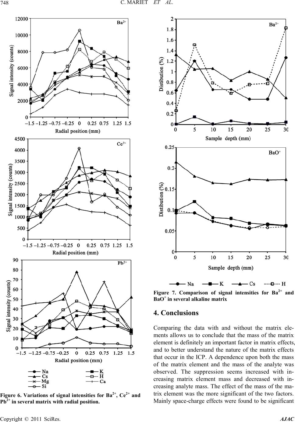
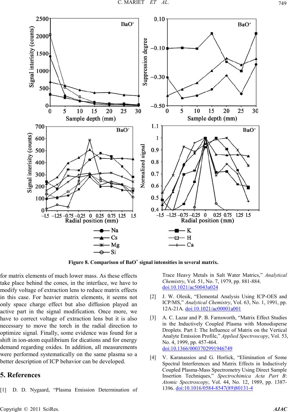
America n Journal of Analy tic al Chemistry, 2011, 2, 739-751
doi:10.4236/ajac.2011.27085 Published Online November 2011 (http://www.SciRP.org/journal/ajac)
Copyright © 2011 SciRes. AJAC
Effect of Sample Matrix on Radial and Axial Profiles of
Ion Abundance in Inductively Coupled Plasma
Mass Spectrometry
Clarisse Mar ie t1*, Francine Carrot2, Mélanie Moskura1
1Laboratoire Interdisciplinaire sur lOrganisation Nanométrique et Supramoléculaire (LIONS),
CEA Saclay Gif-sur Yvett e, France
2UMR 3299, CEA Sacla y Gif-su r Y vette, France
E-mail: *clarisse.mariet@cea .fr
Received April 30, 2011; revised June 26, 201 1; accepted July 8, 2011
Abstract
In inductively coupled plasma mass spectrometry (ICP-MS) analysis, only a few options are available to deal
with non-spectroscopic interferences. Considering that diluting the sample is impractical for traces analysis,
other alternatives must be employed. Traditionally, the method of standard additions is used to correct the
matrix effect but it is a time consuming method. Others methods involves separation techniques. Another
way to overcome matrix interferences is to understand the mechanism involved and adjust plasma viewing
conditions to reduce or eliminate the effect. In this study, the effect of various concomitant elements in
ICP-MS was assessed by measuring the distribution of selected singly charged analyte ions (Al, V, Cr, Mn,
Ni, Co, Cu, Zn, As, In, Ba, La, Ce, Pb), doubly charged ions (La, Ce, Ba and Pb) and oxides ions (BaO) in
the presence of concomitant elements spanning a mass range from 23 (Na) to 133 (Cs) u.m.a. and different
ionization energies. Concomitant elements are alkali metals, alkaline earth metals and Si. Analyte ion sup-
pression was observed while moving the ICP across and away from the sampling interface with or without a
single concomitant element. Matrix effect measures were realised, firstly, to highlight the relation between
the signal extinction of an analyte and the masse of the concomitant element, and secondly to highlight the
relation between the removal of the analyte signal and the first ionization energy of the element of matrix. A
dependence upon both the mass of the matrix element and the mass of the analyte was observed. The sup-
pression seems increased with increasing matrix element mass and decreased with increasing analyte mass.
The effect of the mass of the matrix element was the more significant of the two factors. If space-charge ef-
fects were found to be significant for matrix elements of much lower mass, it seems diffusion also played an
active part for heavier matrix elements. Finally, some evidence was found for a shift in ion-ato m equil ibrium
for dications and for energy demand regarding oxides.
Keywords: Matrix Effects, Easily Ionized Elements, Inductively Coupled Plasma Mass Spectrometry, Spatial
Profiling
1. Introduction
Inductively coupled plasma mass spectrometr y (ICP-MS)
is a well-established analytical technique. Ever since the
development of ICP-MS in the mid 1980s, the determi-
nation of trace levels of certain elements in high matrix
samples can be considered a “classical” difficult applica-
tion. As a resul t of the rather invasive sampling process,
ICP-MS is much more susceptible to effects of concomi-
tant elements (also called matrix effect (ME) and non
-spectroscopic interferences) than inductively coupled
plasma atomic emission spectrometry (ICP-AES). These
interferences are usually observed as a suppression of
analyte signals, although enhancements have also been
report. The ME is probably one of the most crucial limi-
tations leadi ng to a po ssible deterio ration of the acc urac y
of analysis. A considerable number of reports have been
dedicated to the study of the ME since 1979 [1-4], nota-
bly with easily ionizable element (EIE) such as sodium
or calcium. The matrix effect due to Ca is more severe
C. MARIET ET AL.
Copyright © 2011 SciRes. AJAC
740
than t hat due t o K or N a and is well-kno wn b ut the exact
mechanism of the ME is still ignored [5]. Because the
matrix effects of Ca and Na are of different origin, it is
easy to eliminate the ME from Na by choosing operating
conditions under which plasma is robust (good radial and
axial view modes) whereas one can only slightly de-
crease the effects of matrix created by Ca [6]. The same
applies with acids or solvents [7-9]. Because of their
origin, only a few options are available to deal with
non-spectroscopic interferences. Considering that reduc-
ing the amount o f material lo aded into the pla sma ( whic h
can be done by diluting the sample) is impractical for
traces analysis, since it would result in less a nalyte atoms
measured, other alternatives must be employed [10].
Traditionally, in plasma spectrometry, the EIE effect is
mitigated either b y matrix matching the calibration solu-
tions with the sample, so mething not alwa ys easily done
[5]. It is also possible to use the method of standard addi-
tions [11,12] but it is a time consuming method. Several
methods have been proposed for alleviating these inter-
ferences and can be classified into two groups depending
on whether separation techniques are involved or not.
The first group involves separation techniques [13,14].
Second group applies physical techniques like electro-
thermal vapor ization [15-16], which had limitations such
as non-spectroscopic interference, which might occur
when a high salt content sample wa s analyzed .
In our point of view, the best way to overcome matrix
interferences is to understand the mechanism involved
and adjust plasma viewing conditions to reduce or elimi-
nate the effect. Studies relating to ICP-AES [5,6] or
ICP-MS propo se several possible origins with the effects
of matrix related to plasma. The most commonly cited
origins are the ion-electron recombinations [5,6,18,19],
the energy demand [5,6,18], increase in the collisions [6],
the space charge effect [19] and change the spatial dis-
tribution o f the species in the source with latera l [5,6,18 ]
or ambipolar diffusion [5,6,18,19].
So as to bring out the parameters influencing ME, we
suggest describing each of the non-spectroscopic inter-
ferences origins listed here. A displacement of the ioni-
zation equilibrium is the simplest explanation of the sig-
nal extinction. Although the presence of EIE does not
modify significantly the total number of electrons in the
plasma, it causes an important increase of the charge
density along the axis of the plasma [19] and a tempera-
ture decrease. The higher charge density moves the equi-
librium towards the atomic form. T he modificatio n of the
ion-atom equilibrium for an analyte translates by the
maxi mum i nte nsit y of the s ig nal along the central axis of
the plasma. Ions are formed in zones of higher tempera-
tures, closer to the sampler. There is a delay in the ioni-
zation. To observe the maximum intensity of the signal,
it is then necessary to increase the distance
torch-sampler .
According to thermodynamic calculations, there shou-
ld not be refractory oxides in plasma. However, as the
drops size is not always identical, oxides are formed.
Oxides exist mainly in the coldest zones of plasma,
closer to the torch and on the edges of the flame. For an
analyte M, signals of the ions MO+ and MOH+ are ob-
served while moving away plasma from the sampler. If
the formation of oxide is related to a reduction in the
temperature, the maximum of intensity of the MO+ signal
will be observed a t a longer distance from the sampler. If,
indeed, the formation of oxide is related to a problem of
atomizing, the peak of maximum intensity does not var y
with the position of observation. Thus the formation of
oxides on the edges of plasma can be highlighted by an
abrupt modification of the signal when the position of
observation is changed.
The addition of a concomitant element modifies the
energy demand in plasma according to the concentration
and the energ y o f ionization of this element.
The first ones to have mentioned the space charge ef-
fect are Olivares and Houk [20]. This ME involves the
positively char ged lenses that collect the positi ve ions, at
the exit of the skimmer, by confining them in a beam.
According to them, the strong ionic currents in ICP ex-
ceed the maximum current allowing neglecting the
charge effect. The ionic current is inversely proportional
to the ion mass [19]. However, the ions beam is delim-
ited by the total current in the beam and repulsion forces.
Consequently, there is a broadening of the beam espe-
cially since the ions are light and, simultaneously, trans-
mission of t he light ion s is less effective (p art of them is
lost in way) than that of heavy ones. Lighter ions, with
stronger density of charge and weaker kinetic energy
than heavy ions, are more easily deviated. It is what ex-
plains the greatest sensitivity for the heavy ions that for
the light ones (1 Pb corresponds to approximately
120.000 counts for U and 3000 counts for Li). Thus a
matri x of hea vy ele ments will ext inguis h the si gnal mor e
stro ngly tha n a matr ix of l ight ele ments. The signa l wid-
ens with the addition of a heavy matrix element whatever
is the observation position in plasma since the increase in
the diffusion does not take place in plasma but behind
the co ne s, in t he i nterface. Chen and Ho uk [21] observed
it is possible to attenuate the extinction created per 1000
Cs ppm on 50 Sc ppm with a weak loss of sensitivity by
modi fying the pote ntial of the lenses.
In the same way that the ionization of an analyte can
be delayed by the addition of an element of matrix, the
vaporization of the element can take place in zones of
plasma closer to the sampler. The signal thus decreases,
for the same position of observation and widens because
C. MARIET ET AL.
Copyright © 2011 SciRes. AJAC
741
the delay of vaporization allows a greater diffusio n. One
speaks about later al dif fusio n. Sp atial chan ge distrib utio n
can take place in another way. In plasma, the electrons
and the ions diffuse towards the areas of lower charge
density. By consequent the ions and the electrons move
away from the source of arrival of the sample. The elec-
trons diffuse more quickly than the ions. Consequently,
locally there are zones where neutrality is not respected;
forces of repulsion appear to restore it. Because of the
created electric field the electrons are slow whereas they
are accelerated by the repelling powers. The sum of these
two phenomena leads to a total diffusion and the same
speed of the ions and electrons. The larger charge density
is, the more the diffusion increases causing a beam wid-
ening. Moreover, the effect of matrix moves the peak of
maximum intensity towards the greatest sampler torch
distances. Since simultaneously with the ambipolar dif-
fusion, the charge effect and the displacement of atom-
ion equilibrium cause signal extinction along the central
axis of plasma, the broadening of the signal is visible
only far from this area, where the element of matri x has a
weaker concentration.
Thus, the description of possible non-spectroscopic
interferences allows to define parameters highlighting
the origin of the interference and controllable by the ex-
perimenter. These parameters are the nebulizer flow, the
addition of a transport modifier, observation position in
the plasma, mass and electronegativity of elements.
The most studied matrices are Na, K and Ca not only
for historical reasons, since they were largely known in
the techniques with flame, but also because they are most
frequent in the environment (sea water, river or biologi-
cal fluids) [6 ]. To be able to bring clo ser our results those
of the literature and to validate our methodology, our
study will also relate to Na, K and Ca. Since samples
analyzed by ICP-MS in our laboratory being ores, slags,
basalts, slags of re fining, matrix mainly consi st of Si and
Mg, we decided to study alakali metals, rare earth metals
and Si.
2. Experimental
2.1. Instrumentation
The icp-mass spectrometer used in this study is a quad-
rupole Thermo fisher Scientific X7 equipped with a
Meinhard concentric nebulizer. The operation conditions
are shown in Table 1. The parameters were chosen in
such a way as to produce maximum analyte ion count
rate thanks a mass calibration over the whole mass range
performed each day and to provide minimal values of the
ratios CeO+/Ce+ and Ba2+/B a+. This optimization also
established the optimum compromise axial Z (sample
depth), X (horizontal) and radial Y (vertical) positions
according to the nomenclature of Holliday et al. [22].
The axial position is a measured of the relative distance
between the load coil and the sampling cone. All spatial
profiling was carried out under the best compromise
multi-elemental conditions. One requirement for repre-
sentative sampling of the plasma is the absence of a sec-
onda ry disc harge. T hen we use some sort of torch shield
(metal plate inserted between the torch and the load c oil)
in an attempt to mitigate such a discharge.
2.2. Reagents and Solutions
All reagents were of analytical grade. All multielemental
solutions were p repared by diluting certi fied stock multi-
elemental solution (SPEX, suprapur, Jobin Yvon). The
solution containing the analyte elements only is a 10
µg/L Al, V, Cr, Mn, Ni, Co, Cu, Zn, Ga, As, Ba, La, Ce
and Pb in 2% HNO3. It was used for mass calibration and
all analyte elements measures without concomitant.
These elements were chosen because they described the
wall mass range, they have differe nt ionization po tentials
and oxidation formation abilities (Table 2). Height ma-
trix containing 1000 mg/L alkaline salts, alkaline earth
metal salts or inorganic acids were prepared each time by
diluting with 2% HNO3: Na NO3, KNO3, CsNO3, Ca( NO 3)2,
Mg(NO3)2, elementary Si, HCl and HI (Table 3). These
matrix were taken as representative of the different con-
comitants pre sent in liquid samples.
Table 1. Experimental conditions.
ICP sourc e
plasma gas
RF power
Aerosol carrier gas flow rate
Auxiliary gas flow rate
Plasma gas flow rate
argon
1350 W
0.74 L·min–1
0.90 L·min–1
13.8 L·min–1
Ma ss spectrometer
Interface vacuum
Analyser vacuum
Ion lens 1
Ion lens 2
Ni made Xi sampler (1 mm φ) and
skimmer (0.7 mm φ)
1.9 × 1 0 h P a
3.6 × 10⁻⁷ hPa
3.4
30.2
Acquisitio n par ame ters
Full quantitative scan mode
Dwell time
Integration time
Rep l icates
Ion collection m ode
Measuring tim e
10 ms/elem ent
90 s
4
Pulse counting
90 s
C. MARIET ET AL.
Copyright © 2011 SciRes. AJAC
742
Table 2. Anal ytes properties.
Element m/z
ratio Isotope aboundance
(%)d First ionization
energy (eV )a Second ionization
energy (eV )a I o niz ation degree
(%)b Strengh of oxide bond
(kJ/mol)c
Al
V
Cr
Mn
Ni
Co
Cu
Zn
As
In
Ba
La
Ce
Pb
Pb2+
Ce2+
Ba2+
VO+
BaO+
27
51
52
55
58
59
63
64
75
115
138
139
140
208
104
70
69
145
154
100
99.76
83.764
100
67.77
100
69.17
48.6
100
95.7
71.7
99.911
88.48
52.4
5.984
6.74
6.764
7.435
7.635
7.876
7.726
9.394
9.814
5.786
5.577
5.466
7.415
18.83
14.65
16.5
15.64
18.17
17.06
20.29
17.96
18.63
18.87
11.06
10.85
15.03
98
99
98
95
91
93
52
90 (10)e
96 (2)e
97 (0.01)e
512.1
626.8
429.3
402.9
382
384,5
481
552
799
797
382
a[24]; b[21]; c[25]. It de als with the bond strength of neutral molecu les; d[26]; eSeco n d io niza tion deg r ee.
Table 3. Matrix el ements properti es .
Element Atomic mass First ionization energy
(eV)a Sec ond ionization
energy (eV)a Ionization degree
(%)b Strengh of oxide bond
(kJ/mol)c
Na
Mg
Si
K
Ca
Cs
22.9898
24.3050
28.0855
39.0983
40.078
132.9054
5.14
7.64
8.15
4.34
6,11
3.89
47.28
15.03
16.34
31.62
11.87
25.08
100
100
100
256.1
277.8
295.8
a[24]; b[21]; c[25]; It deals with the bond st rength of neutral molecule s.
2.3. Procedure
The following procedure is used in order to determine
the effect of concomitant elements on analyte ion count
rates. The same set of four experiments was performed
for each of the concomitant elements. Each experiment
consisted o f four repe titions: first with a blank cons isting
only of 2% HNO3 t; the second with the multielement
solution; the third with a blank 2% HNO3 solution con-
taining the concomitant element; and the fourth with a
multielement solution containing the concomitant ele-
ment. The multielement solution not containing the con-
comitant element was repeated for every experiment in
order to account for day-to-day variability in sensitivity.
Five replicates were taken at each measurement incre-
ment before advancing to the next point of interest.
Thanks the computer-controlled translation stage of the
instrument four experiments were performed: (1) axial
profiles by moving the torch away from the sampler
along the central axis of the plasma to get a measurement
of the signal intensity at a variety of sampling depths; (2)
radial signal profile of the ion distribution across the
central channel of the plasma by moving the torch across
the sampler at each of three fixed sampling depths (Z =
optimum, Z = optimum + 50 and Z = optimum + 100),
respectively.
2.4. Matrix Effect Measures
ME may be defined as matrix-induced signal variations,
C. MARIET ET AL.
Copyright © 2011 SciRes. AJAC
743
either enhancement or suppression of the analyte signal.
In this p ap er, like so me ot her a uthor s [23 -26], we use the
suppression degree defined as the variation of net inten-
sities using the following formula:
() ()
Suppression degree1
() ()
mm
IAI blank
IAI blank
= −
(1)
In this relation (1) , we compare the analyte signals inten-
sities I(A)m with and without matrix I(A), after correction
of the b lank wa s i nvo l ved si nce we s ubt ra ct ed b la n k with
I(blank)m and without matrix I(blank). We can tell there
is exti ncti on wh en t he supp re ssio n de gree is lo wer than 0,
and there i s enhancement if it i s higher than 0.
3. Results and Discussion
Error bars are not included in the figures below in order
to avoid confusion in the already congested plots. An
average of five measurements was taken for each run and
triplicate runs were used to determine the relative stan-
dard deviation (R.S.D.). Here, a measurement consists of
single integration of the value, which was repeated five
times for each run. The R.S.D. of the runs, obtained from
triplicates taken on a given day, was less than 5%. The
only exceptions to this situation are the plots for barium
ion emission and number density. These plots and the
reasons for the discrepancy will be discussed in detail
later.
3.1. Comparison of Signal of Analytes from Mul-
ti-Elemental Solutions in Different Con-
comitant Elements
First, to highlight the relation between the signal extinc-
tion of an analyte and the masse of the concomitant ele-
ment, we studied the influence of different masse ele-
ments but in t he same chemical family. We compared the
effects of the alkaline (Na, K, Cs) on the one hand and
alkaline earth metal (Mg, Ca) and Si on the other hand.
Fro m this way, there is almost no influe nce of energy of
ionization and only the space charge effect will be high-
lighted.
The analyte signal with and without the element of
matrix will be recorded for several sampling depths. The
evolution of the degree of suppression according to sam-
pling depth for each matrix shows the same trend for
several analytes. Thus, we obtained practically the same
curves for V+, Cr+, Mn+, Ni+, Cu+, Zn+ and Co+ for the
one hand, and the nearly same curves Ce+, Ba+ and La+
on the other hand, like illustrated Figures 1and 2. The
addition of an EIE contributes a large number of elec-
tro ns low in the plas ma (where the electron number den-
sity is low) and results in a shift in the analyte ion-atom
equilibrium taking place. Cs releases more electrons than
Figure 1 . Comparison of signal intensities for Al+, Co+ and La+ in several alkaline matrix.
C. MARIET ET AL.
Copyright © 2011 SciRes. AJAC
744
Figure 2. S uppr es si o n deg ree fro m mul ti -el emental s o lutions in di fferent concomitant elements.
Na, hence reducing the ion population in this region. It
explains that the signal intensities for Cs is always lower
than for Na (Figure 1). Cs suppresses more than Na be-
cause it is heavier and has a lower ionization potential,
thus exacerbating space charge effects. For one mass, for
example Al or Pb, the suppression degree in Na is lower
when the sample depth is 0 because the electron density
is more important where the ionization of Na takes place.
When the torch is moved away from the sampler, the
suppression degree increases, the electron density due to
the EIE is lower because of the diffusion of the electron.
And for very high sample depth (or for lighter analytes
like Al), the suppression degree decreases but it is be-
cause of there are very few analyte ions in this place of
the plasma.
For the weak masses analytes (m/z ≤ 50), the suppres-
sion degree follows the first ionization energy of matrix
element (Tab le 1) while there is an inversion between K
C. MARIET ET AL.
Copyright © 2011 SciRes. AJAC
745
and Cs when analyte mass increases (Figure 2). For m/z
≥ 50, the influence of the ionization energy is combined
with the influence of the mass of the EIE, the space
charge effect takes place and is all the more stronger
since the analyte is heavier. We can note that it is there is
no influence of first ionization energy of the analyte
since Al, La and Ce (Table 2 ) have almost the same one
but don’t present the same suppression degree. As we
obtained the same curves for analyte in the same mass
range, we can think the suppression degree for a given
concomitant element only depends on the analyte mass.
For the analytes studied, it was found that the greater the
atomic mass of the concomitant element, the greater was
the analyte ion count rate suppression. These results al-
lowed us to conclude that in the alkali metals and in the
rare earth metal series, we observed the influence of the
space charge effect.
Figures 3 and 4 illustrate the radial distribution of
elements within the plasma. From these data it can be
seen that despite large variatio ns in absolute signal inten-
sity, the radial distribution is nearly identical for all ana-
lytes. The graphs of the radial distribution of the ions in
the presence EIE reveal that most of the elements, like
As, In, La are slightly suppressed within 1 mm of the
central axis a part with Si.
Figure 3 . Comparison of signal intensity for several elements in different matrix.
C. MARIET ET AL.
Copyright © 2011 SciRes. AJAC
746
Figure 4 . Comparison of no rmalized si gnal for seve ral elements in different matrix .
In some cases, the signals appear to be slightly en-
hanced outside of this region (also referred to as broa-
dening). This broadening is more apparent in the graph
illustrating t he normalized signal (Figure 4).
The heaviest matrix element examined was cesium. Its
mass is considerably heavier than the others EIE ex-
amined, so we expected the suppression in its presence
was much greater than that due to the others EIE ex-
amined. Furthermore Cs also has the lowest first ioniza-
tion energy of any of the matrix elements examined. Fi-
nally these two combined factors don’t make the largest
matrix effect, then the matrix effect of Cs origins in
many phenomenon.
To highlight the relation between the removal of the
C. MARIET ET AL.
Copyright © 2011 SciRes. AJAC
747
analyte signal and the first ionization energy of the ele-
ment of matrix, we studied the influence of elements of
close masses but different chemical families. So, we
compared effects of the couples (K, Ca) and (Na, Mg). In
this time there is no influence of the mass of the matrix
element, and it is the displacement of ion-atom equilib-
rium which will be highlighted.
Ca and K have very close masses; only theirs ioniza-
tion energies are different. Apart from Al, Ca involves a
greater extinction than K. In the same way Mg leads to a
larger broa dening of signal s than N a.
The “bell” shaped ion distribution seen for the ana-
lytes is totally suppressed in the presence of Si matrix.
The signal profiles are almost flat with a slight local
minimum along the central axis. The minimum is more
marked when analyte is heavier. Examining the norma-
lized signal intensity for all the analytes (Figure 4) re-
veals that despite the overall flattened appearance of the
analyte signal, there is still some variation in signal in-
tensity with radial position. The same results are ob-
tained for Pb in K or Cs matrix.
Such broadening of signals indicates that ions are dif-
fusing more quickly than normal in the plasma which
would be a logical consequence of increasing the density
of electrons i n the pla sma, it is ambipolar d iffusion.
3.2. Comparison of Signal of Dications and
Oxides fro m Multi-Elemental Solutions
in Different Concomitant Elements
In a second time we focused on the doubly charged ions.
Signal and suppression degree have the same variations
for dications than for mono cations ( Figure 5). We chose
represent Ba2+, but the same trends were observed for
Ce2+ and P b2+. Examining rad ial profiles of the dications
(Figure 6) reveals that they appear to be broadened
slightly more than the other analytes in the presence of
the same matrix element at the same sample depth. This
broadening increases with decreasing matrix element
ionization energy. There is no dependence on matrix or
analyte element rnasses. Figure 7 shows the dications
are formed at great sample depth, where the plasma is
hottest and t he electr on densit y is lo west. These observa-
tions are in agree ment with a d isplacement of ion atom
equilibrium of the doubly ionized elements.
To study the matrix effect on oxides formation and
where these processes take place, we profile analyte
oxides (Figur e s 7 and 8). Three of the analytes (V, La,
Ba and Ce) form oxides in the plasma, and have dis-
tinctly different behavior than the remainder of the ele-
ment s examined. Note t hat in both of the above tables the
oxide bond strengths are for diatomic molecules, not io ns.
The signal intensity of CeO+ is not representative of the
true distribution within the plasma because the number
of co unts is not high e nou gh. T he d istri butio n o f the io ns
is in agree ment with what was expected (Figure 7) . The
oxides are formed at low sample depth where the plasma
is somewhat cooler. As they are carried to greater depths,
they are then dissociated into their respective analytes
which results in the analyte signals intensity increasing
with sampling distance. The radial profiles of BaO+ are
usually very similar to those of Ba+. Oxides’ formation
seems mainly depends on energy demand in the plasma
(Figure 8).
Figure 5. Variations of signal intensities for Ba2+ in several
matrix with sample dept h.
C. MARIET ET AL.
Copyright © 2011 SciRes. AJAC
748
Figure 6. Variations of signal intensities for Ba2+, Ce2+ and
Pb2+ in several matrix with radial position.
Figure 7. Comparison of signal intensities for Ba2+ and
BaO+ in sev eral alkali ne matrix
4. Conclusions
Comparing the data with and without the matrix ele-
ments allows us to conclude that the mass of the matrix
element is definitel y an important factor in matrix effects,
and to better understand the nature of the matrix effects
that occur in the ICP. A dependence upon both the mass
of the matrix element and the mass of the analyte was
observed. The suppression seems increased with in-
creasing matrix element mass and decreased with in-
creasing analyte mass. The effect of the mass of the ma-
trix element was the more significant of the two factors.
Mainly space-charge effects were found to be significant
C. MARIET ET AL.
Copyright © 2011 SciRes. AJAC
749
Figure 8 . Comparison of BaO+ signal intensities in several matrix.
for matrix elements of much lower mass. As these effects
take place behind the cones, in the interface, we have to
modify voltage of extr action lens t o re duce matrix effects
in this case. For heavier matrix elements, it seems not
only space charge effect but also diffusion played an
active part in the signal modification. Once more, we
have to correct voltage of extraction lens but it is also
necessary to move the torch in the radial direction to
optimize signal. Finally, some evidence was found for a
shift i n i o n-ato m eq uilibriu m fo r d ications and for energy
demand regarding oxides. In addition, all measurements
were performed systematically on the same plasma so a
better description of ICP behavior can be developed.
5. Referen ces
[1] D. D. Nygaard, “Plasma Emission Determination of
Trace Heavy Metals in Salt Water Matrics,Analytical
Chemistry, Vol. 51, No. 7, 1979, pp. 881-884.
doi:10.1021/ac50043a024
[2] J. W. Olesik, “Elemental Analysis Using ICP-OES and
ICP/MS,” Analytical Chemistry, Vol. 63, No. 1, 1991, pp.
12A-21A. doi:10.1021/ac00001a001
[3] A. C. Lazar an d P. B. Farnswort h, “Matrix Effect Stu dies
in the Inductively Coupled Plasma with Monodisperse
Droplets. Part I: The Influence of Matrix on the Vertical
Analyte Emission Profile,” Applied Spectroscopy, Vol. 53,
No. 4, 1999, pp. 457-464.
doi:10.1366/0003702991946749
[4] V. Karanassios and G. Horlick, “Elimination of Some
Spectral Interferences and Matrix Effects in Inductively
Coupled Plasma-Mass Spectrometry Using Direct Sample
Insertion Techniques,” Spectrochimica Acta Part B:
Atomic Spectroscopy, Vol. 44, No. 12, 1989, pp. 1387-
1396. doi:10.1016/0584-8547(89)80131-4
C. MARIET ET AL.
Copyright © 2011 SciRes. AJAC
750
[5] J. L. Venzie an d R. K. Marcus, “Effects of Easily Ionisa-
ble Elements on the Liquid SamplingAtmospheric
Pressure Glow Discharge,” Spectrochimica Acta Part B,
Vol. 61, No. 6, 2006, pp. 715-721.
doi:10.1016/j.sab.2006.02.005
[6] G. C. -Y. Chan and G. M. Hieftje, “Investigation of
Plasma-Related Matrix Effects in Inductively Coupled
Plasma-Atomic Emission Spectrometry Caused by Ma-
trices with Low Second Ionization Potentials Identi-
fication of the Secondary Factor,” Spectrochimica Acta
Part B, V ol. 61, No. 6, 2006, pp . 642 -659.
doi:10.1016/j.sab.2005.09.007
[7] G. Gamez, S. A. Lehn, M. Huang and G. M. Hieftje,
“Effect of Mass Spectrometric Sampling Interface on the
Fundamental Parameters of an Inductively Coupled
Plasma as a Function of Its Operating Conditions Part I.
Applied r.f. Power and Vacuum,Spectrochimica Acta
Part B, V ol. 62, N o. 4, 2007, pp . 357 -369.
doi:10.1016/j.sab.2007.03.015
[8] G. Gamez, S. A. Lehn, M. Huang and G. M. Hieftje,
“Effect of Mass Spectrometric Sampling Interface on the
Fundamental Parameters of an Inductively Coupled
Plasma as a Function of Its Operating Conditions Part II.
Central-Gas Flow Rate and Sampling Depth,” S pectro-
chimica Acta Pa r t B, Vol. 62, No. 4, 2007, pp. 37 0-377.
doi:10.1016/j.sab.2007.03.016
[9] S. A. Lehn, K. A. Warner, M. Huang and G. Hieftje,
“Effect of Sample Matrix on the Fundamental Properties
of the Inductively Coupled Plasma,” S pect rochi mica Acta
Part B, V ol. 58, No. 10, 2003, pp. 1786-1806.
doi:10.1016/S0584-8547(03)00159-9
[10] D. Lariviere, V. F. Taylor, R. D. Evans and R. J. Cornett,
“Radionuclide Determination in Environmental Samples
by Inductively Coupled Plasma Mass Spectrometry,”
Spectrochimica Acta Part B, Vol. 61, No. 8, 2006, pp.
877-904. doi:10.1016/j.sab.2006.07.004
[11] A. M. Desaulty, C. Mariet, P. Dillmann, J. L. Joron and P.
Fluzin, “A Provenance Study of IroN Archaeological Ar-
tefacts by ICP-MS Multi-Elemental Ana lysi s,” Spectro-
chimica Acta Part B, Vol. 63, No. 11, 2008, pp. 1253-
1262. doi:10.1016/j.sab.2008.08.017
[12] M. He, B. Hu, Y. Zeng and Z. Jiang, “ICP-MS Direct
Determination of Trace Amounts of Rare Earth Impuri-
ties in Various Rare Earth Oxides with Only One Stan-
dard Series,Alloys and Compounds, Vol. 390, No. 1-2,
2005, pp . 168-174. doi:10.1016/j.jallcom.2004.06.107
[13] S. Kozono and H. Haraguchi, “Determination of Ultra-
trace Impurity Elements in High Purity Niobium Mate-
rials by on-Line Matrix Separation and Direct Injec-
tion/Inductively Coupled Plasma Mass Spectrometry,”
Talanta, Vol. 72, No. 5, 2007, pp. 1791-1799.
doi:10.1016/j.talanta.2007.02.021
[14] T. Duan, X. Song, P. Guo, H. Li, L. Pan, H. Chena and J.
Xu, “Elimination of Matrix Effect and Spectroscopic In-
terference by Two Compactly Combined Separations in
the Determination of Cd in Geological Samples with
High Mo, Zr or Sn Contents by ICP-MS,” Journal of
Analytical and Atomic Spectrometry, Vol. 22, No. 4, 2007,
pp. 403-406. doi:10.1039/b610685d
[15] B. U. Peschel, W. Herdering and J. A. C. Broekaert, “A
Radiotracer Study on the Volatilization and Transport
Effects of Thermochemical Reagents Used in the Analy-
sis of Alumina Powders by Slurry Electroth ermal Vapo-
rization Inductively Coupled Plasma Mass Spectrome-
try,” Spectrochimica Acta Part B, Vol. 62, No. 2, 2007,
pp. 109-115. doi:10.1016/j.sab.2007.01.006
[16] J. Mora, L. Gras, E. H. van Veen and M. T. C. de
Loos-Vollebregt, “Electrothermal Vaporization of Miner-
al Acid Solutions in Inductively Coupled Plasma Mass
Spectrometry: Comparison with Sample Nebulization,”
Spectrochimica Acta Part B, Vol. 54, No. 6, 1999, pp.
959-974. doi:10.1016/S0584-8547(99)00029-4
[17] T. Ka´ntor, S. Maestre and M. T. C. D. Loos-Vollebregt,
“Studies on Transport Phenomena in Electrothermal Va-
porization Sample Introduction Applied to Inductively
Coupled Plasma for Optical Emission and Mass Spec-
tro me try,” Spectrochimica Acta Part B, Vol. 60, No. 9 -10,
2005, pp . 1323-1333. doi:10.1016/j.sab.2005.06.011
[18] D. C. Gregoire, “The Effect of Easily Ionisable Conco-
mitant Elements on Non-Spectroscopic Interferences in
Inductively Coupled Plasma Mass Sp ectrometry,” Spec-
trochimica Acta Part B, Vol. 42, No. 6, 1987, pp. 895-
907. doi:10.1016/0584-8547(87)80100-3
[19] M. M. Fraser and D. Beauchemin, “Effect of Concomi-
tant Elements on the Distribution of Ions in Inductively
Coupled Plasma Mass Spectrometry. Part 1 Elemental
Ions,” Spectrochimi ca Acta Part B, V o l. 55 , No . 11 , 2000,
pp. 1705 -1731. doi:10.1016/S0584-8547(00)00273-1
[20] J. A. Olivares and R. S. Houk,Ion Sampling for Induc-
tively Coupled Plasma Mass Spectrometry,” Analytical
Chemistry, Vol. 57, No. 13, 1985, pp. 2674-2679.
doi:10.1021/ac00290a054
[21] X. Chen and R. S. Houk, “Spatially Resolved Measure-
ments of Ion Density behind the Skimmer of an Induc-
tively Coupled Plasma Mass Spectrometer,Spectrochi-
mica Acta Part B, Vol. 51, No. 1, 1996, pp. 41-54.
doi:10.1016/0584-8547(95)01387-3
[22] A. E. Holliday and D. Beauchemin, “Spatial Profiling of
Analyte Signal Intensities in Inductively Coupled Plasma
Mass Spectrometry,” Spectrochimica Acta Part B, Vol.
59, No. 3, 2004, pp. 291-311.
doi:10.1016/j.sab.2003.12.018
[23] M. M. Fraser and D. Beauchemin, “Effect of Concomi-
tant Elements on the Distribution of Ions in Inductively
Coupled Plasma Mass Spectrometry. Part 2 Polyatomic
Ions,Spect rochi mica Acta Part B, Vol. 56, No . 12 , 2001,
pp. 2479 -2495. doi:10.1016/S0584-8547(01)00346-9
[24] R. S. Houk, “Mass Spectrometry of Inductively Coupled
Plasmas,” Analytical Chemistry, Vol. 58, No . 1 , 19 86, pp.
97A-105A. doi:10.1021/ac00292a003
[25] H. Ying, M. Antler, J. W. Tromp and E. D. Salin, “Sam-
ple Diagnosis Using Non-Anal yte Signals for Inductively
Coupled Mass Spectrometry,” Spectrochimica Acta Part
B, Vol. 57, No. 2, 2002, pp. 277-290.
doi:10.1016/S0584-8547(01)00382-2
C. MARIET ET AL.
Copyright © 2011 SciRes. AJAC
751
[26] M. T. Larrea, B. Zaldivar, J. C. Farinas, L. G. Firgairad
and M. Pomares, “Matrix Effect of Aluminium, Calcium
and Magnesium in Axially Viewing Inductively Coupled
Plasma Atomic Emission Spectrometry,” Journal of
Analytical Atomic Spectrometry, Vol. 23, 2008, pp.
145-151.
doi:10.1039/b709359d
[27] D. F. Schriver, P. W. Atkins and C. H. Langford, “Inor-
ganic Chemistry,” W. H. Freeman and Company, New
York, 1990.
[28] D. R. Lide, CRC Handbook of Chemistry and Physics,
90th Edition, CRC Press, Boca Raton, 2009.
[29] S. H. Tan and G. Horlick, “Background Spectroscopic
Features in Inductively Coupled Plasma/Mass Spectro-
metry,Applied Spectroscopy, Vol. 40, 1986, pp. 445-
460. doi:10.1366/0003702864508944