Open Journal of Urology, 2011, 1, 81-85
doi:10.4236/oju.2011.14017 Published Online November 2011 (http://www.SciRP.org/journal/oju)
Copyright © 2011 SciRes. OJU
Predictors of Spermatic Cord Torsion—Clinical
Presentation and Intraoperative Findings
Axel Hegele1*, Dirk Wappelhorst1*, Zoltan Varga2, Fabian Brüning1, Peter Olbert1,
Carsten Frohme1, Rainer Hofmann1
1Department of Ur ology and Pediatric Urology, University Medical Center, Marburg, Germany
2Department of Ur ology and Pediatric Urology, Kreisklinikum Sigmaringen, Germany
E-mail: hegele@med.uni-marburg.de
Received July 21, 2011; revised August 30, 2011; accepted September 9, 2011
Abstract
Background: To evaluate aetiology of acute scrotum after surgical exploration suspicious for spermatic cord
torsion, to compare surgical with clinical findings and to support the clinician distinguishing spermatic cord
torsion from other diseases mimicking this emergency requiring surgical exploration. Methods: All men with
the diagnosis of an acute scrotum who underwent emergency scrotal exploration between January 1995 and
October 2009 were retrospectively evaluated. Results: 230 patients were analyzed. Torsion of the spermatic
cord (53%) was the most common cause followed by torsion of the testis appendages (25%). Patients with
spermatic cord torsion were significantly older (15.5 y) and haunted hospital faster than others (p < 0.001).
Statistical analysis revealed that high testicular position and reduced/missing blood flow using duplex sono-
graphy are associated with increased probability of spermatic cord torsion. A significant difference in sea-
sonal variation was not seen. Conclusions: Our data demonstrate that spermatic cord torsion is more common
in adolescent. Short pain duration and high intrascrotal position of the testicle are associated with higher
probability of spermatic cord torsion. Duplex sonography plays an important role in the diagnostic workup
but history and physical examination are the crucial parameters. In nebulous clinical cases emergency surgi-
cal exploration has to be recommended.
Keywords: Acute Scrotum, Spermatic Cord Torsion, Emerge n c y, Cli n i c a l Pr e di c t o r
1. Introduction
Acute scrotum represents an emergency situation, that
has to be diagnosed urgently. Also adequate therapy has
to take place without loss of time [1,2]. An acute painful
swelling of the scrotum is often accompanied with local
signs like reddening and general symptoms. The reasons
causing symptom complex of acute scrotum are manifold.
A lot of differential diagnoses have to be taken into ac-
count: spermatic cord torsion, torsion of the appendages
of testis, inflammatory diseases like acute epididym-
orchitis, incarcerated inguinal hernia or testicular tumors
[3,4]. Among these diverse aetiologies spermatic cord
torsion is of special clinical interest. Spermatic cord tor-
sion mainly affects infants/juveniles and requires imme-
diate surgical intervention [5]. The spermatic cord rotates
in longitudinal axis so blood flow is completely or par-
tially disabled. After 6h of complete isch emia the testis is
irreversible damaged. Therefore, efficient and accurately
diagnostic tools are essential for testicular salvage. In
clinical routine medical history, carefully clinical evalua-
tion and duplex sonography are performed. Physical exa-
mination is often not sufficient to form a diagnosis espe-
cially in infants. Duplex sonography is routinely used but
if investigations are not confidently surgical exploration
has to be performed following the principle “if in doubt
check it out”.
The aim of the present study was to evaluate aetiology
of acute scrotum after surgical exploration suspicious for
spermatic cord torsion, to compare surgical with clinical
findings and to support the clinician distinguishing sper-
matic cord torsion from other diseases mimicking this
emergency requiring s u r gi cal ex ploration.
2. Patient and Methods
All boys and men with the diagno sis of an acute scrotum
*These authors contribut e d e
q
uall
y
to this wor
k
.
A. HEGELE ET AL.
82
clinically suspicious for spermatic cord torsion who un-
derwent emergency scrotal exploration at the Department
of Urology and Pediatric Urology (Medical Center,
Philipps-University Marburg) between January 199 5 and
October 2009 were retrospectively reviewed.
Registered data included, beside medical history,
demographic data like patient age, pain duration until
haunting Department of Urology, affected side, swelling
and actual season. Both physical findings like erythema
of the scrotal region, tenderness of the scrotum, uplifted
testis, testicular and/or epididymal pain and results of
preopera tively performed duplex sonography wer e recor-
ded.
Statistical analyses were performed using the nonpa-
rametric Mann-Whitney U-test to compare the results
between different groups. The Kruskal-Wallis ANOVA
test was used to analyze the differences between the dif-
ferent groups and subgroups (Statistical Packages for
Social Sciences SPSS® for Windows, Version 17). Sta-
tistically significance was accepted when p-value reached
< 0.05.
3. Results
In the mentioned time period a total of 237 men clini-
cally suspect for spermatic cord torsion were surgically
explored at the Department of Urology and Pediatric
Urology. 230 patients were evaluable (median age 12.5
years, range 1 day - 46 years).
All patients suffered testicular pain. Torsion of the
spermatic cord was the most common cause of acute
scrotum found in 122 pa tients (Group A: 53%), follo wed
by torsion of the testis appendages in 58 patients (Group
B: 25%), acute epididym-orchitis in 29 patients (Group C:
13%) and other aetiologies in 21 patients (Group D: 9%).
Patients with spermatic cord torsion (mean age 15.5
years, range 1 day - 43 years) were significantly older
than patients of Group B-D (p < 0.001, Figure 1).
Figure 1. Mean age of patients Group A - D (p < 0.001).
Patients with acute epididym-orchitis (Group C) displa-
yed the youngest age group (mean 6.9 years, range 2
months - 46 years).
Side location showed no significant differences: right
testis was affected in 104 (45.2%) and left testis in 126
patients (54.8%).
Group A had mean symptom duration until haunting
hospital of 6h (SD ± 1 14 h, range 1 - 28 days). No statis-
tical significant differences were found concerning left or
right side (53.3% vs. 46.7%). Swelling and reddening of
the scrotal area were documented in 77.1% of the pa-
tients. Scrotal uplifted testicle was found in 78% of the
cases. Duplex sonography revealed decreased or absent
blood flow in 54%.
Group B haunted hospital after 24h of painful symp-
toms (SD ± 35 h, range 1 h - 6.2 days). Left side was
affected in 58.6% of the cases. Swelling and reddening
were seen in 71% of the cases, uplifted testicle was
found in 32% and dup lex sonography showed no or lim-
ited testis perfusion in 19%.
Group C came to hospital after mean symptom dura-
tion of 24 h (SD ± 22h, range 3.5 h - 4 days), presentin g
swelling and reddening in 97%, uplifted testicle in 19%
and abnormal testicular blood flow in 17% of the cases.
Right side was affected in 55.2% of the patients.
Group D had mean symptom duration of 10 h (SD ±
20.1 h, range 1.5 h - 3 days), showing swelling and red-
dening in 70%, uplifted testicle in 18% and suspicious
duplex sonography in 20%. Left side was affected in
about two third of the cases (66.7% vs. 33.3%).
Statistical analysis revealed that high testicular posi-
tion is common in torsion of the spermatic cord with
significant difference compared to other aetiologies cau-
sing acute scrotum (p < 0.001). Swelling and reddening
showed no significant differences between the different
groups (p = 0.071). Duplex sonography revealed signifi-
cant reduced or totally missing testicular blood flow in
spermatic cord torsion compared to other aetiologies (p =
0.015). Additionally, patients with spermatic cord torsion
haunted hospital significant faster than others (p < 0.001,
Figure 2). No significant difference of affected side lo-
cation was detectable in the investigated cohort and the
subgroups. Patient characteristics, presented symptoms
and clinical findings of the different groups are summa-
rized in Table 1.
A significant difference in seasonal variation was not
seen: highest number of patients with acute scrotum and
need of emergency surgical intervention were found in
spring (n = 63) compared to summer, fall and winter (n =
56, n = 58, n = 53 resp.). Also no significant seasonal
differences were detectable within Group A-D (Figure
3).
Copyright © 2011 SciRes. OJU
A. HEGELE ET AL.
Copyright © 2011 SciRes. OJU
83
Table 1. Patient characteristics separated concerning intra-operative findings (Group A: spermatic cord torsion, Group B:
torsion of the testis appendages, Group C: ac ute e pididym-orchitis, Gr oup D: other s).
Affected side
Number Median age
(years) Median time dura-
tion (h ± SD) right left
High testicular
position Swelling/
reddening
Sonography
(no/reduced
blood flow)
Group A 122 15.5 6 (114) 57 65 77.9% 77.1% 54.4%
Group B 58 10.8 24 (34.8) 24 34 31.8% 71.2% 19.1%
Group C 29 6.9 24 (21.8) 16 13 19% 96.6% 16.7%
Group D 21 9.1 10 (20.1) 7 14 16% 65.1% 12,1%
total 230 12.7 104 126
Figure 2. Median time duration of pain until haunting
hospital (p < 0.001).
Figure 3. No seasonal significant variation was seen between
and within the different aetiologies causing acute scrotum.
4. Discussion
Acute scrotum represents one of the most difficult situa-
tions for the clinician. Especially spermatic cord torsion,
showing an incidence about 1:4000, is utmost impor-
tance. Delay of diagnosis and inadequate therapy may
result in loss of the testis. In view of manifold differen-
tial aetiologies causing an acute scrotum, despite careful
clinical evaluation and lack of time surgical exploration
will be performed in cases suspicious for spermatic cord
torsion [1,2]. Thus, some patients will be operated al-
though their disease could be treated conservatively. We
present our findings in 230 patients undergoing surgical
exploration to exclude spermatic cord torsion after clini-
cal evaluation. In our large cohort spermatic co rd torsion
was most common in 53% of patients, followed by tor-
sion of the testicular appendages in 25% and epidi-
dym-orchitis in 13%. Hegarty and co-workers presented
similar data of 100 patients finding spermatic cord tor-
sion most common (33%) [6]. Cavusoglu et al. found
spermatic cord torsion only in 29% but epididym-orchitis
in 37% of 165 surgically explored patients. Additionally
they reported that in neonatal period the most common
pathology was spermatic cord torsion and in prepubertal
period torsion of the appendages [7]. In contrast our data
of 230 consecutive patients showed that epididym-or-
chitis was most common in younger age group (median
6.9 years), patients suffering spermatic cord torsion were
significantly older (median 15.5 years). An explanation
for this findings maybe the ability of the older patien ts to
tell anamnestic details concerning typical clinical fea-
tures: i.e. sudden-onset p ain in sp ermatic cord to rsion.
Our findings were supported by data of Ben-Chaim
and co-workers. In 70% of 171 patients spermatic cord
torsion was present. Dividing their examined population
concerning age spermatic cord torsion was most common
found in adults, followed by adolescents and children
(88%, 86% and 34%, respectively) indicating the impor-
tance of a detailed anamnesis and the ability of the pa-
tients to tell typical clinical features [8]. Similar data
were published by Beni-Israel and co-workers from a
pediatric emergency department. Out of 523 patients
with a mean age of 10 years and 9 months presenting an
acute scrotum only 3.25% suffered spermatic cord tor-
sion [9]. Furthermore, our data concerning clinical pre-
dictors for spermatic cord torsion reveal that the uplifted
A. HEGELE ET AL.
84
testicle is significantly more existent in spermatic cord
torsion compared to other painful entities causing acute
scrotum. Additionally, patients with spermatic cord tor-
sion haunted hospital significantly faster. Other clinical
signs like reddening and swelling seem not to be suffi-
ciently reliable. Our clinical predictors associated with
higher likelihood of spermatic cord torsion are in line
with others [3,9,10,11]. These authors described also that
high position of testicle and short time duration until
haunting hospital are associated with existence of sper-
matic cord torsion.
Our data underline the impact of duplex sonography in
this emergency situation and in the diagnostic workup of
acute scrotum [1,12,13]. In our large cohort we found
significant reduction or loss of testicular perfusion in
spermatic cord torsion. However, the performance and
interpretation are operator-dependent and are supported
by history and physical findings thus in clinical unclear
cases surgical exploration is still indicated [14]. Some
authors described a seasonal variation of spermatic cord
torsion incidence. Lyronis and co-workers described a
significant increased appearance of spermatic cord tor-
sion during greek winter in 140 boys [15]. Srinivasan
and co-workers found, using multivariate analysis, a sig-
nificant correlation between spermatic cord torsion and
decreasing atmospheric temperature in 58 US children
[16]. Malakindiah and co-workers described an increased
occurrence from October to March in India [17]. Wil-
liams and co-workers did not see statistical relevant dif-
ferences between the different seasons but they also de-
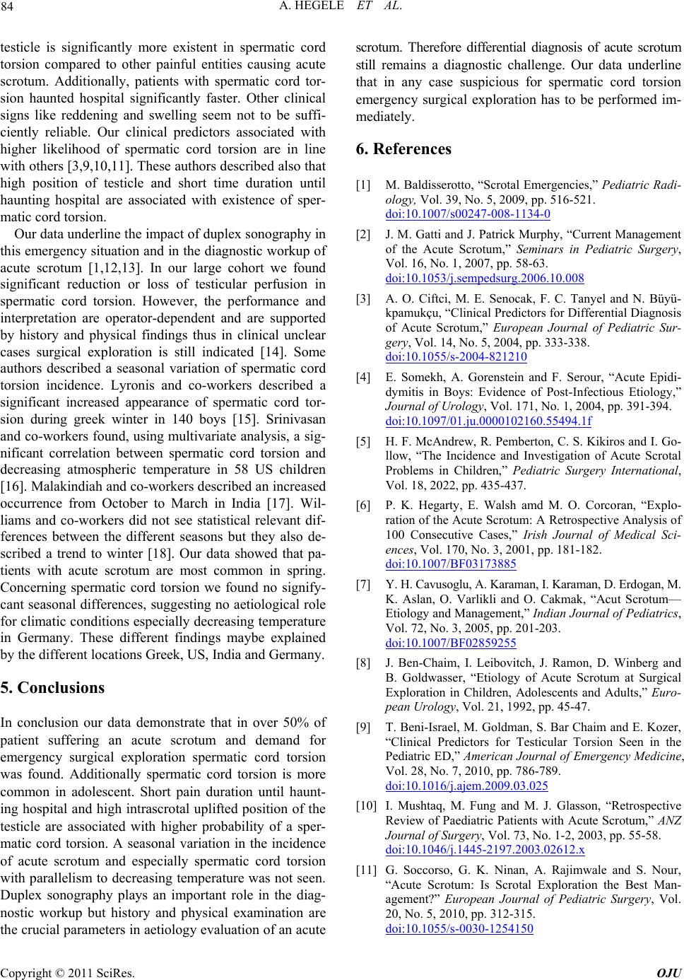
scribed a trend to winter [18]. Our data showed that pa-
tients with acute scrotum are most common in spring.
Concerning spermatic cord torsion we found no signify-
cant seasonal differences, suggesting no aetiological role
for climatic conditions especially decreasing te mperature
in Germany. These different findings maybe explained
by the different locations Greek, US, India and Germany.
5. Conclusions
In conclusion our data demonstrate that in over 50% of
patient suffering an acute scrotum and demand for
emergency surgical exploration spermatic cord torsion
was found. Additionally spermatic cord torsion is more
common in adolescent. Short pain duration until haunt-
ing hospital and high intrascrotal uplifted position of the
testicle are associated with higher probability of a sper-
matic cord torsion. A seasonal variation in the incidence
of acute scrotum and especially spermatic cord torsion
with parallelism to d ecreasing temperature was not seen.
Duplex sonography plays an important role in the diag-
nostic workup but history and physical examination are
the crucial parameters in aetio logy evaluation of an acu te
scrotum. Therefore differential diagnosis of acute scrotum
still remains a diagnostic challenge. Our data underline
that in any case suspicious for spermatic cord torsion
emergency surgical exploration has to be performed im-
mediately.
6. References
[1] M. Baldisserotto, “Scrotal Eme rgencies,” Pediatric Radi-
ology, Vol. 39, No. 5, 2009, pp. 516-521.
doi:10.1007/s00247-008-1134-0
[2] J. M. Gatti and J. Patrick Murphy, “Current Management
of the Acute Scrotum,” Seminars in Pediatric Surgery,
Vol. 16, No. 1, 2007, pp. 58-63.
doi:10.1053/j.sempedsurg.2006.10.008
[3] A. O. Ciftci, M. E. Senocak, F. C. Tanyel and N. Büyü-
kpamukçu, “Clinical Predictors for Differential Diagnosis
of Acute Scrotum,” European Journal of Pediatric Sur-
gery, Vol. 14, No. 5, 2004, pp. 333-338.
doi:10.1055/s-2004-821210
[4] E. Somekh, A. Gorenstein and F. Serour, “Acute Epidi-
dymitis in Boys: Evidence of Post-Infectious Etiology,”
Journal of Urology, Vol. 171, No. 1, 2004, pp. 391-394.
doi:10.1097/01.ju.0000102160.55494.1f
[5] H. F. McAndrew, R. Pemberton, C. S. Kikiros and I. Go-
llow, “The Incidence and Investigation of Acute Scrotal
Problems in Children,” Pediatric Surgery International,
Vol. 18, 2022, pp. 435-437.
[6] P. K. Hegarty, E. Walsh amd M. O. Corcoran, “Explo-
ration of the Acute Scrotum: A Retrospective Analysis of
100 Consecutive Cases,” Irish Journal of Medical Sci-
ences, Vol. 170, No. 3, 2001, pp. 181-182.
doi:10.1007/BF03173885
[7] Y. H. Cavusoglu, A. Karaman, I. Karaman, D. Erdogan, M.
K. Aslan, O. Varlikli and O. Cakmak, “Acut Scrotum—
Etiology and Management,” Indian Journal of Pediatrics,
Vol. 72, No. 3, 2005, pp. 201-203.
doi:10.1007/BF02859255
[8] J. Ben-Chaim, I. Leibovitch, J. Ramon, D. Winberg and
B. Goldwasser, “Etiology of Acute Scrotum at Surgical
Exploration in Children, Adolescents and Adults,” Euro-
pean Urology, Vol. 21, 1992, pp. 45-47.
[9] T. Beni-Israel, M. Goldman, S. Bar Chaim and E. Kozer,
“Clinical Predictors for Testicular Torsion Seen in the
Pediatric ED,” American Journal of Emergency Medicine,
Vol. 28, No. 7, 2010, pp. 786-789.
doi:10.1016/j.ajem.2009.03.025
[10] I. Mushtaq, M. Fung and M. J. Glasson, “Retrospective
Review of Paediatric Patients with Acute Scrotum,” ANZ
Journal of Surgery, Vol. 73, No. 1-2, 2003, pp. 55-58.
doi:10.1046/j.1445-2197.2003.02612.x
[11] G. Soccorso, G. K. Ninan, A. Rajimwale and S. Nour,
“Acute Scrotum: Is Scrotal Exploration the Best Man-
agement?” European Journal of Pediatric Surgery, Vol.
20, No. 5, 2010, pp. 312-315.
doi:10.1055/s-0030-1254150
Copyright © 2011 SciRes. OJU
A. HEGELE ET AL.
Copyright © 2011 SciRes. OJU
85
[12] P. Gunther, J. P. Schenk, R. Wunsch, S. Holland-Cunz, U.
Kessler, J. Trogera and K. L. Waag, “Acute Testicular
Torsion in Children: The Role of Sonography in the Dia-
gnostic Workup,” European Radiology, Vol. 16, No. 11,
2006, pp. 2527-2 532. doi:10.1007/s00330-006-0287-1
[13] P. Pavlica and L. Barozzi, “Imaging of the Acute Scro-
tum,” Eu ropean Radiology, Vol. 11, No. 2, 2001, pp. 220-
228. doi:10.1007/s003300000604
[14] P. Pepe, P. Panella, M. Pennisi and F. Aragona, “Does
Color Doppler Sonography Improve the Clinical Assess-
ment of Patients with Acute Scrotum?” European Journal
of Radiology, Vol. 60, No. 1, 2006, pp. 120-124.
doi:10.1016/j.ejrad.2006.04.016
[15] I. D. Lyronis, N. Ploumis, I. Vlahakis and G. Charissis,
“Acute Scrotum-Etiology, Clinical Presentation and Sea-
sonal Variation,” Indian Journal of Pediatrics, Vol. 76,
2009, pp. 407-410.
[16] A. K. Srinivasan, J. Freyle, J. S. Gitlin and L. S. Palmer,
“Climatic Conditions and the Risk of Testicular Torsion
in Adolescent Males,” Journal of Urology, Vol. 178, No.
6, 2007, pp. 2585-2588. doi:10.1016/j.juro.2007.08.049
[17] G. C. Malakindiah, R. K. Jindal and S. Vaidyanathan,
“Seasonal Variation in the Occurrence of Testicular Tor-
sion at Chandigarh, India,” Indian Journal of Urology,
Vol. 2, 1986, pp. 69-72.
[18] C. R. Williams, K. J. Heaven and D. B. Joseph, “Testicu-
lar Torsion: Is There a Seasonal Predilection for Occur-
rence?” Urology, Vol. 61, No. 3, 2003, pp. 638-641.
doi:10.1016/S0090-4295(02)02498-6