Cordyceps sinensis Acts as an Adenosine A Receptor Agonist on Mouse Melanoma and 267
3
Lung Carcinoma Cells, and Human Fibrosarcoma and Colon Carcinoma Cells
produced by Jiang Men Baode Biological Technology
(Guangdong, China). They were extracted with hot water
(70˚C) for 5 min and the extract was then filtered and ly-
ophilized. The lyophilized powder (WECS) was sealed in
bottles and kept in a refrigerator (4˚C) until use. 3-Ethyl-
5-benzyl-2-methyl-4-phenylethynyl-6-phenyl-1,4-(±)-
dihydropyridine-3,5-di-carboxylate (MRS1191, a selec-
tive A3-R antagonist), cordycepin (3’-deoxyadenosine),
adenosine and fetal bovine serum (FBS) were purchased
from Sigma Chemical Co. (St. Louis, MO). Dulbecco’s
modified Eagle’s medium (DMEM) with L-glutamine,
Earle’s minimum essential medium (EMEM) plus glu-
tamax, RPMI1640 medium and MEM non-essential
amino acids (NEAA) 100 × solution were from Invitro-
gen Co. (Grand Island, NY). Dulbecco’s phosphate-
buffered saline without calcium and magnesium [DPBS
(–)] was from Nissui Pharmaceutical Co., Ltd (Tokyo,
Japan). EDTA trypsin solution (EDTA, 0.02%; trypsin,
0.1%) and penicillin/streptomycin solution (penicillin,
50,000 U/mL; streptomycin, 50 mg/mL) were obtained
from Cosmo Bio Co., Ltd. (Tokyo, Japan).
2.2. Cells
The mouse B16-BL6 epithelial-like melanoma cell line
was kindly provided by Professor Futoshi Okada of Tot-
tori University (Yonago, Japan). The mouse Lewis lung
carcinoma cell line and CW-2 human colon carcinoma
cell line were provided by the RIKEN CELL BANK
(Tsukuba, Japan). The human HT1080 epithelial-like
fibrosarcoma cell line was purchased from DS Pharma
Biomedical Co., Ltd. (Suita, Japan). B16-BL6 cells were
cultured in DMEM containing 10% FBS and a 0.1%
penicillin/streptomycin solution. LLC cells were cultured
in DMEM containing 10% FBS. HT1080 cells were cul-
tured in EMEM containing 10% FBS and a 1% NEAA.
CW-2 cells were cultured in RPMI1640 containing 10%
FBS.
2.3. Growth Curves of B16-BL6 Cells,
LLC Cells, HT1080 Cells and
CW-2 Cells
Sub-confluent cancer cells were harvested with EDTA
trypsin solution and resuspended as 1 × 10⁵ cells/2 mL in
each well of a 12-well culture plate, and then treated with
10 µg/mL WECS. MRS1191 (0.1 and 1 µM) was added
to the wells 30 min before the addition of WECS. To
evaluate the effect of MRS1191 alone on cell growth, 0,
0.1, 1 and 10 μM of MRS1191 were added to each cul-
ture plate at 0 h. After 24, 48 and 72 h incubation at 37˚C,
viable cells were counted with a Coulter counter (Coulter
Z1; Beckman Coulter, Inc., Tokyo, Japan).
2.4. Determination of Cordycepin and Adenosine
Content in WECS
HPLC with electrochemical detection (HPLC-ECD) was
used to quantify cordycepin and adenosine in WECS,
according to a previously described method [4]. Briefly,
20 μL of WECS (1 mg/mL) was injected into the
HPLC-ECD system. The analytical column was a Cos-
mosil 5C18-AR-II (particle diameter, 5 μm; column size,
150 × 4.6 mm I.D.; Nacalai Tesque, Kyoto, Japan) col-
umn equipped with a guard column (10 × 4.6 mm I.D.)
and the mobile phase was 20 mM sodium phosphate
buffer, pH 7.0, including 10% (v/v) methanol. The flow
rate was 1 mL/min and the column temperature was set
at 25˚C. Quantitation of cordycepin and adenosine was
performed by comparison of the peak area with those of
authentic cordycepin and adenosine, respectively.
2.5. Statistical Analyses
The data are expressed as the mean ± S.D. (N = 3). Sta-
tistical analyses were performed by one-way ANOVA
followed by Tukey’s Multiple Comparison Test using
PRISM Version 4 (GraphPad Software, Inc., San Diego,
CA). Differences were considered significant at P < 0.05.
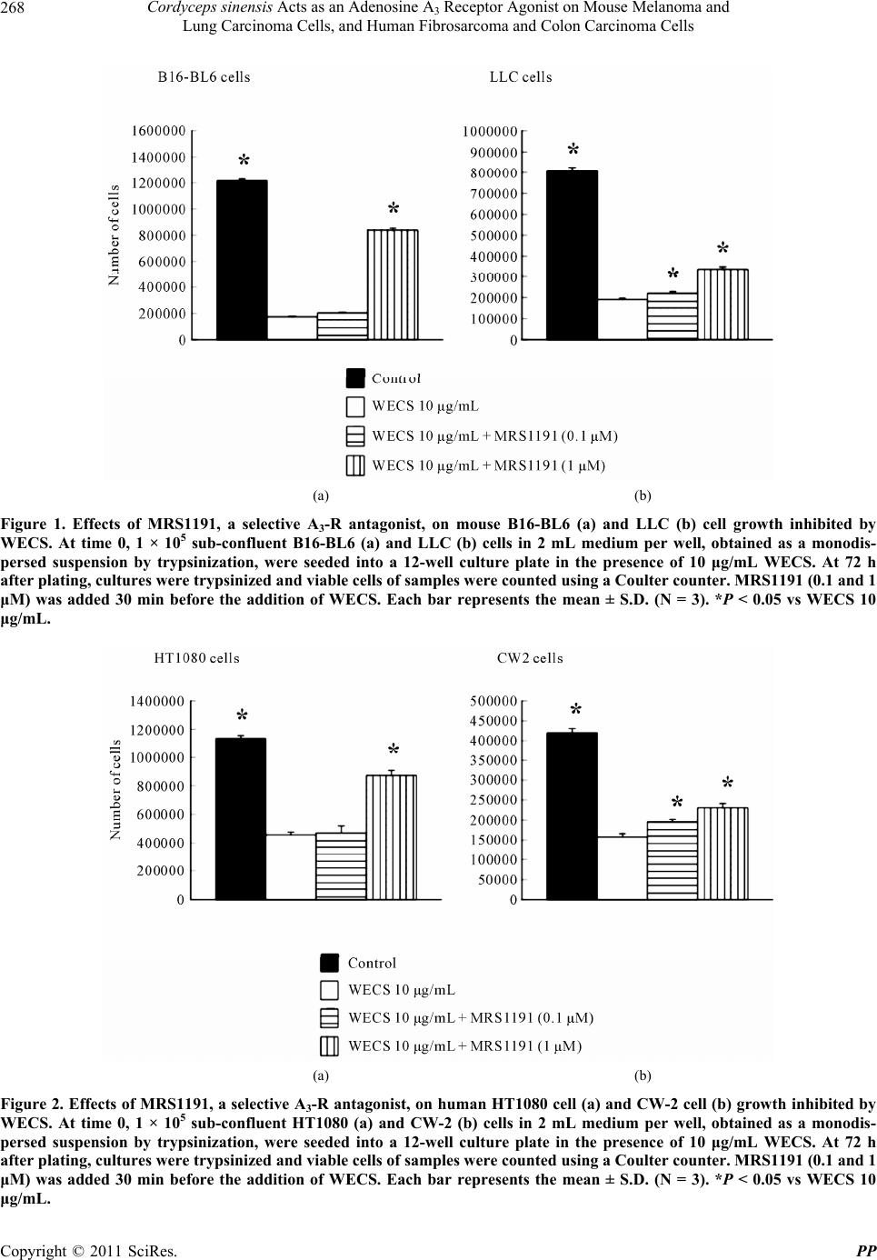
3. Results
In mouse cancer cell lines, WECS (10 µg/mL) inhibited
the growth of B16-BL6 cells and LLC cells potently to
14% and 23% of the non-treated control at 72 h, respec-
tively. When B16-BL6 cells were premixed with 1 µM
MRS1191, a selective A3-R antagonist, 30 min before the
addition of WECS (10 µg/mL), the cell numbers were
significantly antagonized to 69% of the control value.
When LLC cells were premixed with 0.1 and 1 µM
MRS1191, 30 min before the addition of WECS (10
µg/mL), the cell numbers were significantly antagonized
to 27% and 41% of the control value, respectively (Fig-
ure 1).
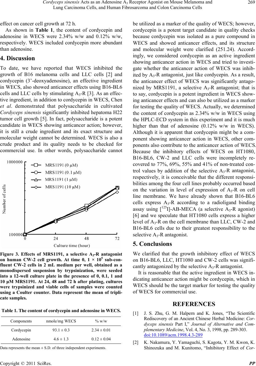
In human cell lines, WECS (10 µg/mL) inhibited the
growth of HT1080 cells and CW-2 cells potently to 40%
and 38% of the non-treated control at 72 h, respectively.
When HT1080 cells were premixed with 1 µM MRS1191,
30 min before the addition of WECS (10 µg/mL), the cell
numbers were significantly antagonized to 77% of the
control value. When CW-2 cells were premixed with 0.1
and 1 µM MRS1191, 30 min before the addition of
WECS (10 µg/mL), the cell numbers were significantly
antagonized to 46% and 55% of the control values, re-
spectively (Figure 2).
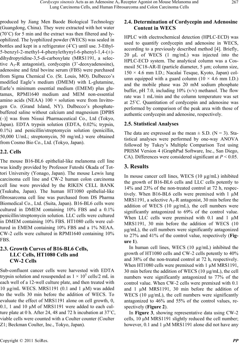
In Figure 3, showing representative data using CW-2
cells, 10 μM MRS1191 slightly reduced the cell number;
however, 0.1 and 1 µM MRS1191 alone did not have any
Copyright © 2011 SciRes. PP