 Vol.1, No.3, 29-43 (2011) doi:10.4236/scd.2011.13004 C opyright © 2011 SciRes. Openly accessible at http://www.scirp.org/journal/SCD/ Stem Cell Discovery Lineage restriction of adult human olfactory-derived progenitors to dopaminergic neurons* Meng Wang, Chengliang Lu, Hong Li, Mengsheng Qiu, Welby Winstead, Fred Roisen# Department of Anatomical Sciences and Neurobiology, University of Louisville, Louisville, USA. #Corresponding Author: fjrois01@louisville.edu Received 11 June 2011; revised 5 July 2011; accepted 30 July 2011. ABSTRACT Human adult olfactory epithelium contains neu- ral progenitors (hONPs) which replace damaged cellular components throughout life. Methods to isolate and expand the hONPs which form neuronspheres in vitro have been developed in our laboratory. In response to morphogens, the hONPs differentiate along several neural line- ages. This study optimized conditions for the differentiation of hONPs towards dopaminergic neurons. The hONPs were treated with Sonic hedgehog (Shh), in the presence or absence of retinoic acid (RA) and/or forskolin (FN). Transcrip- tion factors (Nurr1, Pitx3 and Lmx1a) that pro- mote embryonic mouse or chicken dopaminergic deve lopment w ere e mployed to determine if they would modulate lineage restriction of these adult human progenitors. Four expression vectors (pIRES-Pitx3-Nurr1, pLN-CX2-Pitx3, pLN-CX2-Nurr1 and pLNCX2-Lmx1a) were transfected into the hONPs, respectively. Transcription factor expre- ssion and the rate-limiting enzyme in dopamine synthesis tyrosine hydroxylase (TH) were de- tected in the transfected cells after 4 month-se- lection with G418, indicating transfected hONPs were stably restricted towards a dopaminergic li n ea g e. Fu r th ermore, a dopamine enzyme immu- noassay (EIA) was employed to detect the syn- thesis and release of dopamine. The most efficient transfection paradigm was determined. Several neurotrophic factors were detected in the pre- transfected hONPs which have potential roles in the maintenance, survival and proliferation of dopaminergic neurons. Therefore the effect of transfection on the neurotrophin synthesis was examined. Transfection did not alter synthesis. The use of olfactory progenitors as a cell-based therapy for Parkinson’s disease (PD) would al- low harve st without in vasive surgery, p rovide a n autologous cell population, eliminate need for immunosuppression and avoid the ethical con- cerns associated with embryonic tissues. This study suggests that specific transcription fac- tors and treatment with morphogens can re- strict human adult olfactory-derived progenitors to a dopaminergic neuronal lineage. Future stud- ies will evaluate the utility of these unique cells in cell-replacement paradigms for the treatment of PD like animal models. Keywords: Human Olfactory Epithelium; Progenitors; Dopaminergic Neurons; Parkinson’ s D i s e ase 1. INTRODUCTION Parkinson’s disease (PD) remains one of the leading causes of chronic degenerative neurological disability, which affects more than 6,000,000 people world-wide, with approximately 60,000 new cases diagnosed each year in the United States [1]. The incidence rises with age, being approximately 1:1000 overall and 1% of the population over the age of 60 and 4% in those over 80 years. Unfortunately, the mortality rate of PD has in- creased steadily in recent years [2,3]. PD is characterized by the extensive loss of dopaminergic (DA) neurons in the substantia nigra (SN) in the midbrain [4]. Currently the principle treatment for PD is oral L-3, 4-dihydroxy- phenylalanine (L-dopa) [5], which is the precursor of do- pamine that can pass the blood-brain-barrier [6]. L-dopa promotes symptomatic relief, but with time becomes less effective for two reasons: 1) During the progression of the disease the neurons become less sensitive to the drug [7] and 2) L-DOPA does not prevent or rescue the DA neurons from degeneration [8,9]. Recent research has attempted to find cell populations that can be used to replace lost or degenerating dopaminer- *This work was supported by the Dishman Family Foundation and Funds from RhinoCyte™, Inc. (OICB070316).  M. Wang et al. / Stem Cell Discovery 1 (2011) 29-43 Copyright © 2011 SciRes. Openl y accessible at http://www.scirp.org/journal/SCD/ 30 gic neurons [2,10,11]. The basic concept of cell replace- ment therapy is to restore function lost as a result of the disease in the central nervous system (CNS) by replacing degenerating or lost cells with viable functional cells. Recent studies also suggest that the engraftment of stem cells or progenitors can up-regulate or enhance existing endogenous progenitor populations [12-14]. Studies have employed neural cell grafts obtained from the fetal ventral mesencephalic (VM) dopaminergic neurons [15-20]. However, this frequently resulted in significant dyskinesia [21-24]. Even when clinical improvements were achieved in the absence of dyskinesia, the amount of tissue required for each PD patient necessitated a minimum of 4-5 fetal brains [25]. This requirement in- creased the possibility of viral or bacterial infection and significantly limited the utility of this approach. In addi- tion the number of surviving neurons was highly limited as the majority of the engrafted cells died in the initial days following transplantation [15,20,24]. The limited supply of fetal VM cells coupled with their poor graft viability severely limited the therapeutic utility of this population for the treatment of PD. Therefore, an alter- nate expandable source of dopamine cells has become a major research focus [26-29]. Stem cells are undifferentiated cells with an unlimited capacity for self-renewal and the potential for lineage re- striction (maturation) into one or more specific cell types, depending on their origin and the micro-environmental signals that they receive [28,30]. These characteristics make stem cells an attractive target population for PD cell replacement therapy [31-34]. Human embryonic stem cells (hESCs), lineage-restricted towards dopaminergic neurons when transplanted into a rodent model of PD, provide a significant relief of symptoms. However, with time, animals engrafted with hESCs have frequently de- veloped teratomas [35]. Clearly an alternate approach is warranted. Human olfactory epithelium (OE) is a unique source for neural progenitors that can be harvested by mini- mally invasive endoscopic nasal surgery without a cra- niotomy. Furthermore since no demonstrable olfactory deficits result from OE biopsy [36], the tissue can be used to generate an autologous progenitor population from patients with PD. An autologous cell source pro- vides total histocompatability and thus eliminates the need for immunosuppressive therapy as well as long waiting lists for available matched tissue. Previously our laboratory developed methods for the isolation and cul- ture of a neurosphere forming population [37]. To date more than 150 patient-specific cell lines of human ol- factory neural progenitors (hONPs) have been estab- lished from primary cultures of human adult olfactory epithelium isolated from cadavers [37] and patients un- dergoing endoscopic sinus surgery [36]. Our studies have shown that the hONPs have the potential to differentiate along several neural lineages following exposure to en- vironmental signals in vitro [38]. The objective of this study was to determine if hONPs could be lineage restricted towards dopaminergic neu- rons and if so to optimize the methodology. Molecular techniques were applied for the transfection of Nurr1 [34, 39], Pitx3 [40,41] and Lmx1a [42], transcription factors which promote dopaminergic differentiation. The trans- fection effects of different paradigms were evaluated and compared. Several studies have shown that neurotrophic factors, such as brain-derived neurotrophic factor (BDNF), cil- iary neurotrophic factor (CNTF), neurotrophin-3 (NT-3), etc. are important for the survival and function of dopa- minergic neurons in CNS [43-47]. Recent studies also indicate that the neurotrophins have the potential to op- timize the local micro-environment of the damaged area, and thereby induce endogenous stem cells to replace or rescue degenerating neurons [48,49]. HONPs derived from adult human olfactory epithelium have been shown to produce and release neurotrophins [10,50,51], which could further support their use in a cell-based therapy for PD. Therefore, this study also evaluated the ability of pre and post transfected hONPs to synthesize key neuro- trophins. 2. MATERIAL AND METHODS 2.1. Cell Culture The two patient-specific olfactory progenitor lines used in this study were obtained from adult olfactory epithe- lium harvested from a 42-year-old female patient and a 20-year-old male via endoscopic biopsy [37]. The tissues were cultured to allow the emergence and harvest of hONPs as previously described [36,52]. The hONPs were thawed from frozen stock that was maintained in liquid nitrogen and cultured in minimal essential medium (MEM) with 10% heat inactivated fetal bovine serum (FBS, GIBCO, Grand Island, NY) (10% OE) for one week. The hONPs were adapted to serum-free growth media via serial dilution of serum every day for 4 days until the cells were finally cultured in DFBNM (DMEM/F12 supplemented with 1% B27 and 0.5% N2 and 100 μg/ml gentamycin (GIBCO, Grand Island, NY)) [52]. Parallel independent experiments were performed on hONP lines from the two different patient lines. Since equivalent results were achieved, data from only one line has been presented. 2.2. Construction of Expression Plasmids The mouse Nurr1 cDNA was cloned into the pLNCX2 expression vector (Clontech) between ClaI. Similarly,  M. Wang et al. / Stem Cell Discovery 1 (2011) 29-43 Copyright © 2011 SciRes. http://www.scirp.org/journal/SCD/ 3131 the rat Pitx3 and mouse Lmx1a cDNA were inserted into pLNCX2 vector between ClaI. For the Nurr1 and Pitx3 co-expression vector, Nurr1 cDNA was cloned into pIRES (Clontech) between XbaI and SalI, and pitx3 was inserted between EcoRIs. The pLNCX2 and pIRES ex- pression vectors served as controls (Figure 1). All ex- pression vectors were verified by extensive DNA se- quencing. Openly accessible at 2.3. Transfection and Selection All plasmid constructs were introduced into the hONPs by liposomal transfection. The cells were plated on glass coverslips in six-well plates (5 × 10⁴ cells per 35 mm well) in DFBNM without antibiotics 1 day before transfection. HONPs were transfected with each plasmid (4 μg/well) for 24 hours according to the manufacture’s protocol (Li- pofectamine 2000, Invitrogen, Carlsbad, California). One day after transfection, the cells were fed with 10% FBS in MEM and selected with G418 (400 μg/ml; Invitrogen, Carlsbad, California). The selection pressure was kept for up to 4 months to insure a purified stably transfected cell population. Immunocytochemistry and Western blot analysis were applied to detect several dopaminergic neuronal markers. After a four-month selection, the trans- fected hONPs were frozen in liquid nitrogen for addi- tional four-six months of storage. After removal from cry- ostorage and several days’ recovery in MEM with 10% FBS at 37˚C, the dopaminergic lineage restriction was probed with immunocytochemistry and Western blot analy- sis. 2.4. Treatment with Morphogens The hONPs were treated with Sonic hedgehog (Shh) in the presence or absence of retinoic acid (RA, 1 μM) and/or forskolin (FN, 5 μM) [52]. Highly purified Shh (kindly provided under a Material Transfer Agreement with Curis and Wyeth, Inc.) was applied to hONPs and com- pared to a commercially available control sample obtained from Sigma to determine the extent to which purification of Shh can affect the expression of tyrosine hydroxylase (TH). The hONPs were plated on glass coverslips in six-well plates (5 × 10⁴ cells/35 mm well) in DFBNM and treated with medium containing various concentrations and com- binations of RA, FN, and Shh for 7 days (CO2 atmo- sphere at 5% and temperature of 37˚C). Treatment with Shh included several concentrations: 0.25 mg/ml (Shh0.25), 0.1 mg/ml (Shh0.1), 0.05 mg/ml (Shh0.05), 0.025 mg/ml (Shh0.025) in the presence or absence of Figure 1. Construction of expression plasmids.  M. Wang et al. / Stem Cell Discovery 1 (2011) 29-43 Copyright © 2011 SciRes. Openl y accessible at http://www.scirp.org/journal/SCD/ 32 1 μM retinoic acid (RA1) and/or 5 μM forskolin (FN5). After treatment, the TH expression was determined at 1 - 7 days in vitro by immunocytochemical analysis. Once the optimized environment for inducing dopaminergic neurons was determined, the medium containing the optimized combination was applied to stably transfected hONPs to further improve the yield of these neurons. 2.5. Immunocytochemistry The hONPs (5 × 10⁴ cells/well) were plated on 35 mm round glass coverslips in six-well plates (Becton, Dick- inson and Co.) and incubated at 37˚C in 5% CO2/95% air for 24 hours and treated with RA, FN, and Shh or transfected and selected for different periods of time prior to fixation for immunofluorescence. 4, 6-diamidi- no-2-phenylindole dihydrochloride (DAPI) (1:1000, 2 mg/ml, Molecular Probes, Eugene, OR) was applied in culture for 30 minutes at 37˚C for vital nuclear staining. The coverslips were rinsed with cytoskeletal buffer (CB) twice and fixed in 3% paraformaldehyde in CB (10 min- utes). 0.2% Triton X-100 (10 minutes, Sigma) in tris buf- fered saline (TBS) was applied and cells were incubated (1 hour) in 3% bovine serum albumin (BSA) in TBS. Pri- mary antibodies were applied overnight (4˚C). After 30 minutes washing (10 minutes each, 3 times) in TBS, the cells were incubated with secondary antibodies: Texas- red conjugated goat anti-rabbit immunoglobulin G (IgG), Texas-red-conjugated goat anti-mouse IgG, Cy2-conju- gated goat anti-mouse IgG and/or Cy2-conjugated goat anti-rabbit IgG (all diluted 1:600, Cy2, Jackson Immunol- ogy Research Laboratories; Texas red, Molecular Probes). The coverslips were rinsed in TBS for 30 minutes (10 minutes each, 3 times) and mounted on slides. The slides were examined with confocal microscopy. All experi- ments were repeated a minimum of two times to ensure the specificity of staining; only one set of data has been presented since similar results were obtained. 2.6. Western Blot Analysis Western blot analysis was used to further examine and confirm the immunofluorescence studies. Proteins from hONPs transfected with control vectors, as well as hONPs transfected with the vectors plus each combina- tion of transcriptions factors (pLNCX2-Pitx3, pLNCX2- Nurr1, pLNCX2-Lmx1a, pIRES-Pitx3-Nurr1), cultured in DFBNM, selected in all groups were collected in cell lysis buffer (Sigma, St. Louis, MO). After 15 minutes of incubation on ice, samples were centrifuged for 30 min- utes (4˚C) and the protein concentration of each super- natant was determined. The protein samples (20 μg/well) were electrophoresed on 10% SDS-polyacrylamide gels along with standardized-molecular-size marker proteins in an adjacent lane and transferred from gel to nitrocel- lulose paper. Nonspecific binding was blocked (1 hour) with 5% nonfat dry milk in TBS-Tween (TBST) buffer. Blots were incubated (4˚C overnight) in primary anti- bodies (anti-TH, MAB; anti-actin, MAB). Blots were washed three times for 10 minutes in TBST, after which they were incubated (1 hour, room temperature) mono- clonal horseradish peroxidase-labeled anti-mouse IgG (1:2000). ECL Western blotting detection reagents (A- mersham Biosciences) were used to identify bound an- tibodies. Densitometry of the protein bands was carried out on a high performance chemiluminescence film (A- mersham Biosciences). Data was analyzed using the Image-J software programs supplied by the NIH official website (http://rsb.info.nih.gov/ij/ ). 2.7. Dopamine Assay Stably transfected hONPs were plated into flasks (25 cm2, Corning) at 105 per flask before they were adapt- ed to the absence of serum via serial dilution of serum every day for 4 days until the cells were finally cultured in DFBNM, which was collected daily after the serum was totally eliminated from the medium. The DFBNM collected from each restricted hONP line was then con- centrated to 1/50 volume respectively by centrifugal filters (Amicon Ultra-15, Millipore). The differentiated hONPs were then collected and lysed (lysis buffer, Sigma). Do- pamine expression was analyzed quantitatively in the concentrated medium as well as in the cell lysates with a dopamine enzyme immunoassay kit (Dopamine EIA, Immuno Biological Laboratories, Inc.), according to the manufacture’s protocol. 2.8. Neurotrophin Assay Pre- and post-transfected hONPs were plated into flasks (25 cm2, Corning) at 5 × 10⁵ per flask and cultured in 10% OE media for two days before they were adapted to the absence of serum via serial dilution of serum e- very day until they were finally cultured in DFBNM. The differentiated hONPs were then collected and lysed (lysis buffer, Sigma). Neurotrophins were detected in the extracted protein with different enzyme-linked immu- nosorbent assay (ELISA) kits (BDNF, Chemicon; CNTF, Quantikine; NT-3, Chemicon) respectively, according to the manufacture’s protocol. The ELISA absorbance (OD) was obtained with a microplate spectrophotometer (Spec- tra-max Plus), and the results were plotted and calcu- lated with the compatible software (Softmax Pro). 3. RESULTS Cryopreserved vials of the two representative hONP  M. Wang et al. / Stem Cell Discovery 1 (2011) 29-43 Copyright © 2011 SciRes. Openl y accessible at http://www.scirp.org/journal/SCD/ 3333 lines were obtained from storage and grown for 1 - 2 weeks prior to the start of these experiments to insure equivalent passage (4 - 8) and sufficient cell numbers for the following studies. 3.1. Transfection of Olfactory-Derived Progenitors (hONPs) to Achieve Dopaminergic Lineage Restriction HONPs were obtained from previously frozen stock with low passage number (4 - 8) and maintained in MEM 10 medium during their recovery period. These mitotically active cells divided every 18 - 20 hour which typically required passage three times per week as previ- ously described. The heterogeneous nature of the hONP population prior to transfection was determined by im- munocytochemistry. No reactivity was observed for Pitx3, Nurr1, Lmx1a with pre-transfected hONPs and only a few (5 - 10%) of them were positive for the do- pamine precursor, TH, when treated conditionally [53]. Low passages (Passage 4 - 8) of hONPs from 2 different patient-specific cell lines were employed in parallel trans- fection experiments. To examine the phenotypic expres- sion of hONPs after transfection and selection, repre- sentative cultures as well as their respective pre-trans- fection controls were evaluated. Non-transfected hONPs or those transfected with lipofectamine alone died within 1 week after selection with 400 µg/ml G418. In contrast, 30% of the transfected cells (both with the concerned genes and the control vectors) survived under the selec- tion pressure. Transfection with control vectors, single genes, or Pitx3-Nurr1 combined resulted in no morpho- logic changes compared to the typical pretreated hONPs. However, the transfected hONPs divided more slowly, with a new doubling time of three to four days, which required a feeding schedule of only twice a week and necessitated passage every 9 - 10 days. Immunofluores- cent analysis of the transfected populations demonstrated that hONPs were stably transfected and TH expressed. Human olfactory derived hONPs were transfected by pIRES-Pitx3-Nurr1 to restrict them towards DA neurons. The vector alone was employed as a con- trol. To obtain a purified population of restricted cells the transfected population was maintained in G418 for selection. Although only several weeks of selection produced relatively pure populations, an interval of four months was employed to insure sta- bility and purity. HONPs remained TH positive after transfection of pIRES-Pitx3-Nurr1, whereas the trans- fection of control vectors exhibited no phenotypic changes, demonstrating that hONPs can be restricted towards dopaminergic neurons (Figure 2). HONPs were transfected with pLNCX2-Nurr1, pL- NCX2-Pitx3, pLNCX2-Lmx1a or the vector alone as a control. The transfected cells were exposed to G-418 for selection for periods up to 4 months. HONPs were TH positive after transfection of pLNCX2-Nurr1 and pLNCX2-Pitx3, whereas the transfection of con- trol vectors resulted in no phenotypic changes. There- fore pLNCX2-Nurr1 or pLNCX2-Pitx3 can be em- ployed to lineage restrict the hONPs towards dopa- minergic neurons. In contrast, the hONPs trans- fected with pLNCX2-Lmx1a remained unreactive for TH, although positive of myc, which demonstra- ted the successful incorporation of the plasmid (Fig- ure 2). a b d e f gh c Figure 2. Immunocytochemical analysis. HONPs transfected with pIRES-Pitx3-Nurr1, pLNCX2- Pitx3 or pLNCX2-Nurr1 were tyrosine hydroxylase (TH) positive after 4 months selection with G418 (c, d, f, g), while the lines transfected with pIRES or pLNCX2 were TH negative (b, e). HONPs transfected with pLNCX2-Lmx1a were Myc positive, demonstrating that the plasmid was transfected into the nucleus (h).  M. Wang et al. / Stem Cell Discovery 1 (2011) 29-43 Copyright © 2011 SciRes. Openl y accessible at http://www.scirp.org/journal/SCD/ 34 Western blot analysis was employed to confirm quan- titatively the immunocytochemical studies of the tran- sfected hONP populations. The following transfected lines were analyzed for TH expression: hONPs trans- fected with pIRES-Pitx3-Nurr1, pLNC-X2-Nurr1, pL- NCX2-Ptx3 and pLN-CX2-Lmx1a all of which were TH positive, which indicated their potential to release dopamine. In contrast, the hONP populations’ trans- fected with the control vectors (pIRES and pLNCX2) did not express TH. β-actin, a protein that is widely expressed in all mammalian and avian cells was used as a reference protein for the comparison of TH ex- pression by the various lines. Image-J was applied for the data analysis. Each curve from B to M in Figure 3 illustrates the density of bands evident on the western gel (Figure 3a), and the area that each curve was measured. The bars in picture N represent the ratio of TH expression and ACTIN expression in the cell line. HONPs transfected with pIRES-Pitx3- Nurr1 exhibited the highest ratio for the TH and ACTIN expression, while the cells transfected with the control vector (pLNCX2 or pIRES) had the least TH staining (Figur e 3(b-n)). These results demonstrate that individual tran- scription factors have unique abilities in promoting the dopaminergic restriction of hONPs. 3.2. Transfected hONPs Remain Restricted to Dopaminergic Lineage after Removal from Cryostorage After a 4-month selection, the dopaminergic lineage re- stricted cells were cryopreserved in liquid nitrogen for additional 4 - 6 months. Following their removal from cry- ostorage and several days’ recovery in MEM10 at pIRES- Pitx3-Nurr1 to restrict them towards DA neurons. The vector alone was employed as a 37˚C, all but one of the transfected hONP populations survived under the selec- tion pressure of 400 µg/ml G418, demonstrating that these cells were stably transfected and retained their po- tential for long term storage and clinical application. Immuno-cytochemistry and Western blot analysis was applied to these previously stored populations to exam- ine their TH expression. The hONPs transfected with pLNCX2-Pitx3, pLNCX2-Nurr1 and pIRES-Pitx3-Nurr1 remained healthy and TH positive under the pressure of selection, while the pLNCX2-Lmx1a transfected line did not (Figure 4). 3.3. Lineage Restricted hONPs Produced and Released Dopamine After removal from the cryostorage, dopamine produc- tion was detected in the hONP lines which were stably transfected with concerned genes, while the cells tranfected Figure 3. a. Western blot analysis. b-g. Scanning densitometry demonstrates ACTIN-expression in a hONP line of pL-NCX2, pLNCX2-Pitx3, pLNCX2-Nurr1, pIRES and pIRES-Pitx3- Nurr1 respectively. h-m. Densitometry of TH-expression as shown in A. N. Histogram demonstrating the ratio of TH/ac- tion produced by each population. with control vectors and the non-transfected hONPs didn’t produce dopamine. The dopamine level of each sample was then divided by the concentration of protein in each specific hONP line to calculate the efficiency of dopa- mine production. Among all the 4 gene transfected lines, hONPs transfected with pIRES-Pitx3-Nurr1 exhibited the most efficient dopamine formation (Figure 5(a)). Spent medium was collected 4 days after culturing the lineage restricted hONPs. This medium was then concen- trated to 1/50 volume respectively, and dopamine E. I. A. was applied to detect the dopamine release (extracellular levels). Data were calculated in the same manner as the intracellular dopamine analysis. Lower levels of dopa- mine were detected in the concentrated media compared to the corresponding analysis of the cell lysis. The grea- test level of dopamine release was detected in pIRES- Pitx3-Nurr1 transfected hONPs compared to the other restricted cell lines (Figure 5(b)).  M. Wang et al. / Stem Cell Discovery 1 (2011) 29-43 Copyright © 2011 SciRes. Openl y accessible at http://www.scirp.org/journal/SCD/ 3535 a cd e gh i Figure 4. Immunocytochemistry (a-g) and western blot analysis (i) demonstrating that hONPs transfected with pLNCX2-Pitx3, pLNCX2-Nurr1 and pIRES-Pitx3-Nurr1 remain healthy and TH positive following removal from cryostorage under selection pressure (d, f, g). In contrast, the Lncx2-Lmx1a transfected line no longer expressed TH (h). (a) (b) Figure 5. Histograms demonstrating the ratio of dopamine for- mation (pg/100 µl) to total protein concentration (mg/ml) of cells transfected with pIRES-Pitx3-Nurr1, pLNCX2-Pitx3, pLNCX2- Nurr1, pLNCX2-Lmx1a, pIRES, pLNCX2 and non-transfected hONPs. HONPs transfected with pIRES-Pitx3-Nurr1 exhibited the highest levels of intracellular and extracellular dopamine produc- tion. Dopamine production and release were enhanced in hONPs treated with the morphogens. 3.4. The Effect of Morphogens on Tyrosine Hydroxylase (TH) Expression, Dopamine Formation and Release HONPs were cultured in DFBNM along with RA (1 µM), FN (5 µM) and either of two different sources (puri- ties) of Shh for four days. Both Shh treatments resulted in greater expression than in those cultured solely in DFBNM. TH expression was greater in the cells that were treated with highly purified Shh than the commercial product obtained from SIGMA when applied for same period of time (Figure 6). HONPs treated with RA1FN5 and highly purified Shh expressed seemingly more intensive TH reactivity in the positive cells (Figure 7(a)). Therefore, the concentration of Shh was reduced to determine the lowest concentra- tion of Shh that could drive the hONPs towards dopa- minergic neurons. In contrast to the response when a high level of Shh was applied, the reduction of the Shh to 0.025 mg/ml applied with RA (1 µM) & FN (5 µM) did not produce an immediate response. The hONPs be- came TH positive only after 18 hours of treatment with highly purified Shh; however, they were healthy and maintained TH expression for longer periods. The ap- plication of RA and FN promoted an even greater ex- pression of TH (Figure 6 A). Therefore, the optimal con- ditions for restricting the hONP lineage to dopaminergic neurons (under these defined conditions) was determined to be DFBNM supplemented with RA1FN5Shh0.025 (Fig- ure 7(b)). Stably transfected hONPs were treated with a cock- tail of RA1FN5Shh0.025 to determine if a combination of genetic modification and morphogen exposure would increase intracellular and intercellular dopamine levels. Spent medium was collected four days after morphogenic treatment and concentrated to a 1/50 volume. The treated lineage restricted hONPs were also collected. Dopamine E. I. A. was applied to both cell lysis sample and concentrated medium. Dopamine formation efficiency was calculated as pre- viously described. HONPs transfected with pIRES- Pitx3-Nurr1 were the most efficient population with re- spect to dopamine formation and release after morpho- genic treatment (Figures 5(a)-(b)). Compared to intracel- lular and extracellular dopamine levels of the lineage re- stricted hONPs in the absence of morphogens, dopaminer- gic expression was greatly enhanced in the stably trans- fected hONPs in the presence of the combination of Shh, RA and FN (Figures 5(a)-(b)). These studies suggest that treatment with morphogens can play an important role in dopamine formation and release by the lineage restricted ONPs. h  M. Wang et al. / Stem Cell Discovery 1 (2011) 29-43 Copyright © 2011 SciRes. http://www.scirp.org/journal/SCD/ 36 (a) (b) (c) Figure 6. HONPs treated in DFBNM with a highly purified Shh(c) exhibited greater reactivity to tyrosine hydroxy- lase (TH) than those treated with commercially available Shh (b) for 3 days in the presence of RA and FN. (a) (b) Figure 7. (a) HONPs cultured in DFBNM supplemented with 0.025 mg/ml of Shh, in the presence or absence of reti- noic acid (RA) (1 µM) and forskolin (FN)(5 µm) for days indicated; (b) HONPs were tyrosine hydroxylase (TH) posi- tive following 7 days treatment with RA1FN5Shh. 3.5. Stably Transfected and Pre-transfected hONPs Produce Neurotrophins (BDNF, CNTF and NT-3) at Equivalent Levels The non(pre)-transfected hONPs were found to pro- duce neurotrophic factors such as BDNF (56.09 ± 10.24 pg/ml), CNTF (18.72 ± 1.43 pg/ml) and NT-3 (24.87 ± 6.53 pg/ml). The stably transfected lines were examined to determine if lineage restriction to dopaminergic neurons alters the synthetic capacity and activity of these neuro- trophins; no significant differences in intracellular neuro- trophin (BDNF, CNTF, NT-3) levels between transfected and non-transfected hONP lines were observed (P > 0.01), indicating that transfection did not alter neurotro- phin synthesis (Figure 8). 4. DISCUSSION AND CONCLUSIONS Parkinson’s disease, as a neurodegenerative disease, is characterized by loss of specific dopaminergic neurons in substantia nigra [4]. Although a variety of pharmaco- logical agents have been employed in the treatment of PD their effects are transient. “Proof of Concept Studies” with embryonic adrenal medulla cells [54] although end- ing in failure demonstrated the potential of cell-based re- placement therapy. Recently substantial effort has been Openly accessible at 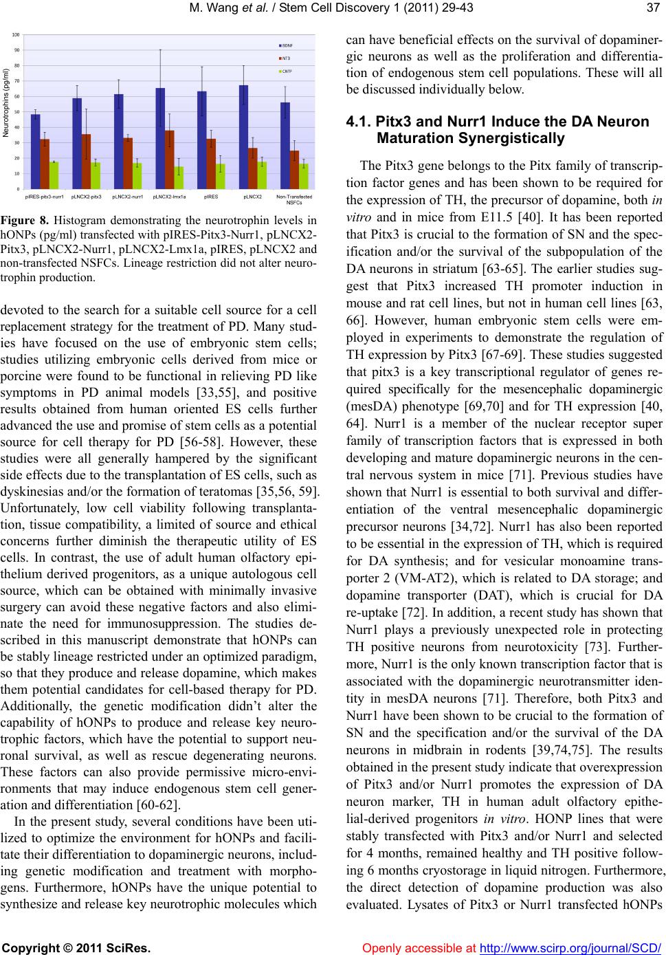 M. Wang et al. / Stem Cell Discovery 1 (2011) 29-43 Copyright © 2011 SciRes. Openl y accessible at http://www.scirp.org/journal/SCD/ 3737 Figure 8. Histogram demonstrating the neurotrophin levels in hONPs (pg/ml) transfected with pIRES-Pitx3-Nurr1, pLNCX2- Pitx3, pLNCX2-Nurr1, pLNCX2-Lmx1a, pIRES, pLNCX2 and non-transfected NSFCs. Lineage restriction did not alter neuro- trophin production. devoted to the search for a suitable cell source for a cell replacement strategy for the treatment of PD. Many stud- ies have focused on the use of embryonic stem cells; studies utilizing embryonic cells derived from mice or porcine were found to be functional in relieving PD like symptoms in PD animal models [33,55], and positive results obtained from human oriented ES cells further advanced the use and promise of stem cells as a potential source for cell therapy for PD [56-58]. However, these studies were all generally hampered by the significant side effects due to the transplantation of ES cells, such as dyskinesias and/or the formation of teratomas [35,56, 59]. Unfortunately, low cell viability following transplanta- tion, tissue compatibility, a limited of source and ethical concerns further diminish the therapeutic utility of ES cells. In contrast, the use of adult human olfactory epi- thelium derived progenitors, as a unique autologous cell source, which can be obtained with minimally invasive surgery can avoid these negative factors and also elimi- nate the need for immunosuppression. The studies de- scribed in this manuscript demonstrate that hONPs can be stably lineage restricted under an optimized paradigm, so that they produce and release dopamine, which makes them potential candidates for cell-based therapy for PD. Additionally, the genetic modification didn’t alter the capability of hONPs to produce and release key neuro- trophic factors, which have the potential to support neu- ronal survival, as well as rescue degenerating neurons. These factors can also provide permissive micro-envi- ronments that may induce endogenous stem cell gener- ation and differentiation [60-62]. In the present study, several conditions have been uti- lized to optimize the environment for hONPs and facili- tate their differentiation to dopaminergic neurons, includ- ing genetic modification and treatment with morpho- gens. Furthermore, hONPs have the unique potential to synthesize and release key neurotrophic molecules which can have beneficial effects on the survival of dopaminer- gic neurons as well as the proliferation and differentia- tion of endogenous stem cell populations. These will all be discussed individually below. 4.1. Pitx3 and Nurr1 Induce the DA Neuron Maturation Synergistically The Pitx3 gene belongs to the Pitx family of transcrip- tion factor genes and has been shown to be required for the expression of TH, the precursor of dopamine, both in vitro and in mice from E11.5 [40]. It has been reported that Pitx3 is crucial to the formation of SN and the spec- ification and/or the survival of the subpopulation of the DA neurons in striatum [63-65]. The earlier studies sug- gest that Pitx3 increased TH promoter induction in mouse and rat cell lines, but not in human cell lines [63, 66]. However, human embryonic stem cells were em- ployed in experiments to demonstrate the regulation of TH expression by Pitx3 [67-69]. These studies suggested that pitx3 is a key transcriptional regulator of genes re- quired specifically for the mesencephalic dopaminergic (mesDA) phenotype [69,70] and for TH expression [40, 64]. Nurr1 is a member of the nuclear receptor super family of transcription factors that is expressed in both developing and mature dopaminergic neurons in the cen- tral nervous system in mice [71]. Previous studies have shown that Nurr1 is essential to both survival and differ- entiation of the ventral mesencephalic dopaminergic precursor neurons [34,72]. Nurr1 has also been reported to be essential in the expression of TH, which is required for DA synthesis; and for vesicular monoamine trans- porter 2 (VM-AT2), which is related to DA storage; and dopamine transporter (DAT), which is crucial for DA re-uptake [72]. In addition, a recent study has shown that Nurr1 plays a previously unexpected role in protecting TH positive neurons from neurotoxicity [73]. Further- more, Nurr1 is the only known transcription factor that is associated with the dopaminergic neurotransmitter iden- tity in mesDA neurons [71]. Therefore, both Pitx3 and Nurr1 have been shown to be crucial to the formation of SN and the specification and/or the survival of the DA neurons in midbrain in rodents [39,74,75]. The results obtained in the present study indicate that overexpression of Pitx3 and/or Nurr1 promotes the expression of DA neuron marker, TH in human adult olfactory epithe- lial-derived progenitors in vitro. HONP lines that were stably transfected with Pitx3 and/or Nurr1 and selected for 4 months, remained healthy and TH positive follow- ing 6 months cryostorage in liquid nitrogen. Furthermore, the direct detection of dopamine production was also evaluated. Lysates of Pitx3 or Nurr1 transfected hONPs  M. Wang et al. / Stem Cell Discovery 1 (2011) 29-43 Copyright © 2011 SciRes. Openl y accessible at http://www.scirp.org/journal/SCD/ 38 were dopaminergic as determined by dopamine E.I.A. These results suggest that the transcription factors, Pitx3 and Nurr1, not only function as a dopaminergic promot- ers in chick, mouse, or human embryonic cells [41,68, 71,76], but also can participate in dopamine production in adult human olfactory-derived progenitors. Based on previous studies which focused on the regulatory func- tion of Pitx3 and Nurr1 in dopaminergic neuron promo- tion [63,68,70,72,74,77] and the studies described in this manuscript, we hypothesized that Pitx3 and Nurr1 may collaborate to induce a higher efficiency of dopamine production in midbrain DA neuron maturation. Previ- ously a synergistic effect between Pitx3 and Nurr1 on TH expression has been reported, which appeared to be spe- cies dependent occurring in human but not in embryonic murine stem cells [66-78]. The current studies demon- strate that the simultaneous transfection of Pitx3 and Nurr1 into the hONPs produced higher levels of TH ex- pression and dopamine production than transfection of either of the individual genes. We evaluated the effect of transfection on the level of the precursor (TH) and final intracellular and extracellular dopamine levels to confirm and compare the efficiency of the different transfected hONP lines. Therefore, our data, in combination with published reports in rodents [79,80] and human embry- onic stem cells [67,81], indicate that Pitx3 and Nurr1 cooperatively induce the maturation of DA neurons. We extend the previous studies to show the feasibility of genetic modification of adult human olfactory-derived progenitors to promote the generation of DA neurons. These studies demonstrate that the co-expression of Pitx3 and Nurr1 will enhance significantly the lineage restric- tion of adult human progenitors toward dopaminergic neurons which can be employed in cell-replacement paradigms for the treatment of PD. 4.2. Treatment of hONPs with Morphogens Enhances Intracellular and Extracellular Dopamine Levels Human adult epithelial derived progenitors have the potential to differentiate along several neural lineages in response to morphogenic signals in vitro [82]. For exam- ple, 11.6 (±1.5) % of hONPs expressed TH following a 7 day treatment of RA1FN5Shh (1 µM RA, 5 µM FN and 15 nM Shh), indicating that a dopaminergic lineage can be driven by exposure to these morphogens [53]. Sonic hedgehog (Shh), (RA) and Forskolin (FN) have all been shown to be crucial developmental factors that regulate neuronal specification and differentiation [83-88]. Shh has been shown to be required for the generation of ven- tral midbrain motor neurons [89,90] as well as dopa- minergic neurons in rodents [56,58,75] and chick em- bryos [59]. This study suggests that Shh increases the expression of TH and that the purity of Shh is an impor- tant determinant of TH expression. RA regulates neu- ronal differentiation in embryonic stem cells [91,92] and adult human neuronal progenitors [93, 94]. RA has sev- eral pathways through which it can effect cellular differ- entiation [95,96]. FN is an adenyl cyclase activator that increases intercellular levels of cAMP that can stimulate axonal elongation [85,86] and induce embryonic rat/mouse motor neuron survival [97,98]. Following the treatment of RA and FN, the progenitor nature of hONPs is dimin- ished, as characterized by a loss of nestin expression, and the presence of more mature neuronal markers. In this study, a combination of highly purified Shh, RA and FN was applied to the lineage restricted hONPs. The intra- cellular level of dopamine was demonstrated to be sig- nificantly increased by this treatment. This result con- firms and extends the published data by showing that these morphogens can increase TH expression by pro- genitors obtained from adult humans [53]. Furthermore, following a 4 day treatment of RA1FN5-Shh, the dopa- mine level of the spent conditioned medium was signifi- cantly enhanced, indicating that the morphogens pro- moted the release of dopamine, which is important for future studies transplanting lineage restricted hONPs into PD animal models. Among all 4 lineage restricted hONP lines, those cells transfected with pIRES-Pitx3-Nurr1 produced and released the highest levels of dopamine in the presence of Shh, RA and FN. This result is consistent with the analysis of the lineage restricted cells in the ab- sence of treatment with the morphogens. This data fur- ther supports the conclusion that hONPs transfected with pIRES-Pitx3-Nurr1 are the most efficient line in dopa- mine production studies to date, and therefore are likely candidates for engraftment into an animal model of PD. Shh is secreted by the notochord and floor plate at early stage of development [99], RA is detectable in the mid- brain of chick and mice embryos [100], and FN is highly concentrated in the rat substantia nigra [101]. The local distribution of these morphogens in situ should influence the engrafted hONPs and may further support their sur- vival and dopamine release following transplantation. The higher level of dopamine released following Shh, RA and FN treatment suggests their potential utility for cell-replacement therapy for PD. Previous studies on the non-human primate PD models, demonstrated that the transplanted responsive human embryonic progenitor cells were still capable of differentiation to DA pheno- type within the micro-environment around the lesioned adult host SN, an unexpected finding was that the en- graftment also up-regulated an endogenous progenitor population [12]. The results of our studies utilizing a paradigm that combines transfection and morphogen in- duced lineage modulation highlight the potential therapeu- tic utility of olfactory epithelial-derived neural progenitors  M. Wang et al. / Stem Cell Discovery 1 (2011) 29-43 Copyright © 2011 SciRes. Openl y accessible at http://www.scirp.org/journal/SCD/ 3939 as an autologous cell source for cell-based replacement and regenerative strategies for patients with Parkinson’s disease. 4.3. Lineage Restricted hONPs Retain Their Capability to Produce Neurotrophic Factors It’s been reported that neurotrophins such as BDNF, CNTF and NT-3 are crucial in the recovery of primate and rodent models of Parkinson’s disease [12,102]. BDNF is a member of the neurotrophin family which supports the maturation and survival of dopaminergic neurons in substantia nigra [44,103]. In the presence of BDNF, more TH positive cells can be found in cultures of ventral mecensephalic tissue than in the absence of the neurotrophin [103,104]. NT-3 belongs to the same family of neurotrophins as BDNF, and has been shown to play a protective role in the degeneration of adult central nora- drenergic neurons in vivo [105,106]. CNTF has been reported to rescue the degenerating striatal neurons in primate and rodent models [45,107]. Furthermore, the absence of CNTF leads to the apoptosis of motor neurons in adult mice [46,108]. Collectively these studies strong- ly suggest an important role for these neurotrophins in future therapeutic strategies for neurodegenerative dis- eases, including PD, Alzheimer’s disease and Huntington disease. Therefore, a cell population that can produce neurotrophins could be an ideal for therapy for these dis- eases. They can provide protective micro-environments in vivo and prevent, rescue and or replace neuronal degen- eration. The pre-transfected hONPs were found to pro- duce several neurotrophins including BDNF, NT-3, and even nerve growth factor (NGF) when in a serum en- riched medium [10]. The stably transfected lines were examined to determine if lineage restriction to dopa- minergic neurons or absence of serum alters the syn- thesis of these neurotrophins since they play a role in neuronal survival, differentiation and maturation. As shown in the results, the transfection of hONPs did not alter neurotrophin production. The post-transfected hONPs produce BDNF, NT-3 and CNTF at equivalent levels with the pre-transfected progenitors. Therefore, genetically modified hONPs can not only serve as replacements of the dead or dysfunctional dopaminergic neurons but also can provide protective micro-environments to help res- cue dying or damaged neurons from further degeneration and to enhance the endogenous progenitor populations. The stably lineage restricted hONPs are unique popula- tions with high potential for cell transplantation for ani- mal models of Parkinson’s disease. The long term goal of this study is to develop restrict- ed hONP lines that will have therapeutic utility in cell replacement strategies for patients with PD. The in vivo viability and stability are important variables, especially considering the likelihood that with time the engrafted population may die and require replacement. Therefore, experiments were undertaken to determine the stability and viability of frozen stocks of transfected cells. HONPs survived under the pressure of selection after removal from cryostorage and retained their ability to express TH, as well as produce and release dopamine and neurotro- phins, which further demonstrates the unique potential of these progenitors to perhaps serve as an autologous cell source for cell-based strategies for the long-term treat- ment of Parkinson’s disease. Human adult olfactory epithelial-derived progenitors may provide a unique autologous cell population for cell- based therapy of Parkinson’s disease, because of their potential to become dopaminergic neurons which pro- duce and release dopamine and their capability to pro- vide neurotrophic factor enriched micro-environments which support cell survival, protect cells from degenera- tion and activate endogenous stem cell populations. In vivo studies are in progress to determine the ability of hONPs to diminish Parkinson like locomotory deficits in a rodent model. REFERENCES [1] National Parkinson Founfdation (2010). http://www.parkinson.org [2] Anderson, L. and Caldwell, M.A. (2007) Human neural progenitor cell transplants into the subthalamic nucleus lead to functional recovery in a rat model of Parkinson’s disease. Neurobiology of Disease, 27, 133-140. doi:10.1016/j.nbd.2007.03.015 [3] De Lau, L.M. and Breteler, M.M. (2006) Epidemiology of Parkinson’s disease. Lancet Neurology, 5, 525-535. doi:10.1016/S1474-4422(06)70471-9 [4] Hornykiewicz, O. (1973) Parkinson’s disease: From brain homogenate to treatment. Fed Proc, 32, 183-190. [5] Bidet-Ildei, C., Pollak, P., Kandel, S., Fraix, V. and Or- liaguet, J.P. (2011) Handwriting in patients with Parkin- son disease: Effect of l-dopa and stimulation of the sub- thalamic nucleus on motor anticipation. Human Move- ment Science, 30, 783-791. [6] Hornykiewicz, O. (1973) Dopamine in the basal ganglia. Its role and therapeutic implications (including the clini- cal use of L-DOPA). British Medical Buletin, 29, 172- 178. [7] Callaway, E. (2011) Gene therapy offers hope for Park- inson’s disease. Nature, Published online 17 March 2011. [8] Lang, A.E. and Lozano, A.M. (1998) Parkinson’s disease. First of two parts. New England Journal of Medicine, 339, 1044-1053. doi:10.1056/NEJM199810083391506 [9] Sheng, D., Qu, D., Kwok, K.H., Ng, S.S., Lim, A.Y., Aw, S.S., Lee, C.W., Sung, W.K., Tan, E.K., Lufkin, T., et al. (2010) Deletion of the WD40 domain of LRRK2 in Ze- brafish causes Parkinsonism-like loss of neurons and lo- comotive defect. PLoS Genetics, 6, e1000914. doi:10.1371/journal.pgen.1000914 [10] Marshall, C.T., Lu, C., Winstead, W., Zhang, X., Xiao, M.,  M. Wang et al. / Stem Cell Discovery 1 (2011) 29-43 Copyright © 2011 SciRes. Openl y accessible at http://www.scirp.org/journal/SCD/ 40 Harding, G., Klueber, K.M. and Roisen, F.J. (2006) The therapeutic potential of human olfactory-derived stem cells. Histology and Histopathology, 21, 633-643. [11] Parish, C.L., Castelo-Branco, G., Rawal, N., Tonnesen, J., Sorensen, A.T., Salto, C., Kokaia, M., Lindvall, O. and Arenas, E. (2008) Wnt5a-treated midbrain neural stem cells improve dopamine cell replacement therapy in par- kinsonian mice. Journal of Clinical Investigation, 11 8, 149-160. doi:10.1172/JCI32273 [12] Redmond, D.E., Jr., Bjugstad, K.B., Teng, Y.D., Ourednik, V., Ourednik, J., Wakeman, D.R., Parsons, X.H., Gon- zalez, R., Blanchard, B.C., Kim, S.U., et al. (2007) Be- havioral improvement in a primate Parkinson’s model is associated with multiple homeostatic effects of human neural stem cells. Proceedings of the National Academy of Sciences of the United States of America, 104, 12175- 12180. doi:10.1073/pnas.0704091104 [13] Abdel-Salam, O.M. (2011) Stem cell therapy for alz- heimer’s disease. CNS & Neurological Disorders-Drug Targets, 10, 459-485. [14] Ruff, C.A., Wilcox, J.T. and Fehlings, M.G. (2011) Cell- based transplantation strategies to promote plasticity fol- lowing spinal cord injury. Experimental Neurology. [15] Borlongan, C.V. (2000) Transplantation therapy for Park- inson’s disease. Expert Opinion on Investigational Drugs, 9, 2319-2330. doi:10.1517/13543784.9.10.2319 [16] Freeman, T.B., Olanow, C.W., Hauser, R.A., Nauert, G.M., Smith, D.A., Borlongan, C.V., Sanberg, P.R., Holt, D.A., Kordower, J.H., Vingerhoets, F.J., et al. (1995) Bilateral fetal nigral transplantation into the postcommissural pu- tamen in Parkinson’s disease. Annals of neurology, 38, 379-388. doi:10.1002/ana.410380307 [17] Lindvall, O., Rehncrona, S., Gustavii, B., Brundin, P., Astedt, B., Widner, H., Lindholm, T., Bjorklund, A., Leenders, K.L., Rothwell, J.C., et al. (1988) Fetal dopa- mine-rich mesencephalic grafts in Parkinson’s disease. Lancet, 2, 1483-1484. doi:10.1016/S0140-6736(88)90950-6 [18] Lindvall, O., Widner, H., Rehncrona, S., Brundin, P., Odin, P., Gustavii, B., Frackowiak, R., Leenders, K.L., Sawle, G., Rothwell, J.C., et al. (1992) Transplantation of fetal dopamine neurons in Parkinson’s disease: One-year clinical and neurophysiological observations in two pa- tients with putaminal implants. Annals of neurology, 31, 155-165. doi:10.1002/ana.410310206 [19] Madrazo, I., Leon, V., Torres, C., Aguilera, M.C., Varela, G., Alvarez, F., Fraga, A., Drucker-Colin, R., Ostrosky, F., Skurovich, M., et al. (1988) Transplantation of fetal sub- stantia nigra and adrenal medulla to the caudate nucleus in two patients with Parkinson’s disease. New England Journal of Medicine, 318, 51. [20] Ganser, C., Papazoglou, A., Just, L. and Nikkhah, G. (2010). Neuroprotective effects of erythropoietin on 6-hydroxy- dopamine-treated ventral mesencephalic dopamine-rich cul- tures. Experimental Cell Research, 316, 737-746. doi:10.1016/j.yexcr.2010.01.001 [21] Freed, C.R., Greene, P.E., Breeze, R.E., Tsai, W.Y., Du- Mouchel, W., Kao, R., Dillon, S., Winfield, H., Culver, S., Trojanowski, J.Q., et al. (2001) Transplantation of em- bryonic dopamine neurons for severe Parkinson’s disease. New England Journal of Medicine, 344, 710-719. doi:10.1056/NEJM200103083441002 [22] Olanow, C.W., Goetz, C.G., Kordower, J.H., Stoessl, A.J., Sossi, V., Brin, M.F., Shannon, K.M., Nauert, G.M., Perl, D.P., Godbold, J., et al. (2003) A double-blind controlled trial of bilateral fetal nigral transplantation in Parkinson’s disease. Annals of Neurology, 54, 403-414. doi:10.1002/ana.10720 [23] Lane, E.L., Bjorklund, A., Dunnett, S.B. and Winkler, C. (2010) Neural grafting in Parkinson’s disease unraveling the mechanisms underlying graft-induced dyskinesia. Progress in Brain Research, 184, 295-309. doi:10.1016/S0079-6123(10)84015-4 [24] Barker, R.A. and Kuan, W.L. (2010) Graft-induced dy- skinesias in Parkinson’s disease: What is it all about? Cell Stem Cell, 7, 148-149. doi:10.1016/j.stem.2010.07.003 [25] Mendez, I., Sanchez-Pernaute, R., Cooper, O., Vinuela, A., Ferrari, D., Bjorklund, L., Dagher, A. and Isacson, O. (2005) Cell type analysis of functional fetal dopamine cell suspension transplants in the striatum and substantia nigra of patients with Parkinson’s disease. Brain: A Jour- nal of Neurology, 128, 1498-1510. [26] Daadi, M.M. (2002) Activation and differentiation of endogenous neural stem cell progeny in the rat Parkinson animal model. Methods in Molecular Biology, 198, 265-271. [27] Doss, M.X., Koehler, C.I., Gissel, C., Hescheler, J. and Sachinidis, A. (2004) Embryonic stem cells: A promising tool for cell replacement therapy. Journal of Cellular and Molecular Medicine, 8, 465-473. doi:10.1111/j.1582-4934.2004.tb00471.x [28] Lindvall, O., Kokaia, Z. and Martinez-Serrano, A. (2004) Stem cell therapy for human neurodegenerative disor- ders-how to make it work. Nature Medicine, 10, S42-S50. doi:10.1038/nm1064 [29] Xiong, N., Zhang, Z., Huang, J., Chen, C., Jia, M., Xiong, J., Liu, X., Wang, F., Cao, X., Liang, Z., et al. (2011) VEGF-expressing human umbilical cord mesenchymal stem cells, an improved therapy strategy for Parkinson’s disease. Gene Therapy, 18, 394-402. doi:10.1038/gt.2010.152 [30] Hwang, D.Y., Kim, D.S. and Kim, D.W. (2010) Human ES and iPS cells as cell sources for the treatment of Par- kinson’s disease: Current state and problems. Journal of Cellular Biochemistry, 109, 292-301. [31] Snyder, B.J. and Olanow, C.W. (2005) Stem cell treat- ment for Parkinson’s disease: An update for 2005. Cur- rent Opinion in Neurology, 18, 376-385. doi:10.1097/01.wco.0000174298.27765.91 [32] Sonntag, K.C., Simantov, R. and Isacson, O. (2005) Stem cells may reshape the prospect of Parkinson’s disease therapy. Molecular Brain Research, 134, 34-51. doi:10.1016/j.molbrainres.2004.09.002 [33] Tonnesen, J., Parish, C.L., Sorensen, A.T., Andersson, A., Lundberg, C., Deisseroth, K., Arenas, E., Lindvall, O. and Kokaia, M. (2011) Functional integration of grafted neu- ral stem cell-derived dopaminergic neurons monitored by optogenetics in an in vitro Parkinson model. PLoS One, 6, e17560. doi:10.1371/journal.pone.0017560 [34] Kim, H.J. (2011) Stem cell potential in Parkinson’s dis- ease and molecular factors for the generation of dopamine neurons. Biochimica et Biophysica Acta, 1812, 1-11. [35] Brederlau, A., Correia, A.S., Anisimov, S.V., Elmi, M.,  M. Wang et al. / Stem Cell Discovery 1 (2011) 29-43 Copyright © 2011 SciRes. Openl y accessible at http://www.scirp.org/journal/SCD/ 4141 Paul, G., Roybon, L., Morizane, A., Bergquist, F., Riebe, I., Nannmark, U., et al. (2006) Transplantation of human embryonic stem cell-derived cells to a rat model of Park- inson’s disease: Effect of in vitro differentiation on graft survival and teratoma formation. Stem Cells, 24, 1433- 1440. doi:10.1634/stemcells.2005-0393 [36] Winstead, W., Marshall, C.T., Lu, C.L., Klueber, K.M. and Roisen, F.J. (2005) Endoscopic biopsy of human ol- factory epithelium as a source of progenitor cells. Ameri- can Journal of Rhinology, 19, 83-90. [37] Roisen, F.J., Klueber, K.M., Lu, C.L., Hatcher, L.M., Dozier, A., Shields, C.B. and Maguire, S. (2001) Adult human olfactory stem cells. Brain Research, 890, 11-22. doi:10.1016/S0006-8993(00)03016-X [38] Zhang, X., Cai, J., Klueber, K.M., Guo, Z., Lu, C., Win- stead, W.I., Qiu, M. and Roisen, F.J. (2006) Role of tran- scription factors in motoneuron differentiation of adult human olfactory neuroepithelial-derived progenitors. Stem Cells, 24, 434-442. doi:10.1634/stemcells.2005-0171 [39] Perlmann, T. and Wallen-Mackenzie, A. (2004) Nurr1, an orphan nuclear receptor with essential functions in de- veloping dopamine cells. Cell Tissue Research, 318, 45- 52. doi:10.1007/s00441-004-0974-7 [40] Maxwell, S.L., Ho, H.Y., Kuehner, E., Zhao, S. and Li, M. (2005) Pitx3 regulates tyrosine hydroxylase expression in the substantia nigra and identifies a subgroup of mesen- cephalic dopaminergic progenitor neurons during mouse development. Developmental Biology, 282, 467-479. doi:10.1016/j.ydbio.2005.03.028 [41] Courtois, E.T., Castillo, C.G., Seiz, E.G., Ramos, M., Bueno, C., Liste, I. and Martinez-Serrano, A. (2010) In vitro and in vivo enhanced generation of human A9 do- pamine neurons from neural stem cells by Bcl-XL. Jour- nal of Biological Chemistry, 285, 9881-9897. doi:10.1074/jbc.M109.054312 [42] Andersson, E., Tryggvason, U., Deng, Q., Friling, S., Ale- kseenko, Z., Robert, B., Perlmann, T. and Ericson, J. (2006) Identification of intrinsic determinants of midbrain do- pamine neurons. Cell, 124, 393-405. doi:10.1016/j.cell.2005.10.037 [43] Karamohamed, S., Latourelle, J.C., Racette, B.A., Perl- mutter, J.S., Wooten, G.F., Lew, M., Klein, C., Shill, H., Golbe, L.I., Mark, M.H., et al. (2005) BDNF genetic va- riants are associated with onset age of familial Parkinson disease: GenePD Study. Neurology, 65, 1823-1825. doi:10.1212/01.wnl.0000187075.81589.fd [44] Singh, S., Ahmad, R., Mathur, D., Sagar, R.K., and Kris- hana, B. (2006). Neuroprotective effect of BDNF in young and aged 6-OHDA treated rat model of Parkinson disease. Indian Journal of Experimental Biology, 44, 699- 704. [45] Edalat, H., Hajebrahimi, Z., Movahedin, M., Tavallaei, M., Amiri, S. and Mowla, S.J. (2011) P75NTR suppres- sion in rat bone marrow stromal stem cells significantly reduced their rate of apoptosis during neural differentia- tion. Neuroscience Letters, 498, 9-15. doi:10.1016/j.neulet.2011.04.050 [46] Li, W. and Ding, S. (2010) Small molecules that modulate embryonic stem cell fate and somatic cell reprogramming. Trends in Pharmacological Sciences, 31, 36-45. doi:10.1016/j.tips.2009.10.002 [47] Pessach, I.M. and Notarangelo, L.D. (2011) Gene therapy for primary immunodeficiencies: Looking ahead, toward gene correction. Journal of Allergy and Clinical Immu- nology, 127, 998-1005. doi:10.1016/j.jaci.2011.02.027 [48] Kassis, I., Vaknin-Dembinsky, A. and Karussis, D. (2011) Bone marrow mesenchymal stem cells: Agents of immu- nomodulation and neuroprotection. Current Stem Cell Re- search & Therapy, 6, 63-68. doi:10.2174/157488811794480762 [49] Lindvall, O. and Kokaia, Z. (2010) Stem cells in human neurodegenerative disorders-time for clinical translation? Journal of Clinical Investigation, 120, 29-40. doi:10.1172/JCI40543 [50] Zhang, J., Geula, C., Lu, C., Koziel, H., Hatcher, L.M. and Roisen, F.J. (2003) Neurotrophins regulate prolifera- tion and survival of two microglial cell lines in vitro. Ex- perimental Neurology, 183, 469-481. doi:10.1016/S0014-4886(03)00222-X [51] Zhang, X.D., Guo, Z.F., Liu, N. and Roisen, F.J. (2000) Effects of bFGF and BDNF on the cells of injured adult mouse olfactory epithelium in vitro. Sheng Li Xue Bao, 52, 193-198. [52] Zhang, X., Klueber, K.M., Guo, Z., Lu, C. and Roisen, F.J. (2004) Adult human olfactory neural progenitors cul- tured in defined medium. Experimental Neurology, 186, 112-123. doi:10.1016/j.expneurol.2003.10.022 [53] Zhang, X., Klueber, K.M., Guo, Z., Cai, J., Lu, C., Win- stead, W.I., Qiu, M. and Roisen, F.J. (2006) Induction of neuronal differentiation of adult human olfactory neu- roepithelial-derived progenitors. Brain Research, 1073- 1074, 109-119. doi:10.1016/j.brainres.2005.12.059 [54] Fitzpatrick, K.M., Raschke, J. and Emborg, M.E. (2009) Cell-based therapies for Parkinson’s disease: Past, present, and future. Antioxidants and Redox Signaling, 11, 2189- 2208. doi:10.1089/ars.2009.2654 [55] Yang, J.R., Liao, C.H., Pang, C.Y., Huang, L.L., Lin, Y.T., Chen, Y.L., Shiue, Y.L. and Chen, L.R. (2010) Directed differentiation into neural lineages and therapeutic poten- tial of porcine embryonic stem cells in rat Parkinson’s disease model. Cell Reprogramming, 12, 447-461. [56] Arenas, E. (2010) Towards stem cell replacement the- rapies for Parkinson’s disease. Biochemical and Bio- physical Research Communications, 396, 152-156. doi:10.1016/j.bbrc.2010.04.037 [57] Tatard, V.M., Sindji, L., Branton, J.G., Aubert-Pouessel, A., Colleau, J., Benoit, J.P. and Montero-Menei, C.N. (2007) Pharmacologically active microcarriers releasing glial cell line-derived neurotrophic factor: Survival and differentiation of embryonic dopaminergic neurons after grafting in hemiparkinsonian rats. Biomaterials, 28, 1978- 1988. doi:10.1016/j.biomaterials.2006.12.021 [58] Blandini, F., Cova, L., Armentero, M.T., Zennaro, E., Le- vandis, G., Bossolasco, P., Calzarossa, C., Mellone, M., Giuseppe, B., Deliliers, G.L., et al. (2010) Transplantation of undifferentiated human mesenchymal stem cells pro- tects against 6-hydroxydopamine neurotoxicity in the rat. Cell Transplantation, 19, 203-217. doi:10.3727/096368909X479839 [59] Brundin, P., Barker, R.A. and Parmar, M. (2010) Neural grafting in Parkinson’s disease Problems and possibilities. Progress in Brain Research, 184, 265-294. doi:10.1016/S0079-6123(10)84014-2 [60] Torp, R., Singh, P.B., Sorensen, D.R., Dietrichs, E. and  M. Wang et al. / Stem Cell Discovery 1 (2011) 29-43 Copyright © 2011 SciRes. Openl y accessible at http://www.scirp.org/journal/SCD/ 42 Hirschberg, H. (2006) Growth factors as neuroprotective treatment in Parkinson disease? Tidsskr Nor Laegeforen, 126, 899-901. [61] Hess, D.C. and Borlongan, C.V. (2008) Stem cells and neurological diseases. Cell Proliferation, 41, 94-114. doi:10.1111/j.1365-2184.2008.00486.x [62] Bao, X., Wei, J., Feng, M., Lu, S., Li, G., Dou, W., Ma, W., Ma, S., An, Y., Qin, C., et al. (2011) Transplantation of human bone marrow-derived mesenchymal stem cells promotes behavioral recovery and endogenous neuro- genesis after cerebral ischemia in rats. Brain Research, 1367, 103-113. doi:10.1016/j.brainres.2010.10.063 [63] Cazorla, P., Smidt, M.P., O’Malley, K.L. and Burbach, J.P. (2000) A response element for the homeodomain transcrip- tion factor Ptx3 in the tyrosine hydroxylase gene promoter. Journal of Neurochemistry, 74, 1829-1837. doi:10.1046/j.1471-4159.2 000.0741829.x [64] Reddy, S.D., Rayala, S.K., Ohshiro, K., Pakala, S.B., Ko- bori, N., Dash, P., Yun, S., Qin, J., O’Malley, B.W. and Kumar, R. (2011) Multiple coregulatory control of tyrosine hydroxylase gene transcription. Proceedings of the Na- tional Academy of Sciences of the United States of America, 108, 4200-4205. doi:10.1073/pna s.1101193108 [65] Haubenberger, D., Reinthaler, E., Mueller, J.C., Pirker, W., Katzenschlager, R., Froehlich, R., Bruecke, T., Daniel, G., Auff, E. and Zimprich, A. (2011) Association of transcrip- tion factor polymorphisms PITX3 and EN1 with Parkin- son’s disease. Neurobiology of Aging, 32, 302-307. doi:10.1016/j.neurobi olaging.2009.02.015 [66] Lebel, M., Gauthier, Y., Moreau, A. and Drouin, J. (2001). Pitx3 activates mouse tyrosine hydroxylase promoter via a high-affinity binding site. Journal of Neurochemistry, 77, 558-567. doi:10.1046/j.1471-41 59.2001.00257.x [67] Martinat, C., Bacci, J.J., Leete, T., Kim, J., Vanti, W.B., Newman, A.H., Cha, J.H., Gether, U., Wang, H. and Abe- liovich, A. (2006) Cooperative transcription activation by Nurr1 and Pitx3 induces embryonic stem cell maturation to the midbrain dopamine neuron phenotype. Proceedings of the National Academy of Sciences of the United States of America, 103, 2874-2879. doi:10.1073/pna s.051115310 3 [68] Hwang, D.Y., Ardayfio, P., Kang, U.J., Semina, E.V. and Kim, K.S. (2003) Selective loss of dopaminergic neurons in the substantia nigra of Pitx3-deficient aphakia mice. Molecular Brain Research, 114, 123-131. doi:10.1016/S0169-328 X(03)00162-1 [69] Liu, S., Tian, Z., Yin, F., Zhao, Q. and Fan, M. (2009) Generation of dopaminergic neurons from human fetal me- sencephalic progenitors after co-culture with striatal-con- ditioned media and exposure to lowered oxygen. Brain Re- search Bulletin, 80, 62-68. doi:10.1016/j.brainresbull.20 09.05.007 [70] Smidt, M.P., van Schaick, H.S., Lanctot, C., Tremblay, J.J., Cox, J.J., van der Kleij, A.A., Wolterink, G., Drouin, J. and Burbach, J.P. (1997) A homeodomain gene Ptx3 has highly restricted brain expression in mesencephalic dopaminergic neurons. Proceedings of the National Academy of Sciences of the United States of America, 94, 13305-13310. doi:10.1073/pnas.94.24.13 305 [71] Saucedo-Cardenas, O., Kardon, R., Ediger, T.R., Lydon, J.P. and Conneely, O.M. (1997) Cloning and structural organi- zation of the gene encoding the murine nuclear receptor transcription factor, NURR1. Gene, 187, 135-139. doi:10.1016/S0378-1119(96)00736-6 [72] Smits, S.M., Ponnio, T., Conneely, O.M., Burbach, J.P. and Smidt, M.P. (2003) Involvement of Nurr1 in specifying the neurotransmitter identity of ventral midbrain dopaminergic neurons. European Journal of Neuroscience, 18, 1731- 1738. doi:10.1046/j.1460-9568.2 003.02885.x [73] Winner, B. (2008) The impact of Nurr1 to antagonizes neurotoxicity of activated microglia in Parkinson’s disease (PD) model. Neuroscience, Program #832.17/L3. [74] Simeone, A. (2005) Genetic control of dopaminergic neu- ron differentiation. Trends Neuroscience, 28, 62-65. doi:10.1016/j.tins.2004.11.007 [75] Krasnova, I.N., Ladenheim, B., Hodges, A.B., Volkow, N. D. and Cadet, J.L. (2011) Chronic methamphetamine ad- ministration causes differential regulation of transcription factors in the rat midbrain. PLoS One, 6, e19-179. [76] Katunar, M.R., Saez, T., Brusco, A. and Antonelli, M.C. (2010) Ontogenetic expression of dopamine-related tran- scription factors and tyrosine hydroxylase in prenatally stressed rats. Neurotoxicity Research, 18, 69-81. doi:10.1007/s12640-009- 9132-z [77] Vazin, T., Becker, K.G., Chen, J., Spivak, C. E., Lupica, C.R., Zhang, Y., Worden, L. and Freed, W.J. (2009) A novel combination of factors, termed SPIE, which promotes do- paminergic neuron differentiation from human embryonic stem cells. PLoS One, 4, e6606. doi:10.1371/journal.pone.00066 06 [78] Messmer, K., Remington, M.P., Skidmore, F. and Fishman, P.S. (2007) Induction of tyrosine hydroxylase expression by the transcription factor Pitx3. International Journal of Developmental Neuroscience, 25, 29-37. doi:10.1016/j.ijdevneu.2006.11.003 [79] Nunes, I., Tovmasian, L.T., Silva, R.M., Burke, R.E., and Goff, S.P. (2003) Pitx3 is required for development of sub- stantia nigra dopaminergic neurons. Proceedings of the Na- tional Academy of Sciences of the United States of America, 100, 4245-4250. doi:10.1073/pna s.0230529100 [80] Zetterstrom, R.H., Solomin, L., Jansson, L., Hoffer, B.J., Olson, L. and Perlmann, T. (1997) Dopamine neuron age- nesis in Nurr1-deficient mice. Science, 276, 248-250. doi:10.1126/science.276.5310.24 8 [81] Jacobs, F.M., van Erp, S., van der Linden, A.J., von Oerthel, L., Burbach, J.P. and Smidt, M.P. (2009) Pitx3 potentiates Nurr1 in dopamine neuron terminal differentiation through release of SMRT-mediated repression. Development, 136, 531-540. doi:10.1242/dev.029769 [82] Zhang, X., Cai, J., Klueber, K.M., Guo, Z., Lu, C., Qiu, M. and Roisen, F.J. (2005) Induction of oligodendrocytes from adult human olfactory epithelial-derived progenitors by transcription factors. Stem Cells, 23, 442-453. doi:10.1634/stemcells.2004-0274 [83] Ericson, J., Rashbass, P., Schedl, A., Brenner-Morton, S., Kawakami, A., van Heyningen, V., Jessell, T.M. and Bri- scoe, J. (1997) Pax6 controls progenitor cell identity and neuronal fate in response to graded Shh signaling. Cell, 90, 169-180. doi:10.1016/S0092-8674(00)80323-2 [84] Novitch, B.G., Wichterle, H., Jessell, T.M. and Sockana- than, S. (2003) A requirement for retinoic acid-mediated transcriptional activation in ventral neural patterning and motor neuron specification. Neuron, 40, 81-95. doi:10.1016/j.neuron.2003.08.006 [85] Roisen, F.J., Murphy, R.A. and Braden, W.G. (1972) Di-  M. Wang et al. / Stem Cell Discovery 1 (2011) 29-43 Copyright © 2011 SciRes. http://www.scirp.org/journal/SCD/Openly accessible at 4343 butyryl cyclic adenosine monophosphate stimulation of colcemid-inhibited axonal elongation. Science, 177, 809- 811. doi:10.1126/science.177.4051.809 [86] Roisen, F.J., Murphy, R.A., Pichichero, M.E. and Braden, W.G. (1972) Cyclic adenosine monophosphate stimula- tion of axonal elongation. Science, 175, 73-74. doi:10.1126/science.175.4017.73 [87] Kurauchi, Y., Hisatsune, A., Isohama, Y., Sawa, T., Akai- ke, T., Shudo, K. and Katsuki, H. (2011) Midbrain do- paminergic neurons utilize nitric oxide/cyclic GMP sig- naling to recruit ERK that links retinoic acid receptor stimulation to up-regulation of BDNF. Journal of Neuro- chemistry, 116, 323-333. doi:10.1111/j.1471-4159.2010.06916.x [88] Trzaska, K.A. and Rameshwar, P. (2011) Dopaminergic neuronal differentiation protocol for human mesenchymal stem cells. Methods in Molecular Biology, 698, 295-303. doi:10.1007/978-1-60761-999-4_22 [89] Wichterle, H., Lieberam, I., Porter, J.A. and Jessell, T.M. (2002) Directed differentiation of embryonic stem cells into motor neurons. Cell, 110, 385-397. doi:10.1016/S0092-8674(02)00835-8 [90] Ko, J.Y., Lee, H.S., Park, C.H., Koh, H.C., Lee, Y.S. and Lee, S.H. (2009) Conditions for tumor-free and dopamine neuron-enriched grafts after transplanting human ES cell-derived neural precursor cells. Molecular Therapy, 17, 1761-1770. doi:10.1038/ mt.2009.148 [91] Bibel, M., Richter, J., Schrenk, K., Tucker, K.L., Staiger, V., Korte, M., Goetz, M. and Barde, Y.A. (2004) Differentia- tion of mouse embryonic stem cells into a defined neuronal lineage. Nature Neuroscience, 7, 1003-1009. doi:10.1038/nn1301 [92] Moliner, A., Enfors, P., Ibanez, C.F. and Andang, M. (2008) Mouse embryonic stem cell-derived spheres with distinct neurogenic potentials. Stem Cells and Development, 17, 233-244. doi:10.1089/scd.2 007.0211 [93] Hsieh, J., Nakashima, K., Kuwabara, T., Mejia, E. and Gage, F.H. (2004) Histone deacetylase inhibition-medi- ated neuronal differentiation of multipotent adult neural progenitor cells. Proceedings of the National Academy of Sciences of the United States of America, 10 1, 16659- 16664. doi:10.1073/pnas.04076 43101 [94] Cooper, O., Hargus, G., Deleidi, M., Blak, A., Osborn, T., Marlow, E., Lee, K., Levy, A., Perez-Torres, E., Yow, A., et al. (2010) Differentiation of human ES and Parkinson’s disease iPS cells into ventral midbrain dopaminergic neu- rons requires a high activity form of SHH, FGF8a and spe- cific regionalization by retinoic acid. Molecular and Cel- lular Neuroscience, 45, 258-266. doi:10.1016/j.mcn.2010. 06.017 [95] Canon, E., Cosgaya, J.M., Scsucova, S. and Aranda, A. (2004) Rapid effects of retinoic acid on CREB and ERK phosphorylation in neuronal cells. Molecular Biology of the Cell, 15, 5583-5592. doi:10.1091/ mbc.E04-05-043 9 [96] Fathi, F., Altiraihi, T., Mowla, S.J. and Movahedin, M. (2010) Transplantation of retinoic acid treated murine em- bryonic stem cells & behavioural deficit in Parkinsonian rats. Indian Journal of Medical Research, 131, 536-544. [97] Hanson, M. G., Shen, S., Wiemelt, A.P., McMorris, F.A. and Barres, B.A. (1998) Cyclic AMP elevation is sufficient to promote the survival of spinal motor neurons in vitro. Journal of Neuroscience, 18, 7361-7371. [98] Kobayashi, K., Umeda-Yano, S., Yamamori, H., Takeda, M., Suzuki, H. and Hashimoto, R. (2011) Correlated altera- tions in serotonergic and dopaminergic modulations at the hippocampal mossy fiber synapse in mice lacking dys- bindin. PLoS One, 6, e18113. doi:10.1371/journal.pone.0018 113 [99] Echelard, Y., Epstein, D.J., St-Jacques, B., Shen, L., Mohler, J., McMahon, J.A. and McMahon, A.P. (1993) Sonic hed- gehog, a member of a family of putative signaling mole- cules, is implicated in the regulation of CNS polarity. Cell, 75, 1417-1430. doi:10.1016/0092 -8674(93)90627- 3 [100] Maden, M. (2002) Retinoid signalling in the development of the central nervous system. Nature Reviews Neurosci- ence, 3, 843-853. doi:10.1038/nrn963 [101] Gehlert, D.R. (1986) Regional modulation of [3H] forsko- lin binding in the rat brain by guanylyl-5’-imidodiphos- phate and sodium fluoride: Comparison with the distribu- tion of guanine nucleotide binding sites. The Journal of pharmacology and experimental therapeutics, 239, 952- 958. [102] Yoneyama, M., Kawada, K., Shiba, T. and Ogita, K. (2011) Endogenous nitric oxide generation linked to ryanodine receptors activates cyclic GMP/protein kinase G pathway for cell proliferation of neural stem/progenitor cells derived from embryonic hippocampus. Journal of Pharmacologi- cal Sciences, 115, 182-195. doi:10.1254/jphs.10290 FP [103] Maia, J., Santos, T., Aday, S., Agasse, F., Cortes, L., Malva, J.O., Bernardino, L. and Ferreira, L. (2011) Controlling the neuronal differentiation of stem cells by the intracellular delivery of retinoic acid-loaded nanoparticles. ACS Nano, 5, 97-106. doi:10.1021/n n101724r [104] Braun, A.A., Herring, N.R., Schaefer, T.L., Hemmerle, A.M., Dickerson, J.W., Seroogy, K.B., Vorhees, C.V. and Williams, M.T. (2011) Neurotoxic (+)– methamphetamine treatment in rats increases brain-derived neurotrophic fac- tor and tropomyosin receptor kinase B expression in multi- ple brain regions. Neuroscience, 184, 164-171. [105] He, B.C., Chen, L., Zuo, G.W., Zhang, W., Bi, Y., Huang, J., Wang, Y., Jiang, W., Luo, Q., Shi, Q., et al. (2010) Syner- gistic antitumor effect of the activated PPARgamma and retinoid receptors on human osteosarcoma. Clinical Can- cer Research, 16, 2235-2245. doi:10.1158/1078-0432.C CR-09-2499 [106] Sadan, O., Bahat-Stromza, M., Barhum, Y., Levy, Y.S., Pisnevsky, A., Peretz, H., Ilan, A.B., Bulvik, S., Shemesh, N., Krepel, D., et al. (2009) Protective effects of neurotro- phic factor-secreting cells in a 6-OHDA rat model of Park- inson disease. Stem Cells and Development, 18, 1179-1190. doi:10.1089/scd.2008.0 411 [107] Fu, Q., Song, X.F., Liao, G.L., Deng, C.L. and Cui, L. (2010) Myoblasts differentiated from adipose-derived stem cells to treat stress urinary incontinence. Urology, 75, 718- 723. doi:10.1016/j.urology.2009.10.003 [108] von Bohlen und Halbach, O. and Unsicker, K. (2009) Neurotrophic support of midbrain dopaminergic neurons. Advances in Experimental Medicine and Biology, 651, 73-80. doi:10.1007/978-1-4419-0322-8_7
|