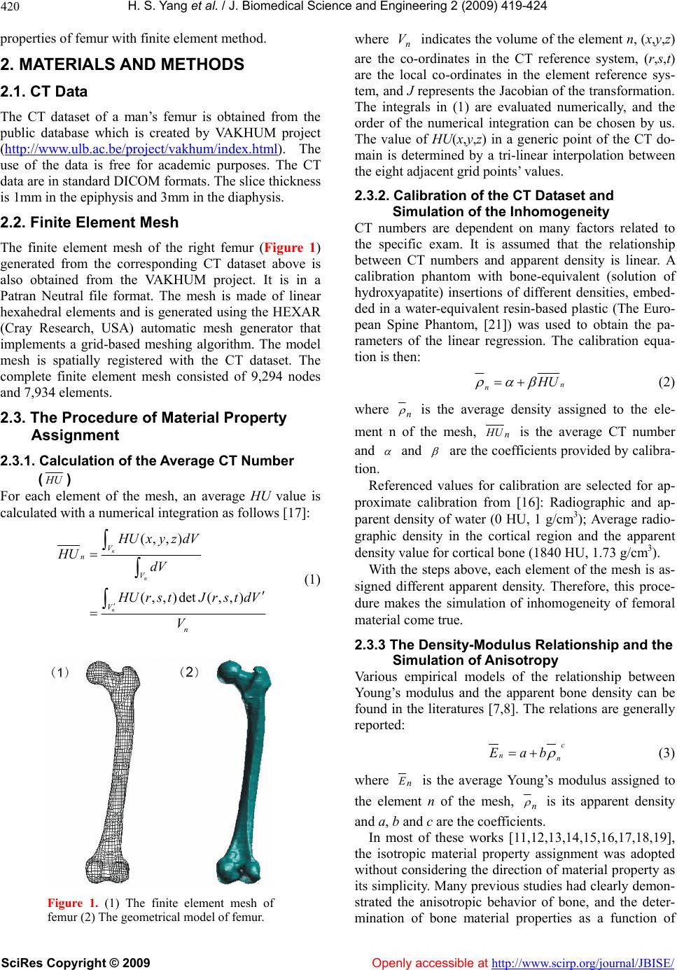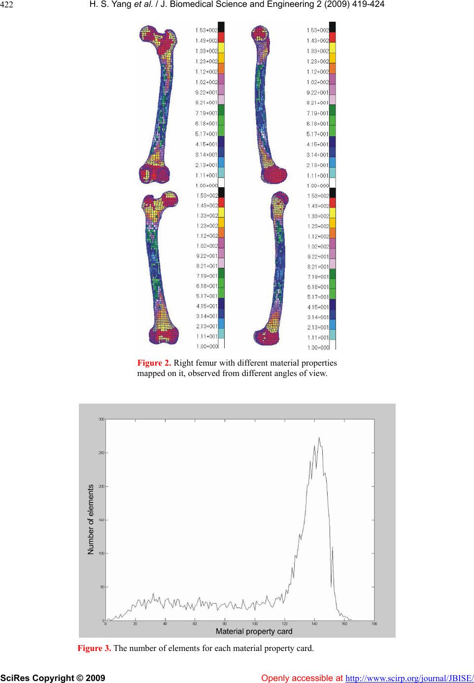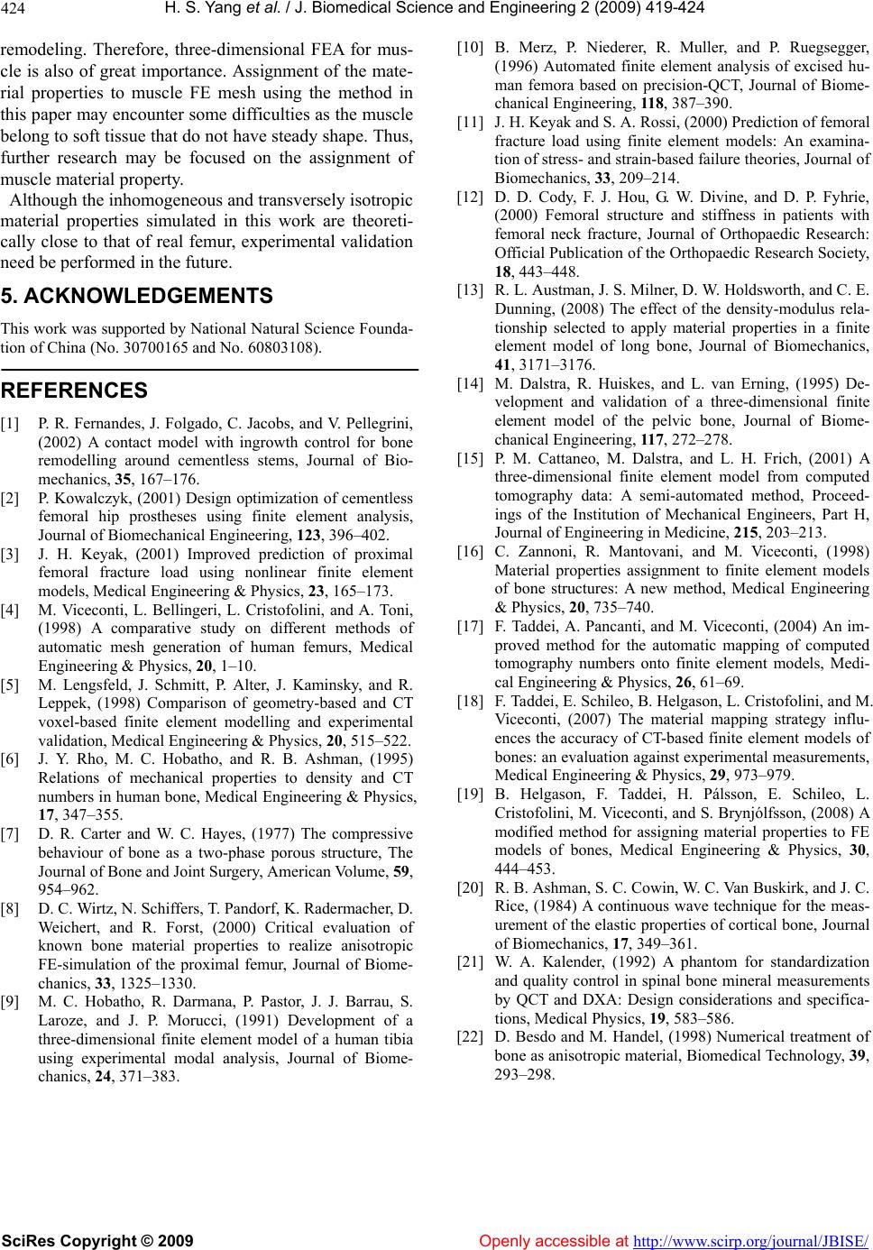
H. S. Yang et al. / J. Biomedical Science and Engineering 2 (2009) 419-424
SciRes Copyright © 2009 Openly accessible at http://www.scirp.org/journal/JBISE/
424
remodeling. Therefore, three-dimensional FEA for mus-
cle is also of great importance. Assignment of the mate-
rial properties to muscle FE mesh using the method in
this paper may encounter some difficulties as the muscle
belong to soft tissue that do not have steady shape. Thus,
further research may be focused on the assignment of
muscle material property.
Although the inhomogen eous and transv ersely iso tropic
material properties simulated in this work are theoreti-
cally close to that of real femur, experimental validation
need be performed in the future.
5. ACKNOWLEDGEMENTS
This work was supported by National Natural Science Founda-
tion of China (No. 30700165 and No. 60803108).
REFERENCES
[1] P. R. Fernandes, J. Folgado, C. Jacobs, and V. Pellegrini,
(2002) A contact model with ingrowth control for bone
remodelling around cementless stems, Journal of Bio-
mechanics, 35, 167–176.
[2] P. Kowalczy k, (2001) Design optimization of cementless
femoral hip prostheses using finite element analysis,
Journal of Biomechanical Engineering, 123, 396–402.
[3] J. H. Keyak, (2001) Improved prediction of proximal
femoral fracture load using nonlinear finite element
models, Medical Engineering & Physics, 23, 165–173.
[4] M. Viceconti, L. Bellingeri, L. Cristofolini, and A. Toni,
(1998) A comparative study on different methods of
automatic mesh generation of human femurs, Medical
Engineering & Ph ysics, 20, 1–10.
[5] M. Lengsfeld, J. Schmitt, P. Alter, J. Kaminsky, and R.
Leppek, (1998) Comparison of geometry-based and CT
voxel-based finite element modelling and experimental
validation, Medical Engineering & Physics, 20, 515–522.
[6] J. Y. Rho, M. C. Hobatho, and R. B. Ashman, (1995)
Relations of mechanical properties to density and CT
numbers in human bone, Medical Engineering & Physics,
17, 347–355.
[7] D. R. Carter and W. C. Hayes, (1977) The compressive
behaviour of bone as a two-phase porous structure, The
Journal of Bone and Joint Surgery, American Volume, 59,
954–962.
[8] D. C. Wirtz, N. Schiffers, T. Pandorf, K. Radermacher, D.
Weichert, and R. Forst, (2000) Critical evaluation of
known bone material properties to realize anisotropic
FE-simulation of the proximal femur, Journal of Biome-
chanics, 33, 1325–1330.
[9] M. C. Hobatho, R. Darmana, P. Pastor, J. J. Barrau, S.
Laroze, and J. P. Morucci, (1991) Development of a
three-dimensional finite element model of a human tibia
using experimental modal analysis, Journal of Biome-
chanics, 24, 371–383.
[10] B. Merz, P. Niederer, R. Muller, and P. Ruegsegger,
(1996) Automated finite element analysis of excised hu-
man femora based on precision-QCT, Journal of Biome-
chanical Engineering, 118, 387–390.
[11] J. H. Keyak and S. A. Rossi, (2000) Prediction of femoral
fracture load using finite element models: An examina-
tion of stress- and strain-based failure theories, Journal of
Biomechanics, 33, 209–214.
[12] D. D. Cody, F. J. Hou, G. W. Divine, and D. P. Fyhrie,
(2000) Femoral structure and stiffness in patients with
femoral neck fracture, Journal of Orthopaedic Research:
Official Publication of the Orthopaedic Research Society,
18, 443–448.
[13] R. L. Austman, J. S. Milner, D. W. Holdsworth, and C. E.
Dunning, (2008) The effect of the density-modulus rela-
tionship selected to apply material properties in a finite
element model of long bone, Journal of Biomechanics,
41, 3171–3176.
[14] M. Dalstra, R. Huiskes, and L. van Erning, (1995) De-
velopment and validation of a three-dimensional finite
element model of the pelvic bone, Journal of Biome-
chanical Engineering, 117, 272–278.
[15] P. M. Cattaneo, M. Dalstra, and L. H. Frich, (2001) A
three-dimensional finite element model from computed
tomography data: A semi-automated method, Proceed-
ings of the Institution of Mechanical Engineers, Part H,
Journal of Engineering in Medicine, 215, 203–213.
[16] C. Zannoni, R. Mantovani, and M. Viceconti, (1998)
Material properties assignment to finite element models
of bone structures: A new method, Medical Engineering
& Physics, 20, 735–740.
[17] F. Taddei, A. Pancanti, and M. Viceconti, (2004) An im-
proved method for the automatic mapping of computed
tomography numbers onto finite element models, Medi-
cal Engineering & Physics, 26, 61–69.
[18] F. Taddei, E. Schileo, B. Helgason, L. Cristofolini, and M.
Viceconti, (2007) The material mapping strategy influ-
ences the accuracy of CT-based finite element models of
bones: an evaluation against experimental measurements,
Medical Engineering & Physics, 29, 973–979.
[19] B. Helgason, F. Taddei, H. Pálsson, E. Schileo, L.
Cristofolini, M. Viceconti, and S. Brynjólfsson, (2008) A
modified method for assigning material properties to FE
models of bones, Medical Engineering & Physics, 30,
444–453.
[20] R. B. Ashman, S. C. Cowin, W. C. Van Buskirk, and J. C.
Rice, (1984) A continuous wave technique for the meas-
urement of the elastic properties of cortical bone, Journal
of Biomechanics, 17, 349–361.
[21] W. A. Kalender, (1992) A phantom for standardization
and quality control in spinal bone mineral measurements
by QCT and DXA: Design considerations and specifica-
tions, Medical Physics, 19, 583–586.
[22] D. Besdo and M. Handel, (1998) Numerical treatment of
bone as anisotropic material, Biomedical Technology, 39,
293–298.