Surgical Science, 2011, 2, 414-417
doi:10.4236/ss.2011.28090 Published Online October 2011 (http://www.SciRP.org/journal/ss)
Copyright © 2011 SciRes. SS
Repeated Hemorrhage after Repair of Orbital Floor
Fracture with a Silicone Implant
Hirohiko Kakizaki1*, Akihiro Ichinose2, Yasuh iro Takahashi1, Nobutada Katori3,
Alejandra A. Valenzuela1, Masayoshi Iwaki1
1Department of Ophthalmology, Aichi Medical University, Nagakute, Japan
2Department of Plastic Surgery, Kobe University, Kobe, Japan
3Department of Oculoplastic and Orbital Surgery, Seirei Hamamatsu General Hospital,
Hamamatsu, Japan
E-mail: *cosme@d1.dion.ne.jp
Received January 19, 2011; revised August 30, 2011; accepted September 16, 2011
Abstract
A 16-year-old girl was accidentally kicked in her right eye by her cheerleading teammate in an exercise.
Upward gaze ability of her right eye was severely impaired and computed tomography (CT) showed a trap-
door fracture of the right orbital floor. After surgical exploration, a silicone implant was inserted. No bleed-
ing was confirmed at this time. The next day, CT detected a hematoma on the right orbital floor. The hema-
toma was drained and meticulous cautery was used to control any potential bleeding. The same silicone im-
plant was re-inserted. Irrespective of attempts to avoid hemorrhage, this occurred twice after the respective
evacuations. During a fourth operation, we removed the silicone implant simultaneously with hematoma
evacuation. No hematoma has occurred since, and the patient’s ocular movement has dramatically improved
to a normal binocular single vision field. When repeated hemorrhages occur after an orbital floor fracture
repair with insertion of a silicone implant, removal of the implant is an effective strategy to resolve the hem-
orrhage.
Keywords: Trapdoor Fracture, Orbital Floor, Silicone Implant, Hematoma, Hemorrhage, Removal
1. Introduction
Orbital hemorrhage in relation to alloplastic implants
after repair of a blowout fracture is an uncommon entity
[1-5]. Although this has been mainly reported as a late
complication [3,4], acute hemorrhage also occurs [1,2];
however, the relationship between hemorrhaging and
alloplastic implants has not been clarified. We report
repeated hemorrhage after repair of an orbital floor frac-
ture using a silicone implant, and the hemorrhage was
resolved after removal of the implant.
2. Case Report
A 16-year-old girl was accidentally kicked in her right
eye by her cheerleading teammate in an exercise. She
had diplopia in upgaze at the time. She had an ophthal-
mological examinatio n on the follo wing da y of the injur y.
Although the globe was not impaired, the upward gaze of
the right eye was severely restricted. A right floor trap-
door fracture was confirmed by computed tomography
(CT) (Figure 1).
Figure 1. Sagittal computed tomography (CT) shows a
trapdoor fracture of the right orbital floor (arrow).
H. KAKIZAKI ET AL.415
Surgical repair of the fracture was performed on the
day of examinations via the swinging eyelid approach [6],
releasing all the entrapped tissues followed by the inser-
tion of a silicone implant on the floor to p revent prolapse
of the orbital tissues. No hemorrhage was confirmed at
the end of the operation.
On the following day of the operation, the patient de-
veloped right proptosis with normal optic nerve function.
CT images de monstrated a hematoma on t he right orbital
floor. We evacuated the hematoma that day and the
bleeding points were thoroughly cauterized. At the same
time, the silicone i mplant was re-inserted and a drain was
positioned bet ween the implant and the floo r .
The day after t he second operation, no obvio us hema-
toma was observed on CT (Figure 2(A)), and blood
drainage was not evident on the second day after this
operation. Three days later, however, right proptosis and
severe chemosis developed (Figure 2(B)), and CT
showed a large hematoma in the same location (Figure
2(C)). Although the hematoma had pushed the inferior
rectus muscle towards the optic nerve, it did not induce
any optic nerve dysfunction. Orbital exploration to re-
move the hematoma (Figure 2(D)) was carried out with
meticulous cauter y, and an additio nal two holes 3 m m in
diameter each were made in the silicone implant to allow
drainage of any orbital collection through the implant
into the maxillary sinus.
On the day after the third operation, the patient had a
hema toma aga in as sho wn by CT imagin g. Altho ugh her
right ocular movement was severely restricted, as her
visual acuity and pupil reflex were not impaired, the he-
matoma was observed for 3 days with intravenous ster-
oid therapy (dexamethasone 8 mg, 4 mg, and 2 mg/day,
respectively). Since the symptoms were not resolved, we
evacuated the hematoma again, and this time, the sili-
cone implant was removed. There were no obvious
bleeding points at any time. We did not explore the or-
bital contents to avoid further disturbance of the stable
anatomy and to avoid the orbital tissues being entrapped
again.
Figure 2. (A) Sagi ttal CT shows no he matoma on t he d ay after the seco nd ope ration; (B ) Rig ht propt osis an d seve re c he mosis
are shown 3 days after the second operation; (C) Sagittal CT shows a large volume of hematoma (asterisk) displacing the
inferior rectus muscle and almost abutting the optic nerve; (D) The hematoma and silicone implant are shown during the
hird surgic al exploration. t
Copyright © 2011 SciRes. SS
H. KAKIZAKI ET AL.
Copyright © 2011 SciRes. SS
416
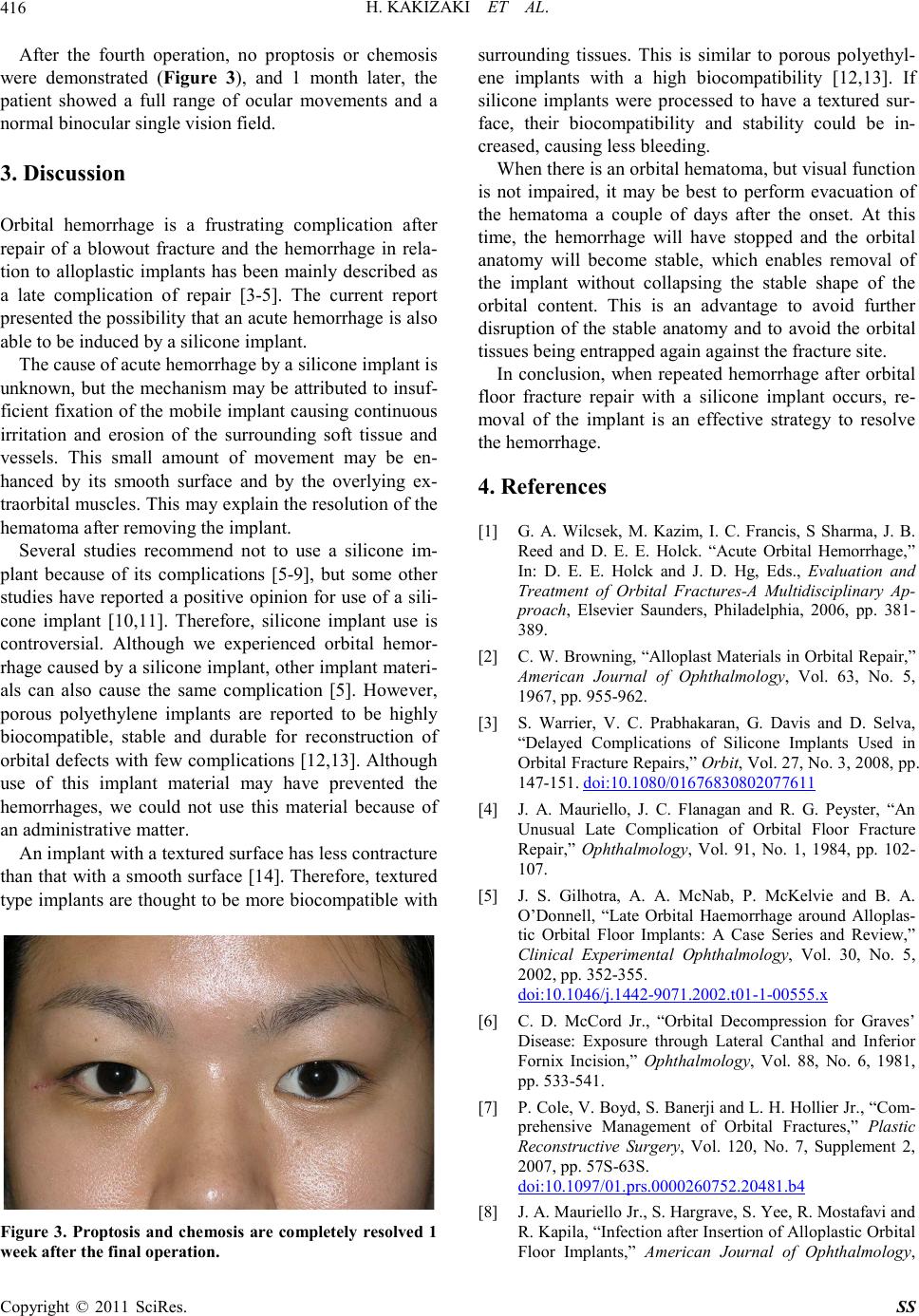
After the fourth operation, no proptosis or chemosis
were demonstrated (Figure 3), and 1 month later, the
patient showed a full range of ocular movements and a
normal binoc ular single visi on field.
3. Discussion
Orbital hemorrhage is a frustrating complication after
repair of a blowout fracture and the hemorrhage in rela-
tion to alloplastic implants has been mainly described as
a late complication of repair [3-5]. The current report
presented the possibility that an acute hemorrhage is also
able to be induced by a silicone implant.
The cause of acute hemorrhage by a silicone implant is
unknown, but the mec hanism may be attributed to insuf-
ficient fixation of the mobile implant causing continuous
irritation and erosion of the surrounding soft tissue and
vessels. This small amount of movement may be en-
hanced by its smooth surface and by the overlying ex-
traorbital muscles. This may explain the resolution of the
hematoma after removing the implant.
Several studies recommend not to use a silicone im-
plant because of its complications [5-9], but some other
studies have reported a positive opinion for use of a sili-
cone implant [10,11]. Therefore, silicone implant use is
controversial. Although we experienced orbital hemor-
rhage caused by a silicone implant, other implant materi-
als can also cause the same complication [5]. However,
porous polyethylene implants are reported to be highly
biocompatible, stable and durable for reconstruction of
orbital defects with few complications [12,13]. Although
use of this implant material may have prevented the
hemorrhages, we could not use this material because of
an administrative matter.
An implant with a textured surface has less contracture
than that with a smooth surface [14]. Therefore, textured
type implants are thought to be more biocompatible with
Figure 3. Proptosis and chemosis are completely resolved 1
week after the fin al operation.
surrounding tissues. This is similar to porous polyethyl-
ene implants with a high biocompatibility [12,13]. If
silicone implants were processed to have a textured sur-
face, their biocompatibility and stability could be in-
creased, causing less bleeding.
When there is an orbital hematoma, but visual function
is not impaired, it may be best to perform evacuation of
the hematoma a couple of days after the onset. At this
time, the hemorrhage will have stopped and the orbital
anatomy will become stable, which enables removal of
the implant without collapsing the stable shape of the
orbital content. This is an advantage to avoid further
disruption of the stable anatomy and to avoid the orbital
tissues being entrapped again against the fracture site.
In conclusion, when repeated hemorrhage after orbital
floor fracture repair with a silicone implant occurs, re-
moval of the implant is an effective strategy to resolve
the hemorrhage.
4. References
[1] G. A. Wilcsek, M. Kazim, I. C. Francis, S Sharma, J. B.
Reed and D. E. E. Holck. “Acute Orbital Hemorrhage,”
In: D. E. E. Holck and J. D. Hg, Eds., Evaluation and
Treatment of Orbital Fractures-A Multidisciplinary Ap-
proach, Elsevier Saunders, Philadelphia, 2006, pp. 381-
389.
[2] C. W. Browning, “Alloplast Materials in Orbital Repair,”
American Journal of Ophthalmology, Vol. 63, No. 5,
1967, pp. 9 55-962.
[3] S. Warrier, V. C. Prabhakaran, G. Davis and D. Selva,
“Delayed Complications of Silicone Implants Used in
Orbital Fracture Repairs,” Orbit, Vol. 27, No. 3, 2008, pp.
147-151. doi:10.1080/01676830802077611
[4] J. A. Mauriello, J. C. Flanagan and R. G. Peyster, “An
Unusual Late Complication of Orbital Floor Fracture
Repair,” Ophthalmology, Vol. 91, No. 1, 1984, pp. 102-
107.
[5] J. S. Gilhotra, A. A. McNab, P. McKelvie and B. A.
O’Donnell, “Late Orbital Haemorrhage around Alloplas-
tic Orbital Floor Implants: A Case Series and Review,”
Clinical Experimental Ophthalmology, Vol. 30, No. 5,
2002, pp. 352-355.
doi:10.1046/j.1442-9071.2002.t01-1-00555.x
[6] C. D. McCord Jr., “Orbital Decompression for Graves’
Disease: Exposure through Lateral Canthal and Inferior
Fornix Incision,” Ophthalmology, Vol. 88, No. 6, 1981,
pp. 533- 541 .
[7] P. Cole, V. Boyd, S. Banerji and L. H. Hollier Jr., “Com-
prehensive Management of Orbital Fractures,” Plastic
Reconstructive Surgery, Vol. 120, No. 7, Supplement 2,
2007, pp . 57S-63S.
do i:10. 1097/01.prs.000026 0752.20481.b4
[8] J. A. Mauriello Jr., S. Hargrave, S. Yee, R. Mostafavi and
R. Kapila, “Infection after Insertion of Alloplastic Orbital
Floor Implants,” American Journal of Ophthalmology,
H. KAKIZAKI ET AL.417
Vol. 117, No. 2, 1994, pp. 246-252.
[9] D. R. Jordan, P. St Onge, R. L. Anderson, J. R. Patrinely
and J. A. Nerad, “Complications Associated with Al-
loplastic Implants Used in Orbital Fracture Repair,”
Ophthalm ology, Vol. 99, No. 10, 199 2, pp . 160 0-1608.
[10] S. J. Prowse, P. M. Hold, R. F. Gilmour, U. Pratap, E.
Mah and F. W. Kimble, “Orbital Floor Reconstruction: A
Case for Silicone. A 12 Year Experience,” Journal of
Plastic, Reconstructive & Aesthetic Surgery, Vol. 63, No.
7, 2010, pp. 1105 -1109 .
do i:10.1016 /j.bjps.2009.05. 057
[11] J. C. Mwanza, D. K. Ngoy and D. L. Kayembe, “Recon-
struction of Orbital Floor Blow-out Fractures with Sili-
cone Implant,” Bulletin Society Belge Ophtalmology, Vol.
280, 2001, pp. 57-61.
[12] J. J. Xu, L. Teng, X. L. Jin, Y. Ji, J. J. Li and B. Zhang,
“Porous Polyethylene Implants in Orbital Blow-out Frac-
tures and Enophthalmos Reconstruction,” The Journal of
Craniofacial Surgery, Vol. 20, No. 3, 2009, pp. 918-920.
doi:10.1097/SCS.0b013e3181a2d728
[13] P. A. Rubin, J. R. Bilyk and J. W. Shore, “Orbital Recon-
struction Using Porous Polyethylene Sheets,” Ophthal-
mology, Vol. 101, No. 10 , 199 4, pp. 16 97 - 17 08 .
[14] G. P. Barnsley, L. J. Sigurd son and S. E. Barnsley. “Tex-
tured Surface Breast Implants in the Prevention of Cap-
sular Contracture among Breast Augmentation Patients:
A Meta-analysis of Randomized Controlled Trials,” Plas-
tic Reconstructive Surgery, Vol. 117, No. 7, 2006, pp.
2182-2190.
do i:10. 1097/01.prs.000021 8184.47372.d5
Copyright © 2011 SciRes. SS