 Journal of Biomaterials and Nanobiotechnology, 2011, 2, 337-346 doi:10.4236/jbnb.2011.24042 Published Online October 2011 (http://www.SciRP.org/journal/jbnb) Copyright © 2011 SciRes. JBNB 337 Blood Compatibility of Amphiphilic Poly(N-α-acrylamide-L-lysine-b-dimethylsiloxane) Block Copolymers Kazuo Sugiyama, Nobuyuki Tanigawa, Kohei Shiraishi Cluster of Biotechnology and Chemistry Systems, Program in Systems Engineering, Graduate School of Systems Engineering, Kinki University, Higashihiroshima-City, Japan. Email: sugiyama@hiro.kindai.ac.jp Received June 21st, 2011; revised July 22nd, 2011; accepted August 20th, 2011. ABSTRACT Amphiphilic block copolymers poly(LysAA-b-DMS) consisting of a hydrophilic poly(N-α-acrylamide-L-lysine) [poly(LysAA)] segment with different molecular weights and a hydrophobic polydimethylsiloxane (PDMS ) segment were prepared as follows. The precursor copolymer poly(Bo c-L ys AA- Ot Bu-b -PD MS) was obtained from radical polymerization of N-α-acrylamide-N-ε-tert-butoxycarbonyl-L-lysine-tert-butylester (Boc-LysAA-OtBu) initiated with 4,4’-azobis(polydi- methylsiloxane 4-cyanopentanoate) (azo-PDMS) with the molecular weight of PDMS Mw = 4.3 × 10³ in the presence of 2-mercaptoethanol (2-ME) as a chain-transfer agent. Removal of the protecting groups of the precursor copolymer was carried out in 80% trifluoroacetic acid aqueous solution to give poly(LysAA-b-DMS)-1-3. The weight average molecu- lar weight of poly(LysAA-b-DMS)-1-3 was Mw = 1.02 × 10⁴ - 2.52 × 10⁴. From the 1H-NMR and fluorescence spectra measurements, poly(LysAA-b-DMS)-1-3 was determined to self-organize and form core-shell micelles in water. The critical micelle concentration (CMC) increased to 1000 - 4000 mg·L–1 with increasing molar ratio of the poly(LysAA) segment from 0.42 to 0.65. From morphological analysis with a scanning probe microscope (SPM), poly(LysAA-b-DMS) has microphase-separated structures made up of hydrophilic and hydrophobic regions with the domain size ranging from several tens to several hundreds of nanometers. Inhibition of thrombin activity of poly(LysAA-b-DMS) was evalu- ated from the Michaelis constant (KM) and catalytic activity (kcat) for the enzymatic reaction of thrombin and synthetic substrate S-2238 in the presence of poly(LysAA-b-DMS). The KM and kcat were 0.10 - 0.11 mM and 4.04 × 10⁵ - 4.26 × 10⁵ min–1, respectively. Fibrinolytic activity was also verified from the transformation of plasminogen to plasmin by tissue plasminogen activator (t-PA) using synthetic substrate S-2251 in the presence of poly(LysAA-b-DMS). The KM and kcat were 0.07 mM and 5.73 × 10⁶ - 5.95 × 10⁶ min–1, respectively. Keywords: Poly(N-α-acrylamide-L-lysine), Polydimethylsiloxane, Block Copolymer, Molecular Assembly, Blood Compatibility, S-2238/S-2251, Biomedical Polymer Material 1. Introduction Some graft and block copolymers containing the polydi- methylsiloxane (PDMS) segment have been synthesized in order to improve the mechanical properties and bio- compatibility of silicone rubber as a useful biomedical material [1-3]. In poly(etherurethaneurea)s including va- rious molar ratios of the tetramethyldisiloxane moiety in the main chain, the siloxane moiety was located on the surface. As the hydrophobicity was increased with in- creasing siloxane content, the surface was able to adsorb bovine serum albumin [4]. On the other hand, a series of block copolymers consisting of PDMS and hydrophilic polymethacrylates, such as poly(2-hydroxyethyl metha- crylate), poly(2,3-dihydroxypropyl methacrylate), and poly (2,3,4,5,6-pentahydroxyhexyl methacrylate) suppressed the adsorption of albumin and -globulin as well as platelets, to a level less than PDMS [5]. The microphase-separated structure of a polymer is considered to exhibit effective suppression of the adsorp- tion of fibrinogen, an important blood coagulation factor, as well as inhibition of adhesion and activation of human platelets. Plasma protein adsorption, which is the initial event in blood–material interaction, influences subse- quent platelet adhesion and activation. The amphiphilic poly(MPC-b-PDMS) obtained from introduction of the  Blood Compatibility of Amphiphilic Poly(N-α-acrylamide-L-lysine-b-dimethylsiloxane) Block Copolymers 338 2-(methacryloyloxy)ethyl phosphorylcholine (MPC) mo- iety into PDMS has a microphase-separated structure and shows no adhesion or activation of human platelets [6,7]. In our work on biocompatible polymeric materials [4- 9], we found that incorporation of L-serine or L-lysine residues into polymers as side groups, for example, poly- (O-methacryloyl-L-serine) [poly(SerMA)] and poly(N- α-methacrylamide-L-lysine) [poly(LysMA)] [10], was useful for creating blood compatible materials. The am- phiphilic block copolymer poly(LysMA-b-DMS) con- sisting of poly(LysMA) as the hydrophilic segment and a PDMS segment with almost monodispersed molecular weight as the hydrophobic segment has also been re- ported [11]. The poly(LysMA-b-DMS) also exhibits an- tithrombotic properties. Blood compatibility of poly(Lys- MA) and poly(LysMA-b-DMS) may arise from the strong interaction of the L-lysine group in the polymer and the L-lysine-binding sites (LBS) of plasminogen, the precursor of plasmin [12]. Plasminogen is converted to the fibrinolytic active plasmin by the tissue plasminogen activator (t-PA), thus showing increased fibrinolytic ac- tivities. It is well known that differences in the glass transition temperatures (Tg) of polymers reflect the flexibility of the main chain and side chain. For instance, the micro- brownian motion of poly(methyl acrylate) and poly(N- tert-butylacrylamide) begins at Tg = 10˚C and Tg = 128˚C, respectively, whereas that of poly(methyl metha- crylate) and poly(N-tert-butylmethacrylamide) begins at Tg = 105˚C and Tg = 160˚C, respectively [13]. The for- mer polymers are more flexible due to the difference in the substituent group (H or methyl) at the α-position of the monomer units in the vinyl polymers. The more flexible main chain is therefore expected to be the origin of the stronger interaction between the polymer L-lysine moiety and the LBS of plasminogen. In the course of our study on biomedical polymeric materials, our interest has been focused on the preparation of amphiphilic block copolymers poly(LysAA-b-DMS) consisting of the hy- drophilic poly(N-α-acrylamide-L-lysine) [poly(LysAA)] segment with different molecular weights and a hydro- phobic PDMS segment. Poly (LysAA-b-DMS) is ex- pected to exhibit stronger fibrinolytic activity than poly (LysMA-b-DMS). There has also been growing interest in the self-orga- nization phenomena of water soluble polymers contain- ing a few hydrophobic substituents per chain and hydro- phobic groups attached to the polymer end [8,9], as well as amphiphilic block copolymers consisting of hy- dro- philic and hydrophobic segments [11,14-17]. These am- phiphilic polymers and copolymers are expected to play a role as carrier molecules for drug delivery systems (DDS), because they form a core-shell polymer micelle composed of a hydrophobic core and a hydrophilic shell, making it possible to stably hold hydrophobic medicines in the core when placed in water. In this article, it is confirmed that poly(LysAA-b-DMS) can self-assemble to form a core-shell polymer micelle composed of a hydrophobic core and a hydrophilic shell in water, which can be used for the intravenous injection of DDS carrier macromolecules. It is also confirmed that its polymer film has a microphase-separated structure useful for biomedical materials such as artificial veins. The blood compatibility of poly (LysAA-b-DMS) was evaluated by the Michaelis constant (KM) and catalytic activity (kcat) for the enzymatic reaction of thrombin and a synthetic substrate S-2238, as well as by the transfor- mation of plasminogen to plasmin by tissue plasminogen activator (t-PA) using synthetic substrate S-2251 in the presence of poly(LysAA-b-DMS). 2. Materials and Methods 2.1. Materials 4,4’-Azobis (4-cyanopentanoic acid) was kindly supplied by Wako Pure Chemical Co. (Japan) and purified by re- crystallization from methanol. 4,4’-Azobis(4-cyano-pen- tanoic acid chloride) was prepared according to the me- thod of Smith [18]. Reactive silicone oil (PDMS-OH; X- 22-170BX, OH value: 21.0 KOH mg·g–1, Mw = 4.3 × 10³, Mn = 3.9 × 10³, Mw/Mn = 1.09) was used as obtained from Shinetsu Chemicals Ind. (Tokyo, Japan). 1-(6-Di- methylamino-2-naphthyl)-1-dodecanone (DMAND; Mo- lecular ProbesTM, invitrogen) was used as obtained. Hu- man thrombin was purchased from Mochida Pharmaceu- tical CO., LTD. (Tokyo, Japan), whereas the synthetic chromogenic substrates S-2238 and S-2251 were pur- chased from Sekisui medical CO. LTD. (Tokyo, Japan), and used without any purification. Distilled and deio- nized water was used for all experiments. 2.2. Synthesis of 4,4’-Azobis(polydimethyl-siloxane 4-cyanopentanoate) (azo-PDMS) To a CHCl3 solution (150 mL) of reactive silicone oil (PDMS-OH: 19 mmol) and triethylamine (10 mmol) was added dropwise a dichloromethane solution (30 mL) of 4,4’-azobis(4-cyanopentanoic acid chloride) (9.5 mmol) at 25˚C. The reaction mixture was washed with water, and then the solution was evaporated under reduced pressure. The crude azo-PDMS was purified by frac- tional precipitation with a solvent system of tetrahydro- furan-methanol. The molecular weight distribution was found to be Mw/Mn = 1.12 by means of gel permeation chromatography analysis conducted in CHCl3 solution with TSK gel (column: TOSOH G4000HXL, G3000HXL, C opyright © 2011 SciRes. JBNB  Blood Compatibility of Amphiphilic Poly(N-α-acrylamide-L-lysine-b-dimethylsiloxane) Block Copolymers339 G2000HXL), using TOSOH LC-8020 GPC. Yield: 68.3%. 1H-NMR (CDCl3, TMS): δ = 0.14 [s, 420H, -Si(CH3)2], 0.79 - 0.82 [m, 6H, -CH3], 1.23 - 1.72 [m, 12H, -CH2-], 2.32 - 2.52 [m, 4H, -CH2-], 3.34 - 3.39 [m, 4H, -OCH2-], 3.55 - 3.58 [m, 4H, -OCH2-], 4.18 - 4.20 [t, 4H, -COOCH2-]. IR(KBr)ν(cm–1): 1255 (Si-CH3), 1735 (-COOR), 2250 (-CN). 2.3. Synthesis of N-α-Acrylamide-N-ε-tert- butoxycarbonyl-L-lysine-tert-butylester (Boc-LysAA-OtBu) To a CHCl3 solution (150 mL) of N-ε-tert-butoxycar- bonyl-L-lysine-tert-butyl ester (15 mmol) and triethyl- amine (23 mmol) was added dropwise a CHCl3 solution (30 mL) of acrylic acid chloride (18 mmol) at 4˚C. The reaction mixture was washed with 1 M HCl and water, and then the solution was evaporated under reduced pressure. The crude Boc-LysAA-OtBu was purified by passage through a basic alumina column with the mixed solvent of dichloromethane and hexane (50/50 vol%). Yield: 64.1%; mp: 62.0˚C - 62.5˚C. [α]D 20 = +20.1°. 1H-NMR (CDCl3, TMS): δ = 1.24 - 1.47(m, 2H, >CH- CH2-CH2-CH2-CH2-), 1.43(s, 9H, -C(CH3)3), 1.47(s, 9H, -C(CH3)3), 1.63 - 1.82 (m, 2H, >CH-CH2-CH2-CH2-CH2-), 1.81 - 2.05 (m, 1H, >CH-CH2-CH2-CH2-CH2-), 2.99 - 3.24 (m, 2H, >CH-CH2-CH2-CH2-CH2-), 4.56 - 4.60 (m, 2H, >CH-CH2-CH2-CH2-CH2-), 5.63 - 5.79(m, 1H, H- CH=CH-), 4.58 (br, 1H, -NH-), 6.12 - 6.29 (m, 1H, H- CH=CH-), 6.24 (br, 1H, -NH-), 6.28 - 6.47 (m, 1H, CH2 =CH-). 13C-NMR (CDCl3, TMS): δ = 22.27 [-CH2-], 28.01 [-(CH3)3], 28.43 [-(CH3)3], 29.59 [-CH2-], 32.34 [-CH2], 40.12 [-CH2-], 52.44 [-CH], 79.02 [>C<], 82.21 [>C<], 126.77 [=CH2], 130.43 [=CH], 155.94 [-CONH], 164.95 [-COO-], 171.51 [-COO-]. Elemental Anal.; Calcd for C18H32N2 356.461: C, 60.65%; H, 9.05%; N, 7.86%, Found: C, 60.46%; H, 9.06%; N, 8.08%. 2.4. Synthesis of Poly(LysAA-b-DMS) A mixed solution of tetrahydrofuran and ethanol (40:60 vol%, 15 mL) containing Boc-LysAA-OtBu (10 mM), azo-PDMS (10 mM) as an initiator, and 2-mercapto- ethanol (2-ME: 0.10 - 1.0 mM) as a chain transfer agent in a glass tube was degassed by the freeze-thaw tech- nique with a liquid nitrogen bath and was sealed in a vacuum. After polymerization at 60˚C for 20 h, the con- tents of the tube were poured into a large quantity of hexane to precipitate the precursor block copolymer, poly (Bo c - Ly sAA- O t Bu- b-DMS)-1-3. The number-aver- age molecular weight (Mn) was calculated from gel per- meation chromatography (GPC) with a polystyrene stan- dard. GPC analysis was conducted in a dimethylfor- mamide solution with a TSK gel column (TOSOH HHR 6000, 5000, 4000, 3000, and 2000) with a TOSOH LC- 8020 GPC apparatus. The composition ratio of the Boc- LysAA-OtBu polymer segment (p) and PDMS segment (q) of poly(Boc-LysAA-OtBu-b-DMS) was calculated using the N content of elemental analysis performed with a Silber Hegner elemental analysensysteme GmbH. The removal of the tert-butoxycarbonyl (Boc) and tert-Bu (t-Bu) protecting groups in the Boc-LysAA-OtBu poly- mer segment was carried out as follows. Poly (Boc-Lys- AA-OtBu-b-DMS)-1-3 was dissolved in tetrahydrofuran, 80% trifluoroacetic acid was added, and the mixture was stirred at 25˚C for 48 h. After removing the solvent under reduced pressure and washing with diethyl ether, the resulting copolymer was neutralized with 0.1 M sodium hydroxide, and then purified by dialysis against water using a seamless cellulose membrane (Cell-SepT1TM, Spectra/PorTM Biotech, USA; MWCO 3500). Poly (Ly- sAA-Zb-DMS)-1-3 was obtained from the aqueous solu- tion by freeze drying. 2.5. Measurements of Critical Micelle Concentration (CMC) The fluorescence spectra of an artificial blood plasma solution of poly(LysAA-b-DMS) were recorded with a Shimadzu RF-1500 fluorophotometer [19]. DMAND with a 364 nm excitation wavelength (λEX) was used as a fluorescent probe for hydrophobicity. The concentration of DMAND was adjusted to 1 μM by the addition of 20 μL of a solution containing 500 μM DMAND in metha- nol to 10 mL of each amphiphilic poly(LysAA-b-DMS) solution. The concentration of poly(LysAA-b-DMS) was varied in the range of 34 - 1.0 × 10⁴ mg·L–1. The CMC was determined by the break point in the plot of the fluorescence intensity and poly(LysAA-b-DMS) concen- tration. 2.6. Measurements with Scanning Probe Microscope (SPM) SPM images of poly(LysAA-b-DMS) film were taken by means of a Shimadzu SPM-9500J3 with Budget sensors BS-Tap 300AI (Innovative Solutions Bulgaria, Ltd., USA). The copolymer film for SPM measurements was prepared by casting 20 mL of an aqueous solution con- taining a known amount of poly(LysAA-b-DMS) on a mercury bath of 6.0 cm diameter. The solvent was eva- porated by the freeze-dry method and a film with a thick- ness of 0.06 - 0.08 mm was obtained. 2.7. Measurements of Degree of Thrombin Activity Inhibition Twenty microliters of thrombin (10 unit mL–1) dissolved in a Tris buffer solution (pH 7.4), and 70 μL of poly (LysAA-b-DMS) (2 mg·mL–1) were placed in a 96-well microplate of diameter φ = 6.4 mm (Iwaki brand, Scitech Copyright © 2011 SciRes. JBNB 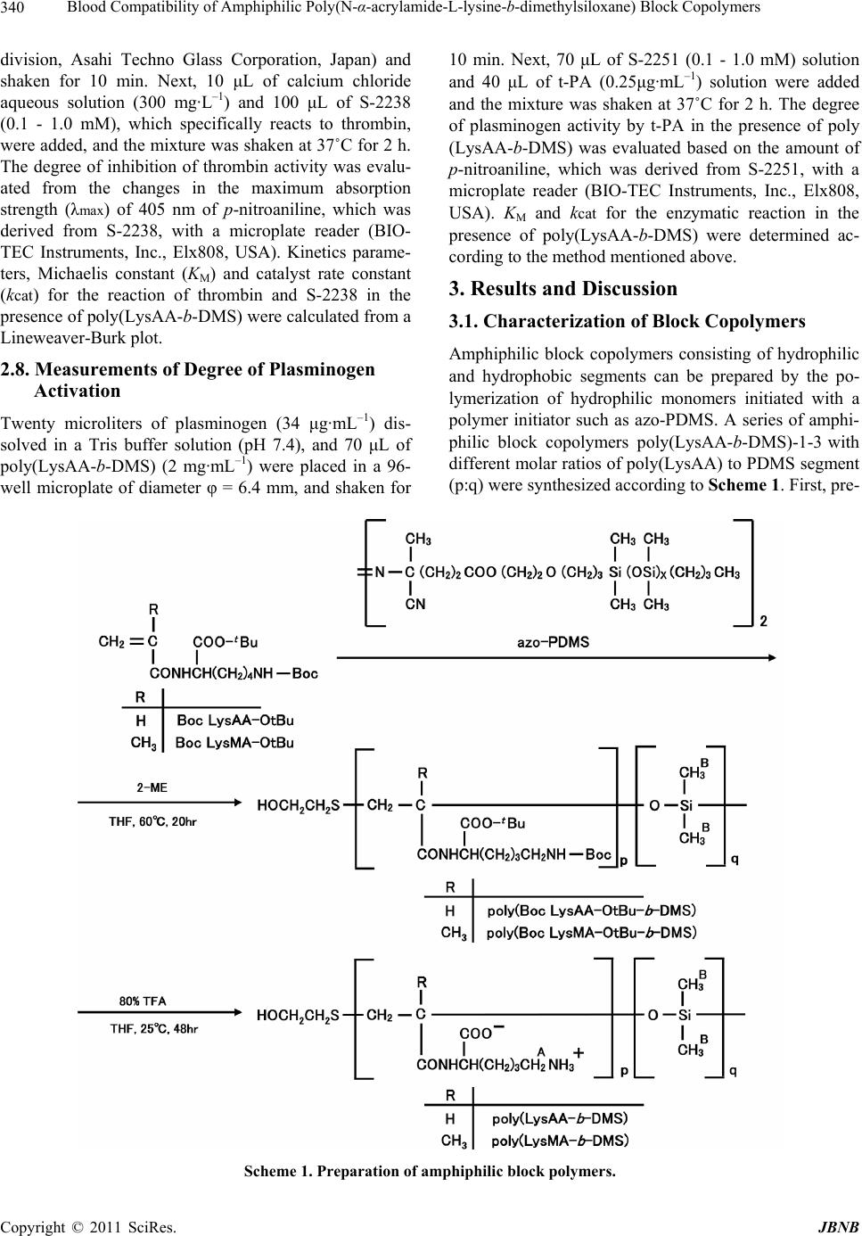 Blood Compatibility of Amphiphilic Poly(N-α-acrylamide-L-lysine-b-dimethylsiloxane) Block Copolymers Copyright © 2011 SciRes. JBNB 340 division, Asahi Techno Glass Corporation, Japan) and shaken for 10 min. Next, 10 μL of calcium chloride aqueous solution (300 mg·L–1) and 100 μL of S-2238 (0.1 - 1.0 mM), which specifically reacts to thrombin, were added, and the mixture was shaken at 37˚C for 2 h. The degree of inhibition of thrombin activity was evalu- ated from the changes in the maximum absorption strength (λmax) of 405 nm of p-nitroaniline, which was derived from S-2238, with a microplate reader (BIO- TEC Instruments, Inc., Elx808, USA). Kinetics parame- ters, Michaelis constant (KM) and catalyst rate constant (kcat) for the reaction of thrombin and S-2238 in the presence of poly(LysAA-b-DMS) were calculated from a Lineweaver-Burk plot. 10 min. Next, 70 μL of S-2251 (0.1 - 1.0 mM) solution and 40 μL of t-PA (0.25μg·mL–1) solution were added and the mixture was shaken at 37˚C for 2 h. The degree of plasminogen activity by t-PA in the presence of poly (LysAA-b-DMS) was evaluated based on the amount of p-nitroaniline, which was derived from S-2251, with a microplate reader (BIO-TEC Instruments, Inc., Elx808, USA). KM and kcat for the enzymatic reaction in the presence of poly(LysAA-b-DMS) were determined ac- cording to the method mentioned above. 3. Results and Discussion 3.1. Characterization of Block Copolymers Amphiphilic block copolymers consisting of hydrophilic and hydrophobic segments can be prepared by the po- lymerization of hydrophilic monomers initiated with a polymer initiator such as azo-PDMS. A series of amphi- philic block copolymers poly(LysAA-b-DMS)-1-3 with different molar ratios of poly(LysAA) to PDMS segment (p:q) were synthesized according to Scheme 1. First, pre- 2.8. Measurements of Degree of Plasminogen Activation Twenty microliters of plasminogen (34 μg·mL–1) dis- solved in a Tris buffer solution (pH 7.4), and 70 μL of poly(LysAA-b-DMS) (2 mg·mL–1) were placed in a 96- well microplate of diameter φ = 6.4 mm, and shaken for Scheme 1. Preparation of amphiphilic block polymers.  Blood Compatibility of Amphiphilic Poly(N-α-acrylamide-L-lysine-b-dimethylsiloxane) Block Copolymers341 cursor block copolymers, poly(Boc-LysAA-OtBu-b- DMS)-1-3, were obtained by radical polymerization of Boc-LysAA-OtBu, in which amino and carbonyl groups of the L-lysine moieties were protected with tert-butoxy- carbonyl (Boc) and tert-butyl (t-Bu) groups, respectively, initiated with azo-PDMS in the presence of chain trans- fer agent 2-ME, and varying the molar ratio of 2-ME to Boc-LysAA-OtBu in the feed from 10:0.1, 10:0.5, and 10:1. For the higher concentration of 2-ME, the smaller molecular weight poly(Boc-LysAA-OtBu) segment was obtained based on the larger chain-transfer constant of 2-ME. Second, the removal of the protecting groups of poly (Bo c - Ly sAA- O t Bu- b-DMS)-1-3 was carried out in 80% trifluoroacetic acid aqueous solution, and then puri- fication by dialysis against water to give poly(LysAA- b-DMS)-1-3. Block copolymer, poly(Boc-LysAA-OtBu- b-DMS) with differing chain lengths of the LysAA polymer segment, was obtained as tabulated in Table 1, along with the results for poly(Boc-LysMA-OtBu- b- DMS) [11], where Boc-LysMA-OtBu represents N-α- methacrylamide-N-ε-tert-butoxycarbonyl-L-lysine-tert- butyl ester. The composition ratio of poly(Boc-LysAA- OtBu) (p) : PDMS (q) was then altered to p:q = 0.35:0.65 - 0.58:0.42. With an increase in the concentration of 2-ME, the number-average molecular weight (Mn) of the poly(Boc-LysAA-OtBu) segment decreased from Mn = 1.56 104 to 0.88 104 and the weight-average molecu- lar weight (Mw) decreased to Mw = 2.52 104 – 1.02 104, whereas the molecular weight of the PDMS seg- ment derived from azo-PDMS had a constant molecular weight of Mn = 3.9 103. Comparing the molecular weight of poly(Boc-LysAA-OtBu-b-DMS) and poly- (Boc-LysMA-OtBu-b-DMS), it can be seen that Boc- LysAA-OtBu without the methyl moiety at the α-posi- tion of the monomer causes a somewhat smaller molecu- lar weight and larger degree of polydispersion (Mw/Mn) than those of Boc-LysMA-OtBu, because the chain transfer constant of Boc-LysAA-OtBu to 2-ME is ex- pected to be larger than that of Boc-LysMA-OtBu based on the higher monomer reactivity of Boc-LysAA-OtBu. The propagating rate constant (kp) of Boc-LysAA-OtBu is also expected to be higher than that of Boc-LysMA- OtBu, though the kp is not known yet. For example, the kp of methyl acrylate and methyl methacrylate are 2090 and 734 L·mol–1·sec–1, respectively [13]. The protecting groups in the Boc-LysAA-OtBu polymer segment were then hydrolyzed in 80% trifluoroacetic acid, converting it to a hydrophilic LysAA polymer segment, and poly- (LysAA-b-DMS)-1-3 was obtained. The removal of the protecting groups (δ = 1.42 ppm, 1.46 ppm) was con- firmed by 1H-NMR spectroscopy. The removal of the protecting group of Boc-LysAA-OtBu resulted in a change in its solubility. Poly(Boc-LysAA-OtBu-b-DMS) is insoluble in water but soluble in acetone, diethyl ether and tetrahydrofuran, yet poly(LysAA-b-DMS) is soluble in water but insoluble in the organic solvents mentioned above. 3.2. Self-Organization The self-organization of poly(LysAA-b-DMS) was first confirmed by 1H-NMR measurements. The spectra of poly(LysAA-b-DMS)-1 in CD3OD and D2O, used as ty- pical examples, are shown in Figure 1. The proton signal (HB) of the methyl moiety in PDMS that appeared in CD3OD weakened gradually in D2O, whereas the signal of HA in the LysAA moiety sharpened gradually with increasing water content. The half-width of the two in- dependent peaks as a function of the D2O content in CD3OD is shown in Figure 2. The half-width of HB (δ = 0 ppm) broadened gradually with an increase in the D2O content. However, the half-width of HA (δ = 2.8 ppm) became narrower with an increase in the water content. The line broadening of the proton signals of the methyl group in PDMS in aqueous medium is ascribed to the restricted molecular motion of the PDMS chains upon self-organ- ization [20,21]. Nevertheless, the mobility of the hydro- philic segments increased when the solvent polarity in- Table 1. Preparationa) and Charactarization of poly(BocLysAA-OtBu-b-DMS). Copolymer 2-ME Yield Elemental analysis P:qb Mnc mmol % C(%) H(%) N(%) ×10–4 ×10⁻⁴ Mwc Mw/Mn poly(Boc LysAA-OtBu-b-DMS) 1 0.10 60.1 58.21 8.56 7.07 0.65:0.35 1.56 2.52 1.62 2 0.50 58.7 55.33 8.48 6.54 0.51:0.49 1.29 1.69 1.31 3 1.00 50.2 54.28 8.41 6.12 0.42:0.58 0.88 1.02 1.16 poly(Boc LysAA-OtBu-b-DMS) 1d 0.10 66.4 58.52 9.02 6.63 0.59:0.41 2.32 3.41 1.47 2 0.50 63.0 56.84 8.95 6.27 0.49:0.51 1.52 1.91 1.26 3 1.00 58.7 54.83 8.63 5.66 0.38:0.62 1.25 1.39 1.11 a) Reaction condition: Boc LysAA-OtBu or Boc LysMA-OtBu: 10.0 mmol. DMS: 100mmol, 2-ME: 0.10 - 1.0 mmol, Mixed solvent of tetrahydrofuran-etha- nol(40:60 vol %) 15 Ml, 60˚C, 20 hr; b) Molar ratio (p:q) in block copolymer was calculated from nitrogen content obtained by elemental analysis; c) Mn and Mw of block copolymers were estimated from GPC; d) Ref.12. Copyright © 2011 SciRes. JBNB 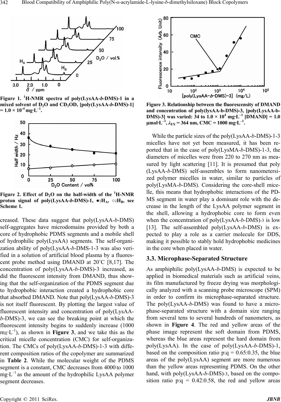 Blood Compatibility of Amphiphilic Poly(N-α-acrylamide-L-lysine-b-dimethylsiloxane) Block Copolymers 342 Figure 1. 1H-NMR spectra of poly(LysAA-b-DMS)-1 in a mixed solvent of D2O and CD3OD, [poly(LysAA-b-DMS)-1] = 1.0 × 10⁻⁴ mg·L–1. Figure 2. Effect of D2O on the half-width of the 1H-NMR proton signal of poly(LysAA-b-DMS)-1, ●:HA, ○:HB, see Scheme 1. creased. These data suggest that poly(LysAA-b-DMS) self-aggregates have microdomains provided by both a core of hydrophobic PDMS segments and a mobile shell of hydrophilic poly(LysAA) segments. The self-organi- zation ability of poly(LysAA-b-DMS-1-3 was also veri- fied in a solution of artificial blood plasma by a fluores- cent probe method using DMAND at 20˚C [8,17]. The concentration of poly(LysAA-b-DMS)-3 increased, as did the fluorescent intensity from DMAND, thus show- ing that the self-organization of the PDMS segment due to hydrophobic interaction created a hydrophobic core that absorbed DMAND. Note that poly(LysAA-b-DMS)-3 is not itself fluorescent. By plotting the largest value of fluorescent intensity and concentration of poly(LysAA- b-DMS)-3, we can see the breaking point at which the fluorescent intensity begins to suddenly increase (1000 mg·L-1), as shown in Figure 3, and we take this as the critical micelle concentration (CMC) for self-organiza- tion. The CMCs of poly(LysAA-b-DMS)-1-3 with diffe- rent composition ratios of the copolymer are summarized in Table 2. While the molecular weight of the PDMS segment is a constant, CMC decreases from 4000 to 1000 mg·L–1 as the amount of the hydrophilic LysAA polymer segment decreases. Figure 3. Relationship between the fluorescensity of DMAND and concentration of poly(lysAA-b-DMS)-3, [poly(LysAA-b- DMS)-3] was varied: 34 to 1.0 × 10⁴ mg·L–1 [DMAND] = 1.0 μmol·L–1, λEX = 364 nm, CMC = 1000 mg·L–1. While the particle sizes of the poly(LysAA-b-DMS)-1-3 micelles have not yet been measured, it has been re- ported that in the case of poly(LysMA-b-DMS)-1-3, the diameters of micelles were from 220 to 270 nm as mea- sured by light scattering [11]. It is presumed that poly (LysAA-b-DMS) self-assembles to form nanometersi- zed polymer micelles in water, similar to particles of poly(LysMA-b-DMS). Considering the core-shell mice- lle, this means that hydrophobic interactions of the PD- MS segment in water play a dominant role with the de- crease in the length of the LysAA polymer segment in the shell, allowing a hydrophobic core to form even when the concentration of poly(LysAA-b-DMS)-3 is low [13]. The self-assembled poly(LysAA-b-DMS) is ex- pected to play a role as a carrier molecule for DDS, making it possible to stably hold hydrophobic medicines in the core when placed in water. 3.3. Microphase-Separated Structure As amphiphilic poly(LysAA-b-DMS) is expected to be applied in biomedical materials such as artificial veins, its film manufactured by freeze drying was morphologi- cally analyzed with a scanning probe microscope (SPM) in order to confirm its microphase-separated structure. The poly(LysAA-b-DMS) was found to have a micro- phase-separated structure with a domain size ranging from several tens to several hundreds of nanometers, as shown in Figure 4. The red and yellow areas of the phase image represent the soft domain from PDMS, whereas the blue areas represent the hard domain from poly(LysAA). In the case of poly(LysAA-b-DMS)-1, based on the composition ratio p:q = 0.65:0.35, the blue areas of the poly(LysAA) segment are more numerous than the yellow areas representing PDMS. On the other hand, with poly(LysAA-b-DMS)-3, based on the compo- sition ratio p:q = 0.42:0.58, the red and yellow areas C opyright © 2011 SciRes. JBNB  Blood Compatibility of Amphiphilic Poly(N-α-acrylamide-L-lysine-b-dimethylsiloxane) Block Copolymers343 Table 2. Kinetics Parameters for the Reaction of Thrombin and S-2238 and for the Reaction of t-PA and S-2251 in the Pres- ence of poly(LysAA-b-DMS). S-2238a S-2238a CMC Vmax K M k cat k cat/KM V max KM kcat kcat/KM Copolymer p:qc mg·L–1 ×10⁻² mM·min–1 mM ×10–5 min–1 ×10⁻⁵ mM·min–1 ×10⁻³ mM·min–1 mM ×10–6 min–1 ×10⁻⁵ mM·min–1 poly(LysAA-b-DMS) 1 0.65:0.35 4000 2.13 0.11 4.26 3.87 8.63 0.07 5.94 8.50 2 0.51:0.49 3000 2.11 0.11 4.22 3.84 8.49 0.07 5.83 8.37 3 0.42:0.58 1000 2.02 0.10 4.04 4.04 8.31 0.07 5.71 8.19 poly(LysAA-b-DMS) 1 0.59:0.41 2000 1.96 0.17 3.92 2.31 6.86 0.09 4.93 5.26 2 0.49:0.51 1000 1.86 0.16 3.72 2.33 6.31 0.09 4.35 4.83 3 0.38:0.62 500 1.82 0.15 3.64 2.43 6.03 0.09 4.02 4.62 Control - - 0.14 1.62 2.68 1.91 0.09 4.01 1.28 1.72 a) [Thrombin] = 10 unit·mL–1, [poly(LysAA-b-DMS)] = 2 mg·mL–1, [S-2238] = 0.1 - 1.0 mM, Ph 7.4, 37˚C, 2 h; b) [plasminogen] = 34 μg·mL–1, [t-PA] = 0.25 μg·mL–1 [poly(LysAA-b-DMS)] = 2 mg·mL–1, [S-2251] = 0.1~1.0 mM pH 7.4, 37˚C, 2 h; c) p:q represents the molar ratio of poly(LysAA) and PDMS seg- ments; d) Ref.12. Figure 4. SPM images of poly(LysAA-b-DMS) films. outnumber the blue. The ratio of poly(LysAA) segment (p) in the copolymer decreased with increasing amounts of 2-ME due to lowering of the molecular weight of poly(LysAA) based on the chain transfer reaction. In this manner, we can see that poly(LysAA-b-DMS) possesses a sea-island structure, made up of domains corresponding to the composition ratio. 3.4. Evaluation of Inhibition of Thrombin Activity In order to use poly(LysAA-b-DMS) in biomedical ma- terials such as DDS carrier molecules for intravenous injection and artificial blood vessels, it must have a high blood compatibility. To confirm this, the inhibition of thrombin activity was evaluated. Thrombus formation in- cludes a process whereby fibrinogen is converted to fi- brin by thrombin. Furthermore, the fibrin forms a cross- linked structure by transglutaminase XIIIa and completes the blood clotting. The level of antithrombotic activity can be verified by the enzymatic reaction of thrombin and a synthetic substrate S-2238 in the presence of poly(LysAA-b-DMS)-1-3 as shown in Scheme 2 [22]. The reaction in the absence of poly(LysAA-b-DMS) was used as a control. The reaction rate [V] is monitored by the absorption change at 405 nm of p-nitroaniline de- rived from S-2238 over time when thrombin causes the C-terminal of the residual arginine to hydrolyze [16]. A Lineweaver-Burk plot of this reaction is shown in Figure 5. [V] is the rate in terms of the amount of p-nitroaniline produced by one unit of thrombin in one minute, while [S] represents the initial concentration of S-2238. By the slope of the line (KM/Vmax) and the in- tercept (1/Vmax), kinetics parameters such as the maxi- mum rate of enzymatic reaction (Vmax), Michaelis con- stant (KM) and the catalyst constant of enzyme (kcat) in the presence of poly(LysAA-b-DMS)-3 were calculated as summarized in Table 2, together with those data ob- tained in the presence of poly(LysMA-b-DMS). The Vmax was 2.02 102 - 2.13 102 mM·min–1 and that of the control was 0.14 102 mM·min–1. With increasing molar ratio of the poly(LysAA) segment, kcat increased from 4.04 105 - 4.26 105 min–1, while KM remained almost constant. kcat/KM decreased from 4.04 105 - 3.87 105 mM–1·min–1 with increasing molar ratio of the poly(LysAA) segment. KM, kcat, and kcat/KM were 0.06 - 0.07, 1.51 - 1.59, and 2.12 - 2.03 times those of the con- trol, respectively. The effect of the molecular weight of the poly(LysAA) segment on the kinetics data cannot be Copyright © 2011 SciRes. JBNB  Blood Compatibility of Amphiphilic Poly(N-α-acrylamide-L-lysine-b-dimethylsiloxane) Block Copolymers 344 Scheme 2. Enzymatic reaction of thrombin/S-2238 and plasminogen /S-2251 in the presence of poly(LysAA-b-DMS). [V] –1 /min·mM –1 Figure 5. Lineweaver-Burk plots for the reaction of throm- bin and S-2238 in the absence (○:control) and in the pre- sence of poly (LysAA-b-DMS)-3 (●) in a Tris buffer solution (pH = 7.4) at 37˚C, [Thrombin] = 1 unit, [S-2238] was varied from 0.05 to 0.5 mM. [poly(LysAA-b-DMS)-3] = 0.7 mg·L–1. regarded as significant. Comparing poly(LysAA-b-DMS) and poly(LysMA-b-DMS), the former shows larger kcat and kcat/KM than the latter with kcat (3.64 105 - 3.92 105 min–1) and kcat/KM (2.43 106 - 2.31 106 mM–1·min–1). From the difference in kcat/KM of the enzymatic reaction in the presence of the two different copolymers, poly (LysAA-b-DMS) shows a somewhat stronger tendency to promote thrombin activity than poly(LysMA-b-DMS). The poly(LysAA) segment can interact with thrombin and promote the enzymatic reaction more efficiently than the poly(LysMA) segment, due to the differences in their flexibility. From the kinetics parameters of the enzymatic reaction, it was found that neither of the two copolymers could inhibit the thrombin activity. 3.5. Evaluation of Activation of Tissue Plasminogen Activator (t-PA) Plasmin, which converts fibrinogen to a fibrin net, exists as a precursor called plasminogen in the blood. Plasmi- nogen is activated by t-PA, becoming the fibrinolytic agent plasmin, and dissolving the fibrin net. At this point, the L-lysine-binding sites (LBS) of the plasminogen or t-PA interact with the L-lysine group of fibrin. The de- gree of activation can be verified using the enzymatic reaction of plasmin and a synthetic substrate S-2251 [23]. It is known that polymeric materials with surfaces modi- fied by L-lysine groups exhibit strong interaction with plasminogen, increasing the fibrinolytic activity [24-26]. We have also reported that plasminogen activation by t-PA using S-2251 in the presence of poly(LysMA) in- creases fibrinolytic characteristics compared to a control in the absence of poly(LysMA) [12]. Based on this finding, fibrinolytic activity was verified by enzymatic reaction of plasminogen and t-PA using S-2251 in the presence of poly(LysMA-b-DMS) as shown in Scheme 2. A Lineweaver-Burk plot of this en- zymatic reaction is shown in Figure 6. Kinetic pa- rame- ters in the presence of poly(LysAA-b-DMS)-3 were cal- culated as summarized in Table 2, together with those parameters obtained in the presence of poly(LysMA-b- DMS). Vmax, KM, and kcat increased with increasing mo- lar ratio of the poly(LysAA) segment. The Vmax of the transformation of plasminogen to plasmin by t-PA in C opyright © 2011 SciRes. JBNB  Blood Compatibility of Amphiphilic Poly(N-α-acrylamide-L-lysine-b-dimethylsiloxane) Block Copolymers345 [V] –1 /min·mM –1 Figure 6. Lineweaver-Burk plots for the reaction of plas- minogen by t-PA and S-2251 in the absence (○: control) and in the presence of poly (LysAA-b-DMS)-3 (●) in a Tris buf- fer solution (pH=7.4) at 37˚C, [Plg] = 34 μL–1, [S-2251] was varied from 0.035 to 3.5 mM. [poly(LysAA-b-DMS)-3] = 0.7 mg·mL–1, [t-PA] = 0.2 μg·mL–1. the presence of poly(LysAA-b-DMS)-1-3 using S-2251 was found to be 92 - 96 times faster than the control, proving its increased fibrinolytic activities. KM was 0.017 times that of the control, while kcat was 4.46 - 4.64 times that of the control. It is suggested that the blood com- patibility of the L-lysine group in poly(LysAA-b-DMS) is due to the strong interaction of the L-lysine residue and the L-lysine-binding sites (LBS) of the plasminogen [12]. With increasing molar ratio of the poly(LysAA) segment, Vmax, kcat, and kcat/KM increased from 8.31 103 - 8.63 103 mM·min–1, 5.71 106 - 5.95 106 min–1, and 8.19 105 - 8.50 105 M–1·min–1, respectively, while KM was constant. Comparing poly(LysAA-b-DMS) and poly (LysMA-b-DMS), the former shows larger kcat and kcat/KM than the latter with kcat (4.02 106 - 4.93 106 min–1) and kcat/KM (4.62 105 - 5.26 105 mM–1·min–1). These data indicate that poly(LysAA-b-DMS) shows a stronger tendency to promote fibrinolytic activity than poly(LysMA-b-DMS). It is explained that the L-lysine residue of poly(LysAA) segment can interact with t-PA easier and accelerate the fibrinolysis better than the poly (LysMA) segment can based on the difference in their flexibilities as mentioned above. It should be emphasized from the difference in kcat/KM of the enzymatic reaction in the presence of different block copolymers that poly (LysAA-b-DMS) exhibits much more tendency to pro- mote fibrinolysis than poly(LysMA-b-DMS) does. 3.6. Surface Plasmon Resonance Measurements From the surface plasmon resonance measurements on the polymer immobilized sensor surface, the association equilibrium constants (KA) for the enzymatic reaction of plasminogen with poly(α-LysAA) and poly(α-LysMA) were determined to be 389 109 and 0.281 109, re- spectively. KA for the reaction of t-PA with poly(α-Lys- AA) and poly(α-LysMA) were found to be 155 109 and 6.24 109, respectively [27]. This means that poly(α- LysAA) binds more strongly to plasminogen and t-PA than poly(α-LysMA). Based on these results, it was found that poly(LysAA-b-DMS) promotes thrombin activity and is a better enhancer of fibrinolytic activity than poly (LysMA-b-DMS) in promoting the conversion of plas- minogen into plasmin. 4. Conclusions From the results obtained here, the following conclusions can be drawn: 1) Because the nanometer-sized micelles derived from self-assembly of poly(LysAA-b-DMS) had microdomains consisting of a hydrophobic core of PDMS segments and a mobile shell of hydrophilic poly(LysAA) segments, lipophilic drugs could be included in the hydrophobic core to form a novel intravenous injection DDS. 2) A film of amphiphilic copolymer poly(LysAA-b- DMS) adopts a microphase-separated structure. The co- polymer is expected to be applied in biomedical poly- meric materials for artificial organs such as blood ves- sels. 3) The inhibition of thrombin activity is promoted by the presence of poly(LysAA-b-DMS) in the enzymatic reaction system more than poly(LysMA-b-DMS). 4) Because it can promote the conversion of plasmi- nogen into plasmin by t-PA, poly(LysAA-b-DMS) en- hances the fibrinolytic activity of t-PA, thus increasing blood compatibility. 5) Both activities of the inhibition of thrombin and fi- brinolysis are influenced by the differences in the molar ratio of poly(LysAA) and the flexibility of the main chain hydrophilic polymer segment. The kinetic parameters of the enzymatic reaction in the presence of poly (LysAA- b-DMS) containing the flexible poly(LysAA) segment showed larger KM and kcat than poly(LysMA-b-DMS). REFERENCES [1] A. Kishida, T. Furuzono, T. Oshige, I. Maruyama, T. Matsumoto, H. Itoh, M. Murakami and M. Akashi, “Study of the Surface Properties of Ultrathin Films of Poly(di- methylsiloxane)-polyamide Multiblock Copolymers,” An- gewante Makromolekulare Chemie, Vol. 220, No. 7, 1994, pp. 89-97. doi:10.1002/apmc.1994.052200108 [2] T. Furuzono, E. Yashima, A. Kishida, I. Maruyama, T. Matsumoto and M. Akashi, “A Novel Biomaterial Poly (dimethylsiloxane)-Polyamide Multiblock Copolymer 1. Synthesis and Evaluation of Blood Compatibility,” Jour- nal of Biomaterals Science-Polymer Edition, Vol. 5, No. 1-2, 1993, pp. 89-98. [3] E. Nyilas, “Polysiloxane-Polyurethane Block Copoly- mers,” US Patent No. 3562352, 1971. [4] K. Sugiyama, K. Isobe and K. Shiraishi, “Introduction of Tetramethyldisiloxane Moiety into Polyetherure Thaneu- Copyright © 2011 SciRes. JBNB  Blood Compatibility of Amphiphilic Poly(N-α-acrylamide-L-lysine-b-dimethylsiloxane) Block Copolymers Copyright © 2011 SciRes. JBNB 346 rea,” Nippon KagakuKaishi, Vol. 1997, No. 11, pp. 816- 820. [5] K. Sugiyama, M. Tanigawa and K. Shiraishi, “Charac- terization of Polysimethylsiloxane Block Copolymers Con- taining Hydrophobic Polymethacrylate Segments”, Nip- pon KagakuKaishi, Vol. 1998, No. 8, 1998, pp. 551-557. [6] K. Sugiyama and H. Aoki, “Surface Modified Polymer Microspheres Obtained by the Emulsion Copolymeriza- tion of 2-Methacryloyloxyethyl Phosphorylcholine with Various Vinyl Monomers,” Polymer Journal, Vol. 26, No. 5, 1994, pp. 561-569. doi:10.1295/polymj.26.561 [7] K. Sugiyama, K. Shiraishi, K. Okada and O. Matsuo, “Biocompatible Block Copoymers Composed of Polydi- methylsiloxane and Poly[2-(methacryloyloxy)ethyl phos- porylcholine] Segments,” Polymer Journal, Vol. 31, No. 10, 1999, pp. 883-886. doi:10.1295/polymj.31.883 [8] K. Sugiyama, K. Shiraishi and T. Matsumoto, “Assem- bly of Amphiphilic Poly[2-(methacryloyloxy)ethyl phos- phorylcholine] with Cholesteryl Moieties as Terminal Groups,” Journal of Polymer Science: Part A: Polymer Chemistry, Vol. 41, 2003, pp. 1992-2000. doi:10.1002/pola.10746 [9] K. Shiraishi, M. Sugiyama, Y. Okamura and K. Sugi- yama, “Cholesteryl Moiety Terminated Amphiphilic Po- lymethacrylates Containing Nucleic Acid Base for Drug Delivery,” Journal of Applied Polymer Science, Vol. 103, 2007, pp. 3064-3075. doi:10.1002/app.25445 [10] K. Shiraishi, K. Miura, G. Asami, M. Kohta and K. Su- giyama, “Analysis of pH Response for the Amphoteric Poly(O-methacrloyl-L-serine and Interaction with Serum Protein by Fluorescence Spectroscopy,” Japanese Jour- nal Polymer Science and Technology, Vol. 60, No. 1, 2003, pp. 30-37. [11] N. Tanigawa, K. Shiraishi and K. Sugiyama, “Blood Compatibility of Self-Assembled Poly(N-α-methacryl- amide-L-lysine-b-dimethylsiloxane) Copolymers,” Japa- nese Journal of Polymer Science and Technology, Vol. 65, No. 2, 2008, pp. 150-156. [12] K. Shiraishi, M. Kohta and K. Sugiyama, “Preparation of Zwitterionic Polyacrylamide Modified with L-Lysine and Its Effect on Fibrinolytic Activity,” Chemistry Letters, Vol. 33, No. 6, 2004, pp. 646-647. doi:10.1246/cl.2004.646 [13] J. Brandrup and E. H. Immergut, “Polymer Handbook,” 2nd Edition, John Willy & Sons, New York, 1975. [14] G. S. Kwon, M. Naito, M. Yokoyama, T. Okano, Y. Sa- kurai and K. Kataoka, “Micelles Based on AB Block Co- polymers of Poly(ethylene oxide) and Poly(β-Benzyl- L-Aspartate),” Langmuir, Vol. 9, No. 4, 1993, pp. 945- 949. doi:10.1021/la00028a012 [15] G. S. Kwon, M. Naito, M. Yokoyama, T. Okano, Y. Sa- kurai and K. Kataoka, “Physical Entrapment of Adriamy- cin in AB Block-Copolymer Micelles,” Pharmaceutical Research, Vol. 12, No. 2, 1995, pp. 192-195. doi:10.1023/A:1016266523505 [16] N. Tanigawa, K. Shiraishi and K. Sugiyama, “Charac- terization of Thermo-Responsive Poly[N-(2-Hydroxypro- pyl)methacrylamide-dimethylsiloxane] Block Copolymers,” Journal of the Society of Material Science Japan, Vol. 55, No. 4, 2006, pp. 391-396. doi:10.2472/jsms.55.391 [17] N. Tanigawa, K. Shiraishi and K. Sugiyama, “Molecular Assembly and Blood Compatibility of Poly[2-(metha-cry- loyloxy)ethyl phosporylcholine-b-dimethylsiloxane] Block Co- polymers,” Japanese Journal of Polymer Science and Technology, Vol. 64, No. 6, 2007, pp. 373- 379. [18] D. A. Smith, “The Thermal Decomposition of Azonitrile Polymers,” Die Makromolekulare Chemie, Vol. 103, No. 1, 1967, pp. 301-303. doi:10.1002/macp.1967.021030134 [19] S. B. Cho, K. Nakanishi, T. Kokubo, N. Soga, C. Ohtsuki, T. Nakamura, K. Kitsugi and T. Yamamuro, “Depend- ence of Apatite Formation on Silica-Gel. on Its Structure Effect of Heat-Treatment,” Journal of the American Che- mical Society, Vol. 78, No. 7, 1995, pp. 1769-1774. [20] J. E. Chung, M. Yokoyama, K. Suzuki, T. Aoyagi, Y. Sakurai and T. Okano, “Reversible Thermo-responsive Alkyl-terminated Poly(N-isopropylacrylamide) Core- Shell Micellar structures,” Colloids and Surfaces B-Bio- interfaces, Vol. 9, No. 1-2, 1997, pp. 37-48. doi:10.1016/S0927-7765(97)00015-5 [21] K. Akiyoshi, S. Deguchi, N. Moriguchi, S. Yamaguchi and J. Sunamoto, “Self-Aggregates of Hydrophobized Polysaccharides in Water-Formation and Characteristics of Nanoparticles,” Macromolecules, Vol. 26, No. 12, 1993, pp. 3062-3068. doi:10.1021/ma00064a011 [22] A. Girolami, L. Saggin and G. Boeri, “Factor X Assays Using Chromogenic Substrate S-2238,” American Jour- nal of Clinical Pathology, Vol. 73, No. 3, 1980, pp. 400-402. [23] H. Fukao, S. Ueshima, T. Takaishi, K. Okada and O. Ma- tsuo, “Enhancement of Tissue-Type Plasminogen Ac- ti- vator (t-PA) Activity by Purified t-PA Receptor Ex- pressed in Human Endothelial Cells,” Biochimica et Bio- physica Acta-Molecular Cell Research, Vol. 1356, No. 1, 1997, pp. 111-120. [24] K. D. Fowers and J. Kopeček, “Development of a Fibri- nolytic surface: Specific and Non-specific Binding of Plasminogen,” Colloids & Surfaces B-Biointerfaces, Vol. 9, No. 6, 1997, pp. 315-330. doi:10.1016/S0927-7765(97)00034-9 [25] P. H. Warkentin, K. Johansen, J. L. Brash and J. Lund- strom, “The Kinetics and Affinity of Binding of GLU- Plasminogen Specific to the -Amino Group of L-Lysine: Its potential Application to Modified Biomaterials,” Jour- nal of Colloid & Interface Science, Vol. 199, No. 2, 1998, pp. 131-139. doi:10.1006/jcis.1997.5210 [26] P. H. Warkentin, “Selective Plasminogen Binding: Cys- tein-Lysine-Dextran Protein Interactions,” Biomaterials, Vol. 19, No. 19, 1998, pp. 1753-1761. doi:10.1016/S0142-9612(98)00086-6 [27] K. Shiraishi, S. Wakisaka, J. Satozaki and K. Sugiyama, “Preparation of Poly(acrylamide) Having L-Lysine Moi- ety and Its Effect on Fibrinolytic Activity,” Japanese Journal of Plomer Science and Technology, 2011, In Press.
|