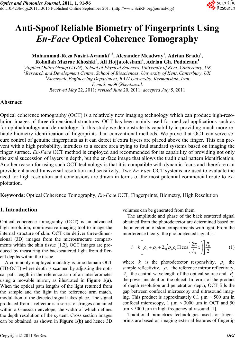
M.-R. NASIRI-AVANAKI ET AL.
Copyright © 2011 SciRes. OPJ
96
ve also shown here, that eF-OCT can also per-
fo
. Acknowledgements
. Meadway, M. R. Nasiri-Avanaki and A. Bradu ac-
. References
] A. Gh. Podoleanu, “Optical Coherence Tomograph
itt, “Principles and Application of Optica
Mao, S. Sherif and C.
. S. Mehta,
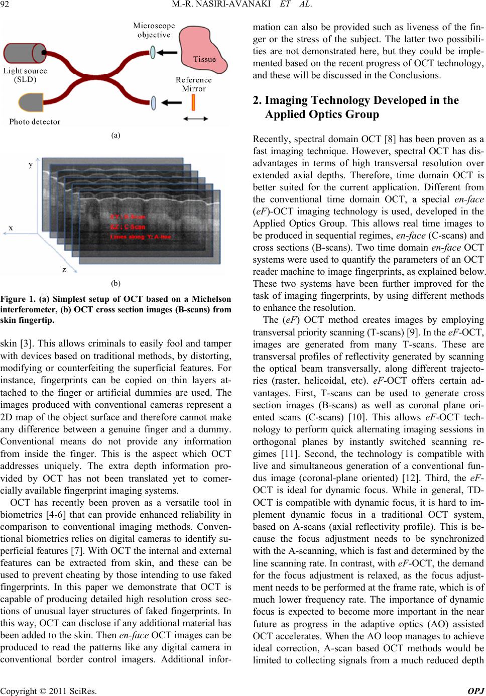
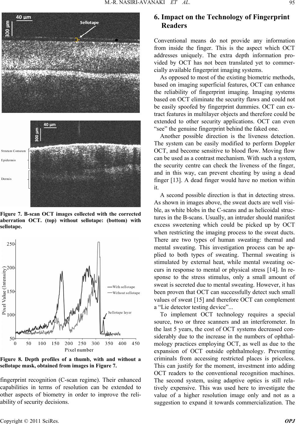
first system however, implementing dynamic focus, is
not more expensive than any other OCT system on the
market.
We ha
rm the task of recognition, as it can provide a real time
en-face image, similar in its orientation to a microscopy
image. Such a system can accomplish booth goals, detect
extra layers in top of the finger (in the B-scan regime)
and be used in the en-face image collection (in the
C-scan regime).
7
A
knowledge respectively support of the Ariba Foundation,
New York, University of Kent and EPSRC EP/H004963/1.
8
[1 y,”
British Institute of Radiology, Vol. 78, No. 935, 2005, pp.
976-988.
[2] A. M. Schml
Coherent Tomography in Dermatology,” Dermatology,
Vol. 217, No. 1, 2008, pp. 12-13.
[3] S. Chang, Y. Cheng, K. Larin, Y.
Flueraru, “Optical Coherence Tomography Used for Se-
curity and Fingerprint-Sensing Applications,” IET Image
Processing, Vol. 2, No. 1, 2008, pp. 48-58.
[4] S. K. Dubey, T. Anna, C. Shakher and D
“Fingerprint Detection Using Full-Field Swept-Source
Optical Coherence Tomography,” Applied Physics Let-
ters, Vol. 91, No. 18, 2007, Article ID: 181106.
doi:10.1063/1.2800823
[5] S. K. Dubey, D. S. Mehta, A. Anand and C. Shakher,
“Simultaneous Topography and Tomography of Latent
Fingerprints Using Full-Field Swept-Source Optical Co-
herence Tomography,” Journal of Optics A: Pure and
Applied Optics, Vol. 10, No. 1, 2008, Article ID: 015307.
doi:10.1088/1464-4258/10/01/015307
[6] R. K. Manapuram, M. Ghosn and K. V. Larin, “Identifi-
Lennard, “Fingerprint detection
r, J. Reynolds,
cation of Artificial Fingerprints Using Optical Coherence
Tomography Technique,” Asian Journal of Physics, Vol.
15, 2006, pp. 15-27.
[7] P. Margot and C.
techniques,” Universite de Lausanne, Institut de Police
Scientifique et de Criminologie and Switzerland, Lau-
sanne, 1994, p. 190. ISBN 2-940098-01-8
[8] R. Leitgeb, C. K. Hitzenberger, A. Schaefe
D. Marks and A. F. Fercher, “Performance of Fourier
domain vs. S. Boppart, Real-Time Domaindigital Signal
Processing-Based Optical Coherence Tomography,”
Optics Express, Vol. 11, No. 8, 2003, pp. 889-894.
doi:10.1364/OE.11.000889
[9] A. Gh. Podoleanu, G. M. Dobre and D. A. Jackson,
. Seeger, G. M. Dobre, D. J. Webb,
o, R. Rosen and A. Po-
“En-Face Coherence Imaging Using Galvanometer
Scanner Modulation,” Optics Letters, Vol. 23, No. 3,
1998, pp. 147-149.
[10] A. Gh. Podoleanu, M
D. A. Jackson and F. Fitzke, “Transversal and Longitudi-
nal Images from the Retina of the Living Eye Using Low
Coherence Reflectometry,” Journal of Biomedical Optics,
Vol. 3, No. 1, 1998, pp. 12-20.
[11] C. C. Rosa, J. Rogers, J. Pedr
doleanu, “Multi-Scan Time Domain OCT for Retina Im-
aging,” Applied Optics, Vol. 46, No. 10, 2007, pp. 1795-
1807. doi:10.1364/AO.46.001795
[12] A. Gh. Podoleanu and D. A. Jackson, “Combined Optical
, J. Reynolds, D. Marks and S. Boppart,
Coherence Tomograph and Scanning Laser Ophthal-
moscope,” Electronics Letters, Vol. 34, No. 11, 1998, pp.
1088-1090.
[13] A. Schaefer
“Real-Time Digital Signal Processing-Based Optical Co-
herence Tomography and Doppler Optical Coherence
Tomography,” IEEE Transactions on Biomedical Engi-
neering, Vol. 51, No. 1, 2004, pp. 186-190.
doi:10.1109/TBME.2003.820369
[14] M. Ohmi, M. Tanigawa, A. Yamada, Y. Ueda and M.
and Y. Yasuno, “Quantitative
Haruna, “Dynamic Analysis of Internal and External
Mental Sweating by Optical Coherence Tomography,”
Journal of Biomedical Optics, Vol. 14, No. 1, 2009, Arti-
cle ID: 014026.
[15] S. Makita, T. Fabritius
Retinal-Blood Flow Measurement with Three-Dimen-
sional Vessel Geometry Determination Using Ultrahigh-
Resolution Doppler Optical Coherence Angiography,”
Optics Letters, Vol. 33, No. 8, 2008, pp. 836-838.
doi:10.1364/OL.33.000836