A. G. Ad e s i yu n et al. / Open Journal of Obstetrics and Gynecology 1 (2 011) 121-123
Copyright © 2011 SciRes. OJOG
ginal deli ver y at a private hos p ital, three d a ys be fore p re-
sentat ion. S he had an une vent ful delive ry of a te rm male
neonate, who had been vomiting, with associated abdo-
minal distention and difficulty in breathing. The baby
was yet to pass meconium. Examination of the baby re-
vealed a body weight of 2.4 kg, with moderate pallor,
dehydration and pyrexia of 38°. The respiratory rate was
140/min with crepitations over the lung field. There was
moderate abdominal distension. A diagnosis of neonatal
intestinal obstruction and aspiration pneumonitis was
made. The baby was optimized for surgery .The intra
operative findings were consistent with intestinal ob-
struction secondary to proximal jejunal atresia. This was
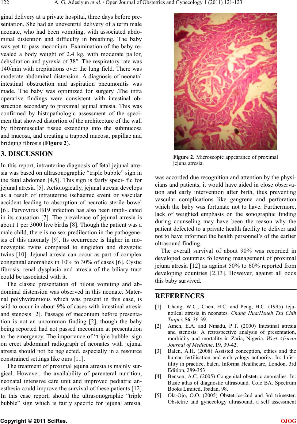
confirmed by histopathologic assessment of the speci-
men that sho wed distor tio n of the architecture of the wall
by fibromuscular tissue extending into the submucosa
and mucosa, and creating a trapped mucosa, papillae and
bridging fibro sis ( Figure 2).
3. DISCUSSION
In this report, intrauterine diagnosis of fetal jejunal atre-
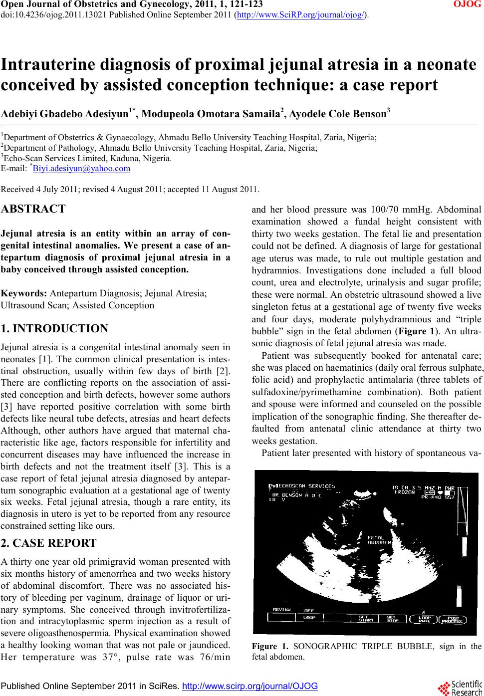
sia was based on ultrasonogra phic “triple b ubble” sign in
the fetal abdomen [4,5]. This sign is fairly speci- fic for
jejunal atresia [5]. Aetiologicall y, jejunal atresia develops
as a result of intrauterine ischaemic event or vascular
accident leading to absorption of necrotic sterile bowel
[6]. Parvovirus B19 infection has also been impli- cated
in its causation [7]. The prevalence of jejunal atresia is
about 1 per 3000 live births [8]. Though the patient was a
male c hild, ther e is no sex p red ilectio n in t he pat hoge ne-
sis of this anomaly [9]. Its occurrence is higher in mo-
nozygotic twins compared to singleton and dizygotic
twins [10]. Jejunal atresia can occur as part of complex
congenital anomalies in 10% to 30% of cases [6]. Cystic
fibrosis, renal dysplasia and atresia of the biliary tract
could be associated with it.
The classic presentation of bilous vomiting and ab-
dominal distension was observed in this neonate. Mater-
nal polyhydramious which was present in this case, is
said to occur in about 9% of cases with intestinal atresia
and stenosis [2]. Passage of meconium before presenta-
tion is not an uncommon finding [2], though the baby
being reported had not passed meconium at presentation
to the emergency. The importance of “triple bubble: sign
on erect abdominal radiograph of neonates with jejunal
atresia should not be neglected, especially in a resource
constrained se ttings like ours [11].
The treatment of proximal jejuna atresia is mainly sur-
gical. However, the availability of parenteral nutrition,
neonatal intensive care unit and improved pediatric an-
esthe sia c ould imp ro ve t he sur viva l of t hese pat ient s [ 12] .
In this case report, should the ultrasonographic “triple
bubble” sign which is fairly specific for jejunal atresia,
Figure 2. Microscop ic appearance of pro ximal
jejuna atresia.
was accorded due r eco gnitio n a nd at tent ion b y the ph ysi -
cians and patients, it would have aided in close observa-
tion and early intervention after birth, thus preventing
vascular complications like gangrene and perforation
which the baby was fortunate not to have. Furthermore,
lack of weighted emphasis on the sonographic finding
during counseling may have been the reason why the
patient defected to a private health facility to deliver and
not to have informed the health personnel’s of the earlier
ultrasound finding.
The overall survival of about 90% was recorded in
developed countries following management of proximal
jejuna atresia [12] as against 50% to 60% reported from
developing countries [2,13]. However, against all odds
this baby survived .
REFERENCES
[1] Chang, W.C., Chen, H.C. and Peng, H.C. (1995) Jeju-
noileal atresia in neonates. Chang Hua/Hsueh Tsa Chih
Taipei, 56, 36-39.
[2] Ameh, E.A. and Nmad u , P.T. (2000) Intestinal atresia
and stenosis: A retrospective analysis of presentation,
morbidity and mortality in Zaria, Nigeria. West African
Journal of Medicine, 19, 39-42.
[3] Balen, A.H. (2008) Assisted conception, ethics and the
human fertilisation and embryology authority. In: Infer-
tility in practice, balen. Informa Healthcare, London. 3rd
Edition, 289-353.
[4] Benson, A.C. (2005) Congenital obstetric anomalies. In:
Basic atlas of diagnostic ultrasound. Cole BA. Spectrum
Books Limited, Ibadan, 98.
[5] Ola-Ojo, O.O. (2005) Obstetrics-2nd and 3rd trimester.
Obstetric and gynecology ultrasound, a self assessment