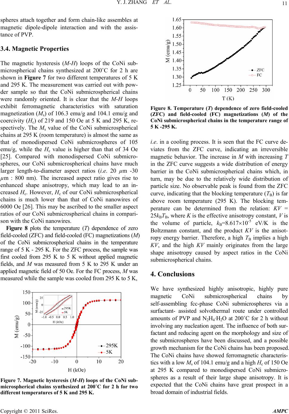
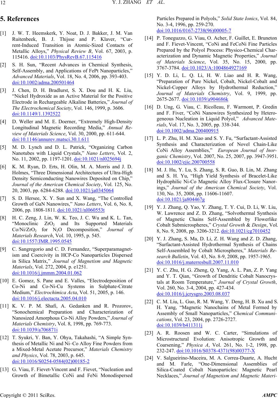
Advances in Materials Physics and Chemistry, 2011, 1, 7-13
doi:10.4236/ampc.2011.12002 Published Online September 2011 (http://www.SciRP.org/journal/ampc)
Copyright © 2011 SciRes. AMPC
Synthesis, Structure and Magnetic Properties of CoNi
Submicrospherical Chains*
Yajing Zhang1, Siu-Wing Or1, Zhidong Zhang2
1Department of Electrical Engineering, Hong Kong Polytechnic University, Hung Hom, Hong Kong, China
2Shenyang National Laboratory for Materials Science, Institute of Metal Research and International Centre for
Materials Physics, Chinese Academy of Sciences, Shenyang, China
E-mail: eeswor@polyu.edu.hk
Received July 1,2011; revised August 14, 2011; accepted August 25, 2011
Abstract
High-purity magnetic CoNi submicrospherical chains, each of length 20 μm - 30 μm and self-assembled
from fcc-phase CoNi submicrospheres of average diameter 800 nm, are synthesized via a surfactant-assisted
solvothermal route without the aid of nucleation agent. The effects of surfactant and reducing agent on the
morphology and size of the CoNi chains are studied, and a possible growth mechanism for the CoNi chains
is proposed. The CoNi chains show ferromagnetic characteristics with a similarly small saturation magneti-
zation of 104.1 emu/g and a larger coercivity of 150 Oe at room temperature compared to the monodispersed
CoNi submicrospheres of 105 emu/g and 34 Oe as a result of the increased shape anisotropy.
Keywords: Chain-Like Assembles, CoNi, Magnetic Properties, Morphology, Solvothermal Route,
Submicrospherical Chains
1. Introduction
Magnetic nanomaterials have received much attention in
recent years because of their unique magnetic properties
and high application potential for data storages, micro-
electronic devices, medical diagnosis, catalysis, etc. [1,2].
In fact, the magnetic properties of magnetic nanomateri-
als depend greatly on their size and shape [3]. Most of
the isolated magnetic nanoparticles with sizes below a
critical value (e.g. diameter < 10 nm for CoNi particles)
are superparamagnetic at room temperature and thus are
inappropriate for many fields of applications [4]. In view
of this, there is an increasing driving force pushing to-
wards the synthesis of magnetic nanomaterials with va-
rious nanostructures in order to increase the shape ani-
sotropy for pos sible fe rr o magnetism [5-7].
Among various magnetic metals, CoNi is regarded as
an important magnetic alloy due to its attractive magne-
tic and catalytic properties as well as improved mechan-
ical properties compared to Co [8]. In the past decade, a
variety of methods have been developed to synthesize
micro- and nanosized CoNi particles such as sol-gel rout-
es, electrodeposition, sonochemical synthesis, decom-
position of organometallic precursors, polyol reduction
and hydrothermal routes [9-15]. Recently, several CoNi
assemblies have also been synthesized, including sea-ur-
chin-like particles prepared using CoNi nanowires as
building block by heterogeneous nucleation in liquid
polyol [16], chain-like CoNi assemblies synthesized by a
surfactant-assisted hydrothermal route [17] and sin-
gle-walled flux-closure CoNi magnetic nanorings syn-
thesized using triethylene glycol as solvent in the pres-
ence of poly (vinylpyrrolidone) [18]. With the prelimi-
nary remarks on CoNi assemblies, continual research
effort is required to explore other possible synthesis
methods, interesting assemblies, the relationship between
structure and properties, etc.
In this work, we present the synthesis and structure of
highly anisotropic, highly pure CoNi submicrospherical
chains self-assembled from fcc-phase CoNi submicro-
spheres via a surfactant-assisted solvothermal route with-
out involving any nucleation agent. The influence of both
surfactant and reducing agent on the morphology and
size of the submicrospheres is discussed, together with a
possible growth mechanism for the CoNi chains. The
magnetic properties of the CoNi chains are also investi-
*This work was supported by the Research Grants Council of the
HKSAR Government (PolyU 5266/08E), The Hong Kong Polytechnic
University (1-ZV7P, 4-ZZ7L and G-U741), and the National Nature
Science Foundation of China (50331 030).
Y. J. ZHANG ET AL.
8
gated by measuring their magnetic hysteresis loops as
well as zero field-cooled and field-cooled magnetizations
at various temperatures.
2. Experimental Details
In the proposed surfactant-assisted solvothermal route,
cobalt (II) chloride hexahydrate (CoCl2·6H2O) and nickel
(II) chloride hexahydrate (NiCl2·6H2O) were used as
precursors, while poly (vinyl pyrrolidone) (PVP), hydra-
zine monohydrate (N2H4·H2O) and ethylene glycol (EG)
were employed as surfactant, reducing agent and base
solution, respectively. These reagents were commercial
acquired with analytical pure and without further purifi-
cation. In a typical synthesis procedure, 0.5 mmol (0.12 g)
of CoCl2·6H2O, 0.5 mmol (0.12 g) of NiCl2·6H2O and
2.1 mmol (0.5 g) of PVP were dissolved in 30 mL of EG,
and the solution was stirred steadily for 30 min at room
temperature. After adding 2 mL of N2H4·H2O, the solu-
tion was further stirred for 30 min. The stirred solution
was transferred into a Teflon-lined stainless steel auto-
clave of volume 50 ml. The autoclave was subsequently
sealed and maintained at 200˚C for 2 h before being
cooled to room temperature naturally. The as-prepared
product was filtered off, washed with distilled water and
ethanol for several times, and dried in a vacuum oven at
60˚C for 4 h. Control experiments were carried out by
adjusting the amounts of PVP (from 0 g to 1 g) and
N2H4·H2O (from 0 mL to 3 mL), while keeping other
reaction parameters unchanged.
The phase and purity of all the as-prepared products
were identified by a Rigaku D/max 2500pc X-ray dif-
fractometer with Cu-Kα radiation (
= 1.54156 Å) at a
scan rate of 0.04˚/s. The morphology and size were
characterized by a JEOL JSM-6335F field emission
scanning electron microscope equipped with an en-
ergy-dispersive X-ray spectrometer and operated at an
emission voltage of 15 kV. The detailed morphology was
investigated using a JEOL2010 transmission electron
microscope with an emission voltage of 200 kV. The
magnetic hysteresis loops were measured at both 5 K and
295 K (room temperature) using a MPMS-7 supercon-
ducting quantum interference device (SQUID) magne-
tometer for magnetic fields up to 20 kOe. The tempera-
ture dependence of zero field-cooled and field-cooled
magnetizations was also measured using the SQUID ma-
gnetometer in the temperature range of 5 K - 295 K.
3. Results and Discussion
3.1. Structure, Morphology and Size
Figure 1 shows a typical X-ray diffraction (XRD)
pattern of the product syn thesized at 200˚C for 2 h. Three
sharp XRD peaks are observed at 44.34˚, 51.65˚ and
76.14˚, corresponding to the (111), (200) and (220)
planes, respectively. The appearance of these peaks in
the XRD pattern indicates the existence of a face cen-
tered cubic (fcc) phase in the product. Apart from these
peaks, the XRD pattern is essentially clean with no other
detectable peaks, especially for those caused by impuri-
ties such as Co(OH)2 or Ni(OH)2. This suggests that the
reduction reaction in our solvothermal route is rather
complete.
Figure 2 plots a typical energy-dispersive X-ray spec-
troscopy (EDS) spectrum of the product synthesized at
200˚C for 2 h. Four different types of elements are de-
tected, including Co, Ni, Au and Si. The presence of Au
element in the EDS spectrum originates from the sput-
tered thin Au layer on the product for the test purpose,
while the occurrence of Si element results from the use
of a silicon wafer to support the product. Excluding th ese
two types of elements, the mean molar ratio of Co to Ni
in the product at different selected locations is approxi-
mately 0.96 (49:51). This value not only agrees with the
starting stoichiometric proportion, but also confirms, in
Figure 1. Typical XRD pattern of the product synthesized
at 200˚C for 2 h.
Figure 2. Typical EDS spectr um of the product synthesized
at 200˚C for 2 h.
Copyright © 2011 SciRes. AMPC
9
Y. J. ZHANG ET AL.
conjunction with the XRD results in Figure 1, the suc-
cess synthesis of high-purity fcc-phase CoNi product.
Figure 3(a) shows a low-magnification field emission
scanning electron microscopy (FESEM) image of the
CoNi product synthesized at 200˚C for 2 h. It is clear that
the product is essentially chain-like CoNi assemblies with
high uniformity of 20 μm - 30 μm i n length. The chain-li ke
assemblies can be classified as a type of hierarchical as-
semblies. Figure 3(b) displays the high-magnification
FESEM image of the CoNi product. Each CoNi chain is
assembled from CoNi submicrospheres with an average
diameter of 800 nm. These submicrospheres serve as
building block for the chains. Moreover, the surface of the
CoNi submicrospheres is quite rough because they are
composed of CoNi nanoparticles. Figure 3(c) illustrates
the transmissio n electron microscopy (TEM) image of the
CoNi product. It i s seen that the product i s chain-li ke CoNi
assemblies of CoNi submicrospheres of average diameter
800 nm. The observation is consistent with those observed
by FESEM in Figures 3(a)-(b). In addition, bright contrast
is not found inside the submicrospheres, indicating that
they are solid rather than hollow submicrospheres. The
inset of Figure 3(c) shows the electron diffraction pattern
of a CoNi submicrosphere, revealing its polycrystalline
nature. It is noted that the CoNi submicrospherical chains
are very stable even subjected to an ultrasonic treatment of
30 min. This reflects that the chain-like CoNi assemblies
are actually integrated and not simply aggregations of
nanoparticles.
Figure 3. (a) Low-magnification FESEM image, (b) high-
magnification FESEM image, and (c) TEM image of CoNi
product synthesized at 200˚C for 2 h.
3.2. Effects of Surfactant and Reducing Agent on
Morphology and Size
We found that the amounts of PVP surfactant and
N2H4·H2O reducing agent in the solution have the pre-
dominant effects on the morphology and size of the re-
sulting CoNi products. In this section, we study the ef-
fects of PVP and N2H4·H2O on the morphology and size
of the CoNi products by keeping other reaction parame-
ters unchanged.
Figure 4 shows scanning electron microscopy (SEM)
images of CoNi products prepared using four different
amounts of PVP at an optimal amount of N2H4·H2O of 2
mL. In the absence of PVP, the CoNi submicrospherical
chains self-assembled by CoNi submicrospheres as sho-
wn in Figure 3 cannot be formed, but monodispersed mi-
crospheres with an average diameter of 1.5 μm are ob-
tained instead (Figure 4(a)). When an increased amount
of PVP of 0.3 g is used, the size of the microspheres is
not uniform; it actually varies from 2 μm to 8 μm in di-
ameter (Figure 4(b)). When the amount of PVP is in-
creased to 0.7 g, chain-like assembles self-assembled
from uniform submicrospheres of average diameter 800
nm are obtained (Figure 4(c)). When the amount of PVP
is further increased to 1 g, chain-like assembles consist-
ing of microspheres of average diameter 5 μm are formed
(Figure 4(d)). The results suggest that the morphology
and size of the CoNi products can be controlled by the
amount of PVP. The optimal PVP is found to be 0.5 g,
and the resulting CoNi submicrospherical chains are
shown in Figure 3.
Figure 5 shows SEM images of CoNi products pre-
pared using three different amounts of N2H4·H2O at an
Figure 4. SEM images of CoNi products prepared using
four different amounts of PVP of (a) 0 g, (b) 0.3 g, (c) 0.7 g,
and (d) 1 g at an optimal amount of N2H4·H2O of 2 mL.
Copyright © 2011 SciRes. AMPC
Y. J. ZHANG ET AL.
10
optimal amount of PVP of 0.5 g. With the addition of 0.5
mL of N2H4·H2O, the average diameter of microspheres
is 10 μm (Figure 5(a)). When the amount of N2H4·H2O
is doubly in creased to 1 mL, th e average d iameter o f mi-
crospheres is half-reduced to 5 μm (Figure 5(b)). A fur-
ther increase the amount of N2H4·H2O to 2 mL leads to
submicrospher es of av erage diamete r 800 nm as repor ted in
Figure 3. A subsequent increase the amount of N2H4·H2O
beyond 2 mL, e.g. 3 mL, cannot further decrease the size
of the submicrospheres (Figure 5(c)). Therefore, the size
of spheres in the CoNi products can be controlled by
adjusting the amount of N2H4·H2O. In general, the size of
spheres decreases with an increase in the amount of
N2H4·H2O up to 2 mL. The change in size of spheres is
likely caused by the change in reaction rate. With in-
creasing the amount of N2H4·H2O, the reaction rate is
accelerated so that more nuclei are formed in the initial
nucleation process and lead to the formation of more
spheres with smaller diameters [19].
3.3. Possible Growth Mechanism
On the basis of the observed reaction phenomena and
results, a possible growth mechanism for the formation
of CoNi submicrospherical chains is addressed in this
section. A similar mechanism for the growth of 1D
magnetic cobalt chains has been presented in our early
works [19,20]. As shown in Figure 6, three consecutive
steps: namely: steps a, b, and c are possibly included in
the growth mechanism. In step a, CoNi nanoparticles are
reduced. This step actually includes three consequent
processes. First, Co2+ and Ni2+ ions can combine with EG to
form pink-mixed complexes [Co(EG)n]2+ and [Ni(EG)n]2+.
Figure 5. SEM images of CoNi products prepared using
three different amounts of N2H4·H2O of (a) 0.5 mL, (b) 1
mL, and (c) 3 mL at an optimal amount of PVP of 0.5 g.
Figure 6. Schematic diagram of a possible growth mecha-
nism for CoNi submicrospherical chains.
After N2H4·H2O is added into the solution, the complexes
react with N2H4·H2O to give complexes of [Co(N2H4)3]2+
and [Ni(N2H4)3]2+ so that the solution turns turbid. The
whole complex process can be expressed by the follow-
ing two equations [21,22]:
 
2
2
24 24
n3
CoEG3N HCoN HnEG


 
 
(1)
 
2
2
24 24
n3
N
iEG3NH NiNHnEG


 
 
(2)
Second, the complexes of [Co(N2H4)3]2+ and [Ni(N2H4)3]2+
can react with excessive hydrazine and thus reduce to Co
and Ni nuclei at high temperature (at the boiling point of
EG of 197˚C) even though they are stable at room tem-
perature. The Co and Ni nuclei then form CoNi alloy
nuclei. This reducing process accompanies the change in
the color of the solution; that is, the solution is pink tur-
bid and colorless before and after the reaction, respec-
tively. The reducing process can be described by the fol-
lowing three equation s:

2
24 24
3
322
CoNHNH
Co 4NHNH2H



  (3)

2
24 24
3
322
NiNHNH
N
i4NH NH 2H



  (4)
Co NiCoNi
(5)
In this reducing process, High reaction temperature is
critical to simultaneously reduce the Co2+ and Ni2+ ions.
If the reducing process is carried out at a lower tempera-
ture, for example, at 160˚C, elemental Co is found in the
final product in addition to CoNi alloy. Third, the CoNi
nuclei grow into CoNi nanoparticles. In step b, the CoNi
nanoparticles diffuse and aggregate into CoNi spheres
through an Ostwald ripening process [23] and the mag-
netic dipole-dipole interaction [24]. In step c, the CoNi
Copyright © 2011 SciRes. AMPC
11
Y. J. ZHANG ET AL.
spheres attach together and form chain-like assembles at
magnetic dipole-dipole interaction and with the assis-
tance of PVP.
3.4. Magnetic Properties
The magnetic hysteresis (M-H) loops of the CoNi sub-
microspherical chains synthesized at 200˚C for 2 h are
shown in Figure 7 for two different temperatures of 5 K
and 295 K. The measurement was carried out with pow-
der sample so that the CoNi submicrospherical chains
were randomly oriented. It is clear that the M-H loops
exhibit ferromagnetic characteristics with saturation
magnetization (Ms) of 106.3 emu/g and 104.1 emu/g and
coercivity (Hc) of 219 and 150 Oe at 5 K and 295 K, re-
spectively. The Ms value of the CoNi submicrospherical
chains at 295 K (room temperature) is almost the same as
that of monodispersed CoNi submicrospheres of 105
emu/g, while the Hc value is higher than that of 34 Oe
[25]. Compared with monodispersed CoNi submicro-
spheres, our CoNi submicrospherical chains have much
larger length-to-diameter aspect ratios (i.e. 20 µm -30
µm : 800 nm). The increased aspect ratio gives rise to
enhanced shape anisotropy, which may lead to an in-
creased Hc. However, Hc of our CoNi submicrospherical
chains is much lower than that of CoNi nanowires of
6000 Oe [26]. This may be ascribed to the smaller aspect
ratios of our CoNi submicrospherical chains in compari-
son with the CoNi nanowires.
Figure 8 plots the temperature (T) dependence of zero
field-coo led (ZFC) and field- cooled (FC) magn etizations (M)
of the CoNi submicrospherical chains in the temperature
range of 5 K - 295 K. For the ZFC process, the sample was
first cooled from 295 K to 5 K without applied magnetic
fields, and M was measured from 5 K to 295 K under an
applied magnetic field of 50 O e. For the FC process, M was
measured while the sample was cooled from 295 K to 5 K,
Figure 7. Magnetic hysteresis (M-H) loops of the CoNi sub-
microspherical chains synthesized at 200˚C for 2 h for two
different temperatures of 5 K and 295 K.
Figure 8. Temperature (T) dependence of zero field-cooled
(ZFC) and field-cooled (FC) magnetizations (M) of the
CoNi submicrospherical chains in the temperature range of
5 K -295 K.
i.e. in a cooling process. It is seen that the FC curve de-
viates from the ZFC curve, indicating an irreversible
magnetic behavior. The increase in M with increasing T
in the ZFC curve suggests a wide distribution of energy
barrier in the CoNi submicrospherical chains which, in
turn, may be due to the relatively wide distribution of
particle size. No observable peak is found from the ZFC
curve, indicating that the blocking temperature (TB) is far
above room temperature (295 K). The blocking tem-
perature can be determined from the relation: KV =
25kBTB, where K is the effective anisotropy constant, V is
the volume of particle, kB=8.61710-5 eV/K is the
Boltzmann constant, and the product KV is the anisot-
ropy energy barrier. Therefore, a high TB implies a high
KV, and the high KV mainly originates from the large
shape anisotropy caused by aspect ratios in the CoNi
submicrospherical chains.
4. Conclusions
We have synthesized highly anisotropic, highly pure
magnetic CoNi submicrospherical chains by
self-assembling fcc-phase CoNi submicrospheres via a
surfactant- assisted solvothermal route under controlled
amounts of PVP and N2H4·H2O at 200˚C for 2 h without
involving any nucleation agent. The influence of both sur-
factant and reducing agent on the morphology and size of
the submicrospheres have been discussed, and a possible
growth mechanism for the CoNi chains has been proposed.
The CoNi chains have showed ferromagnetic characteris-
tics with a low Ms of 104.1 emu/g and a high Hc of 150 Oe
at 295 K compared to monodispersed CoNi submicro-
spheres as a result of their large shape anisotropy. It is
expected that the CoNi chains have great prospect in a
broad domain of industri al fields.
Copyright © 2011 SciRes. AMPC
Y. J. ZHANG ET AL.
12
5. References
[1] J. W. T. Heemskerk, Y. Noat, D. J. Bakker, J. M. Van
Ruitenbeek, B. J. Thijsse and P. Klaver, “Cur-
rent-Induced Transition in Atomic-Sized Contacts of
Metallic Alloys,” Physical Review B, Vol. 67, 2003, p.
115416. doi:10.1103/PhysRevB.67.115416
[2] S. H. Sun, “Recent Advances in Chemical Synthesis,
Self-Assembly, and Applications of FePt Nanoparticles,”
Advanced Materials, Vol. 18, No. 4, 2006, pp. 393-403.
doi:10.1002/adma.200501464
[3] J. Chen, D. H. Bradlurst, S. X. Dou and H. K. Liu,
“Nickel Hydroxide as an Active Material for the Positive
Electrode in Rechargeable Alkaline Batteries,” Journal of
The Electrochemical Society, Vol. 146, 1999, p. 3606.
doi:10.1149/1.1392522
[4] D. Weller and M. E. Doerner, “Extremely High-Density
Longitudinal Magnetic Recording Media,” Annual Re-
view of Materials Science, Vol. 30, 2000, pp. 611-644.
doi:10.1146/annurev.matsci.30.1.611
[5] M. D. Lynch and D. L. Patrick, “Organizing Carbon
Nanotubes with Liquid Crystals,Nano Letters, Vol. 2,
No. 11, 2002, pp. 1197-1201. doi:10.1021/nl025694j
[6] K. M. Ryan, D. Erts, H. Olin, M. A. Morris and J. D.
Holmes, “Three Dimensional Architectures of Ultra-High
Density Semiconducting Nanowires Deposited on Chip,”
Journal of the American Chemical Society, Vol. 125, No.
20, 2003, pp. 6284-6288. doi:10.1021/ja0345064
[7] S. D. Hersee, X. Y. Sun and X. Wang, “The Controlled
Growth of GaN Nanowires,” Nano Letters, Vol. 6, No. 8,
2006, pp. 1808-1811. doi:10.1021/nl060553t
[8] H. C. Zeng, J. Lin, W. K. Teo, J. C. Wu and K. L. Tan,
“Monoclinic ZrO2 and Its Supported Materials
Co/Ni/ZrO2 for N2O Decomposition,” Journal of
Materials Research, Vol. 10, 1995, p. 545.
doi:10.1557/JMR.1995.0545
[9] C. Sangregorio and C. D. Fernandez, “Superparamagnet-
ism and Coercivity in HCP-Co Nanoparticles Dispersed
in Silica Matrix,” Journal of Magnetism and Magnetic
Materials, Vol. 272, 2004, p. e1251.
doi:10.1016/j.jmmm.2004.01.062
[10] E. Gomez, S. Pane and E. Valles, “Electrodeposition of
Co-Ni and Co-Ni-Cu Systems in Sulphate-Citrate
Medium,Electrochimica Acta, Vol. 51, 2005, p. 146.
doi:10.1016/j.electacta.2005.04.010
[11] K. V. P. M. Shafi, A. Gedanken and R. Prozorov,
“Sonochemical Preparation and Characterization of
Nanosized Amorphous Co-Ni Alloy Powders,” Journal of
Materials Chemistry, Vol. 8, 1998, pp. 769-773.
doi:10.1039/a706871i
[12] T. Syukri, Y. Ban, Y. Ohya, Takahashi, “A Simple Syn-
thesis of Metallic Ni and Ni–Co Alloy Fine Powders from
a Mixed-Metal Acetate Precursor,” Materials Chemistry
and Physics, Vol. 78, 2003, p. 645.
doi:10.1016/S0254-0584(02)00185-2
[13] G. Viau, F. Fievet-Vincent and F. Fievet, “Nucleation and
Growth of Bimetallic CoNi and FeNi Monodispersed
Particles Prepared in Polyols,” Solid State Ionics, Vol. 84,
No. 3-4, 1996, pp. 259-270.
doi:10.1016/0167-2738(96)00005-7
[14] P. Toneguzzo, G. Viau, O. Acher, F. Guillet, E. Bruneton
and F. Fievet-Vincent, “CoNi and FeCoNi Fine Particles
Prepared by the Polyol Process: Physico-Chemical Char-
acterization and Dynamic Magnetic Properties,” Journal
of Materials Science, Vol. 35, No. 15, 2000, pp.
3767-3784. doi:10.1023/A:1004864927169
[15] Y. D. Li, L. Q. Li, H. W. Liao and H. R. Wang,
Preparation of Pure Nickel, Cobalt, Nickel-Cobalt and
Nickel-Copper Alloys by Hydrothermal Reduction,”
Journal of Materials Chemistry, Vol. 9, 1999, pp.
2675-2677. doi:10.1039/a904686k
[16] D. Ung, G. Viau, C. Ricolleau, F. Warmont, P. Gredin
and F. Fiver, “CoNi Nanowires Synthesized by Hetero-
geneous Nucleation in Liquid Polyol,” Advanced Mate-
rials, Vol. 17, No. 3, 2005, pp. 338-344.
doi:10.1002/adma.200400915
[17] L. P. Zhu, H. M. Xiao and S. Y. Fu, “Surfactant-Assisted
Synthesis and Characterization of Novel Chain-Like
CoNi Alloy Assemblies,” European Journal of Inor-
ganic Chemistry, Vol. 2007, No. 25, 2007, pp. 3947-3951.
doi:10.1002/ejic.200700558
[18] M. J. Hu, Y. Lu, S. Zhang, S. R. Guo, B. Lin, M. Zhang
and S. H. Yu, “High Yield Synthesis of Bracelet-Like
Hydrophilic Ni-Co Magnetic Alloy Flux-Closure Nanor-
ings,” Journal of the American Chemical Society, Vol.
130, No. 35, 2008, pp. 11606-11607.
doi:10.1021/ja804467g
[19] Y. J. Zhang, Q. Yao, Y. Zhang, T. Y. Cui, D. Li, W. Liu,
W. Lawrence and Z. D. Zhang, “Solvothermal Synthesis
of Magnetic Chains Self-Assembled by Flowerlike
Cobalt Submicrospheres,Crystal Growth & Design, Vol.
8, No. 9, 2008, pp. 3206-3212. doi:10.1021/cg7010452
[20] Y. J. Zhang, S. Ma, D. Li, Z. H. Wang and Z. D. Zhang,
Surfactant-Assisted Hydrothermal Synthesis of Chains
Self-Assembled by Cobalt Microspheres,” Materials Re-
search Bulletin, Vol. 43, No. 8-9, 2008, pp. 1957-1965.
doi:10.1016/j.materresbull.2007.11.010
[21] Y. C. Zhu, H. G. Zheng, Q. Yang, A. L. Pan, Z. P. Yang
and Y. T. Qian, “Growth of Dendritic Cobalt Nanocrys-
tals at Room Temperature,” Journal of Crystal Growth,
Vol. 260, No. 3-4, 2004, pp. 427-434.
doi:10.1016/j.jcrysgro.2003.08.037
[22] C. M. Liu, L. Guo, R. M. Wang, Y. Deng, H. B. Xu and S.
H. Yang, “Magnetic Nanochains of Metal Formed by
Assembly of Small Nanoparticles,” Chemical Communi-
cations, Vol. 23, 2004, pp. 2726-2727.
doi:10.1039/b411311j
[23] A. R. Roosen and W. C. Carter, “Simulations of
Microstructural Evolution: Anisotropic Growth and
Coarsening,” Physica A, Vol. 261, No. 1-2, 1998, pp.
232-247. doi:10.1016/S0378-4371(98)00377-X
[24] V. Salgueirino-Maceira, M. A. Correa-Duarte, A. Hucht
and M. Farle, “One-Dimensional Assemblies of
Silica-Coated Cobalt Nanoparticles: Magnetic Pearl
Necklaces,” Journal of Magnetism and Magnetic Materi-
Copyright © 2011 SciRes. AMPC
Y. J. ZHANG ET AL.
Copyright © 2011 SciRes. AMPC
13
als, Vol. 303, No. 1, 2006, pp. 163-166.
doi:10.1016/j.jmmm.2005.11.003
[25] P. Elumalai, H. N. Vasan, M. Verelst, P. Lecante, V.
Carles and P. Taihades, “Synthesis and Characterization
of Sub-Micron Size Co-Ni Alloys using Malonate as
Precursor,” Materials Research Bulletin, Vol. 37, No. 2,
2002, pp. 353-363. doi:10.1016/S0025-5408(01)00768-1
[26] D. Ung, Y. Soumare, N. Chakroune, G. Viau, M. J. Vau-
lay, V. Richard and F. Fiévet, “Growth of Magnetic
Nanowires and Nanodumbbells in Liquid Polyol,”
Chemistry of Materials, Vol. 19, No. 8, 2007, pp.
2084-2094. doi:10.1021/cm0627387
[27] Z. D. Zhang, J. G. Zheng, I. Skorvanek, G. H. Wen, J.
Kovac, F. W. Wang, J. L. Yu, J. J. Li, X. L. Dong, S. R.
Jin, W. Liu, and X. X. Zhang, “Shell/Core Structure and
Magnetic Properties of Carbon-Coated Fe-Co(C)
Nanocapsules,” Journal of Physics: Condensed Matter,
Vol. 13, No. 9, 2001, p. 1921.
doi:10.1088/0953-8984/13/9/314