Chinese Medicine, 2011, 2, 115-119
doi:10.4236/cm.2011.23019 Published Online September 2011 (http://www.SciRP.org/journal/cm)
Copyright © 2011 SciRes. CM
The Effect of Aqueous Extract of Cecropia glazioui
Snethlage (Embauba) in the Rat Fetal Development
Priscila Randazzo-Moura1,2, Magali Glauzer Silva1, Yoko Oshima-Franco1,
Francisco Carlos Groppo3, Marli Gerenutti1*
1School of Pharm acy, University of Sorocaba, Sorocaba, Brazil
2Pontifical Catholic University of Sorocaba, Sorocaba, Brasil
3Piracicaba Dental School, State University of Campinas, Piracicaba, Brazil
E-mail: *marli.gerenutti@prof.uniso.br
Received May 15, 2011; revised May 29, 2011; accepted June 8, 2011
Abstract
The effect of aqueous extract of Cecropia glazioui Snethlage (Embauba) in the rat fetal development. This
study was to complement previous assays on the physical and neurobehavioral development of rats resulted
from oral administration of 1 g/kg/day Cecropia glazioui Snethlage (C. glazioui) aqueous extract (LD50 > 5
g·kg1) in pregnant rats (peri- and post-natal studies). In the present study, the effect of 2.5 g/kg/week C.
glazioui aqueous extract, administered to pregnant rats during 15 days, was verified in the rat offspring de-
velopment. No acute or chronic toxicity (no effects on mortality or weight average daily gain) were observed.
In addition, no effects on reproductive parameters (offspring vitality, placenta and fetus weight, number of
corpora lutea on each ovary, pre- and post-implantation loss) and on offspring external morphology were
found. We concluded that C. glazioui aqueous extract administered during pregnancy did not cause abnor-
malities in rat offspring.
Keywords: Fetuses’ Abnormalities, Medicinal Plants, Teratogenesis
1. Introduction
Several studies concerning the pharmacological effects
of Cecropia glazioui Snethlage (Cecropiaceae), popu-
larly known as “Embauba” in Brazil, were published in
the last decade. The C. glazioui aqueous extract showed
anxiolytic-like effect in mice 1, probable by blocking
monoamines uptake in the central nervous system 2.
An anti-asthmatic property was confirmed in guinea pigs
treated with C. glazioui purified fraction 3. It also
showed hypotensive activity 4, which is not related to
angiotensin-converter enzyme 5, and it has antisecre-
tory/antiulcer properties 6. Plants are an important
source of new molecules, which result mainly as conse-
quence of their ontogenesis caused by internal and
external forces 7. They produce active primary and
secondary metabolites to defend themselves against preda-
tors, microorganisms, and UV rays; and to attract seed-
dispersive animals and insects 8. Some of C. glazioui
constituents, such as catechins, procyanidins and flavo-
noids, were observed by Tanae et al. 9. Brazilian legal
requirements of phytotherapics’ marketing demand data
on their pharmacological action and therapeutic efficacy.
Safety is other legal aspect regarding phytotherapics
10-15. However, few studies on phytotherapic safety
are available in the literature and data regarding C.
glazioui toxicity is still scarce. A previous study showed
that the daily exposure to 1 g/kg C. glazioui aqueous
extract showed low toxicity to pregnant rats and their
litters 16. The objective of the present study was to
evaluate the influence of weekly oral administration of C.
glazioui aqueous extract (2.5 g/kg) in pregnant rats,
simulating the human sporadic use.
2. Material and Methods
2.1. Plants
The Cecropia glazioui Snethlage specimen were col-
lected in Tapirai city (State of São Paulo, Brazil). A
voucher specimen is deposited in University of Sorocaba
(UNISO) herbarium, being identified by Dr Sérgio
Romaniuc Neto (Botanic Institute of São Paulo, Brazil).
P. RANDAZZO-MOURA ET AL.
116
2.2. Preparation of the Aqueous Extract
Fresh leaves (450 g) of C. glazioui without petiole were
dried, powdered and a 70% hydroalcoholic extract was
obtained by percolation. The extract was concentrated
under reduced pressure and lyophilized providing 102.3
g of powder (efficiency = 22.7%). It was stored at room
temperature without light and humidity until the toxico-
logical assays were performed. The C. glazioui aqueous
extract was freshly prepared in distillated/deionizated
water before oral administration.
2.3. Animals
Wistar rats (160 - 200 g) of both genders were obtained
and kept at UNISO/Pharmacy School facilities according
to “The Guide for the Care and Use of Laboratory
Animal” (National Research Council 1996) and “Euro-
pean Community Guidelines” (EEC Directive of 1986;
86/609/EEC). All animals were maintained in groups (5 -
6 rats/cage) with food and water ad libitum. A twelve-
hour light/dark cycle at constant temperature (23˚C ±
1˚C) was observed. All animals were previously adapted
to laboratory conditions during one week before the
experiments. The study design was approved by the
UNISO Ethical Committee for Experiments.
2.4. Reproductive Ability Evaluation
Twelve sexually-naive rat females were mated with rat
males (five females and one male per cage). Pregnancy
was confirmed through the presence of spermatozoids in
vaginal-washing rubbing observed by microscopy analy-
sis 17. The presence of spermatozoids was considered
as the first day of pregnancy. Pregnant females were kept
in separate cages. At the 1st, 5th, 10th and 15th days of
pregnancy, six females received 2.5 mL/kg of deionized
water by gavage (control group) and six females received
2.5 g/kg of C. glazioui solution by gavage (treated group).
According to Gerenutti et al. 16, this C. glazioui dose is
non-toxic for adult female rats. The posology of C.
glazioui was based on rat-fetus development (in days
from ovulation): blastocyst formation—3 to 5 days; im-
plantation—5 to 6 days; organogenesis—7 to 17 days
18. The weight gain of the pregnant rats exposed to any
chemical agent during a specific period is one of the
most used parameters to determine toxicological effects
19,20. At the 18th day of pregnancy, each rat female
was anesthetized by halothane (Halotano®, Cristalia,
Brazil) inhalation and the uterus was rapidly excised.
The following macroscopic parameters were evaluated in
order to observe the reproductive performance 20,21:
1) Offspring vitality;
2) Placenta and fetus weight (grams);
3) The number of corpora lutea on each ovary;
4) Pre-implantation loss (%) = corpora lutea number –
implantation number/corpora lutea number
5) Post-implantation loss (%) = implantation number –
alive fetus number/implantation number
Afterward, offspring animals were killed by halothane
inhalation, fixed in Bouin’s solution for 24 - 48 h, and
kept in 70% hydroalcoholic solution in order to measure
the following parameters (in cm): antero-posterior (A)
and latero-lateral (B) of cranio; antero-posterior (C) and
latero-lateral (D) of thorax; cranio-caudal (E) and tail (F),
as showed in Figu re 1.
2.5. Statistical Analysis
The data were analyzed by using Student’s t test. The
significance level was set at 5%. The results were ex-
pressed as mean ± standard error mean (SEM).
3. Results
Figure 2 shows the body weight (in grams) gain during
pregnancy and killing period (18th day). At the 1st day of
pregnancy, the females of C. glazioui group showed
lower (p < 0.05) body weight gain when compared to
control group. There was no influence (p > 0.05) of C.
glazioui in body weight in the further periods. No other
changes, such as morbidity or mortality, were registered
during the experimental period.
Figure 3 shows the weight of placenta and fetus. No
statistically significant difference (p > 0.05) was obser-
ved between control and treated groups for both parame-
ters.
The Table 1 shows the reproductive performance of pre-
gnant rats exposed to C. glazioui (n = 6) compared to the
control group (n = 6). C. glazioui did not influence (p >
Figure 1. Measurement of external anatomy aspects: an-
tero-posterior (A) and latero-lateral (B) of cranio; an-
tero-posterior (C) and latero-lateral (D) of thorax; cranio-
caudal (E) and tail (F).
Copyright © 2011 SciRes. CM
P. RANDAZZO-MOURA ET AL.117
Figure 2. Mean (±SEM) body weight gain (in grams) of
both control and C. glazioui treated groups. (*p < 0.05).
Figure 3. Mean (±SEM) of weight gain of placenta and fetus.
No statistically significant changes were observed between
groups (p > 0.05).
Table 1. Reproductive performance of pregnant rats ex-
posed to Cecropia glazioui Snethl aqueous extract.
Parameters Control group C. glazioui 2.5 g/kg
Number of fetus 63 64
Number of corpora lutea 73 67
Pre-implantation loss (%) 0.38 ± 0.31 0.04 ± 0.04
Post-implantation loss (%) 0 0
Offspring vitality (%) 100 100
0.05) any of the five reproductive performance parame-
ters.
Figure 4 shows the measurement (in cm) of offspring
external anatomical parameters. No statistically signifi-
cant difference between C. glazioui and control groups
was observed regarding any of the measurements.
4. Discussion
Health governmental agencies and guidelines usually
Figure 4. Offspring external morphological parameters (in
cm): antero-posterior (A) and latero-lateral (B) of cranio;
antero-posterior (C) and latero-lateral (D) of thorax; cra-
nio-caudal (E) and tail (F). No statistically significant changes
were observed between groups (p > 0.05).
require developmental and reproductive toxicology
(DART) tests for drugs destined for human use. DART
studies require at least one of three segments of repro-
ductive cycle 18: 1) premating and mating through im-
plantation (reproduction and fertility studies); 2) from
implantation through major organogenesis (teratology
and/or development toxicological studies); 3) late preg-
nancy and post-natal development (the perinatal/post-
natal studies). In the present study, the segment 2 was
observed, while segment 3 was previously studied by
Gerenutti et al. 16. Those authors observed the effect of
C. glazioui extract (Cg) on physical and neurobehavioral
development of rats. Female rats received 1 g/kg/day of
Cg during pregnancy and they observed that LD50 was
higher than 5 g/kg. In addition, latency of uprightness
and negative geotaxis reflexes were enhanced by Cg in
comparison to the control group, but the rearing fre-
quency decreased 18. They concluded that Cg showed
low toxicity to the pregnant rats and their litters, and
these effects were similar to the anxiolytic-like effect
observed in mice by Rocha et al. 1. In our study, the
second segment of reproductive cycle, commonly re-
ferred as the major period of organogenesis (6 to 15 days
in rats), was focused in a new experimental design. This
included the period from implantation to closure of the
hard palate, being the Cg extract administered at critical
points (1st, 5th, 10th and 15th days of pregnancy). No signs
of morbidity, mortality or abortion were registered dur-
ing the pregnancy period, and no evidence of chronic
toxicity, food intake changes, or body weight gain (ex-
cept at the 1st day) was induced by Cg, which is similar
to the results observed by Gerenutti et al. 16. Abnor-
malities are usually classified as malformations or de-
velopmental variations. Malformations are structural
anomalies altering general body conformity, causing dis-
Copyright © 2011 SciRes. CM
P. RANDAZZO-MOURA ET AL.
118
ruption or interfering with body function, being generally
incompatible with life. Developmental variations are
defined as anatomical structure alterations having no
significant biological effect on health or body conformity,
usually representing slight deviations from normal 22.
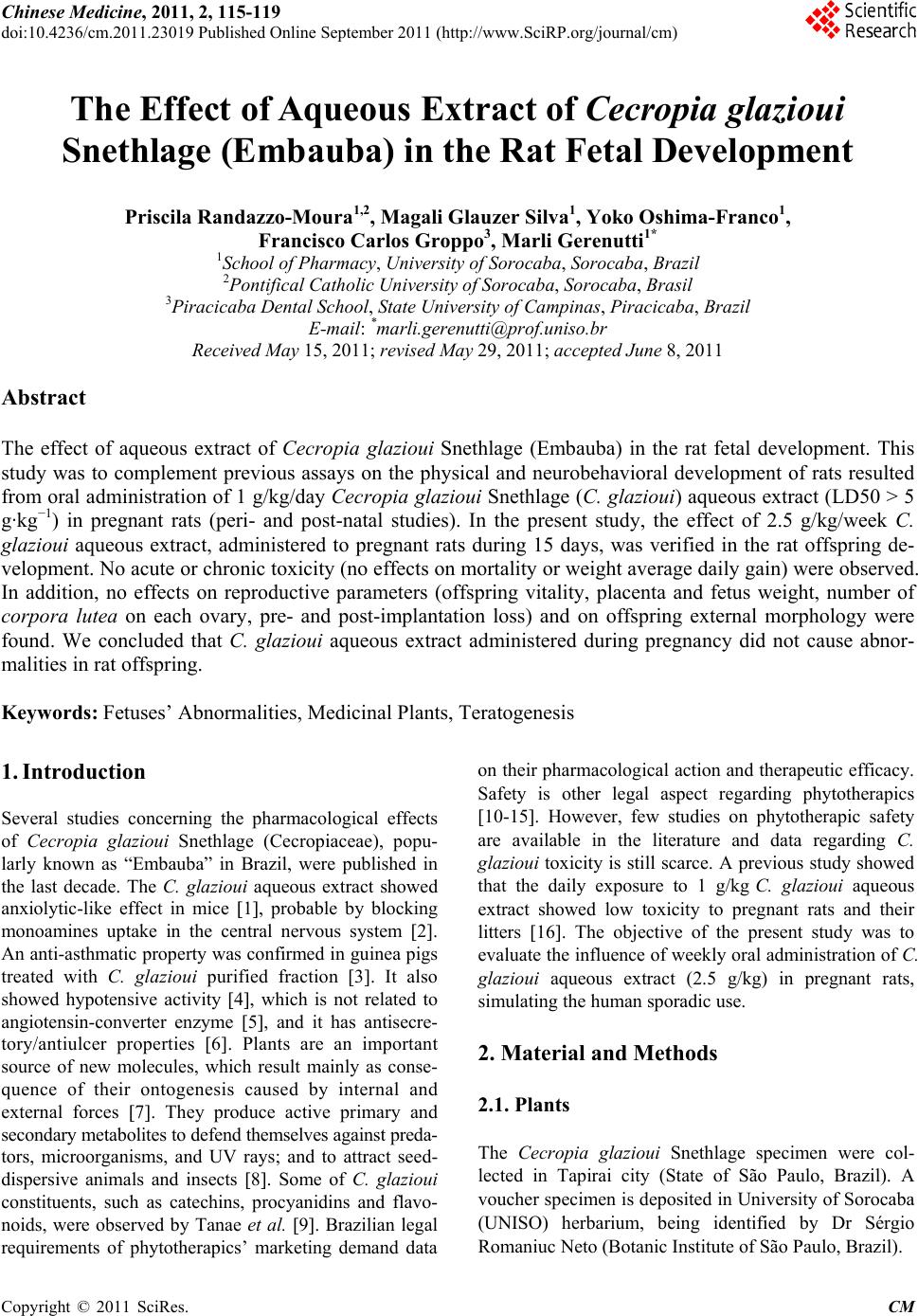
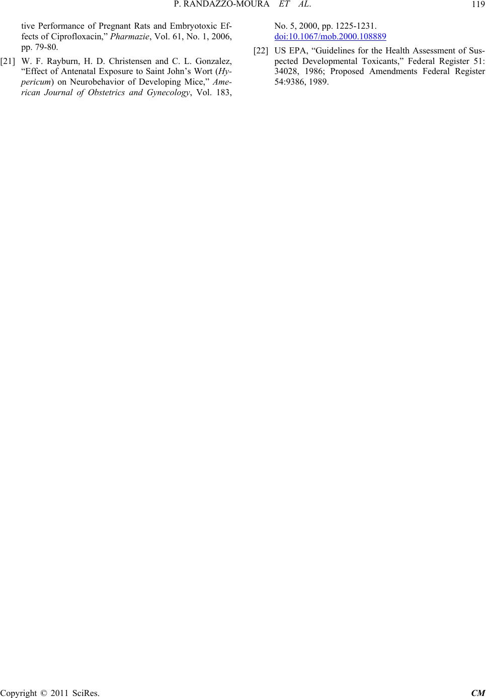
Considering the offspring external morphological mea-
surements in the present study, Cg extract did not induce
significant effects in these measurements when com-
pared with the control group. Studies carried out to es-
tablish the potential ability of drugs to induce adverse
effects on the fetal development, which used acceptable
and rationale experimental design, could provide accu-
rate extrapolation of the potential risk in human beings of
that particular drug. Considering the previous study per-
formed by Gerenutti et al. 16, which used the same
aqueous extract of Cg, it was possible to conclude that
Cecropia glazioui Snethlage presents no toxicity on
morphological development of rat offspring.
5. Acknowledgements
This work was supported by UNISO. R-M, P. was stu-
dent of Scientific Initiation from UNISO.
6. References
[1] F. F. Rocha, A. J. Lapa and T. C. de Lima, “Evaluation of
the Anxiolytic-Like Effects of Cecropia glazioui Sneth in
Mice,” Pharmacology, Biochemistry and Behavior, Vol.
71, No. 1-2, 2002, pp. 183-190.
doi:10.1016/S0091-3057(01)00695-5
[2] F. F. Rocha, M. T. Lima-Landman, M. M. Tanae, T. C.
de Lima and A. J. Lapa, “Antidepressant-Like Effect of
Cecropia glazioui Sneth and Its ConstituentsIn Vivo
and in Vitro Characterization of the Underlying Mecha-
nism,” Phytomedicine, Vol. 14, No. 6, 2007, pp. 396-402.
doi:10.1016/j.phymed.2007.03.011
[3] S. Delarcina, M. T. Lima-Landman, C. Souccar, R. M.
Cysneiros, M. M. Tanae and A. J. Lapa, “Inhibition of
Histamine-Induced Bronchospasm in Guinea Pigs Treated
with Cecropia glaziovi Sneth and Correlation with the in
Vitro Activity in Tracheal Muscles,” Phytomedici ne, Vol.
14, No. 5, 2007, pp. 328-332.
doi:10.1016/j.phymed.2006.12.022
[4] M. T. Lima-Landman, A. C. Borges, R. M. Cysneiros, T.
C. de Lima, C. Souccar and A. J. Lapa, “Antihypertensive
Effect of a Standardized Aqueous Extract of Cecropia
glaziovii Sneth in Rats: An in Vivo Approach to the Hy-
potensive Mechanism,” Phytomedicine, Vol. 14, No. 5,
2007, pp. 314-320. doi:10.1016/j.phymed.2007.03.003
[5] M. F. Ninahuaman, C. Souccar, A. J. Lapa and M. T.
Lima-Landman, “ACE Activity during the Hypotension
Produced by Standardized Aqueous Extract of Cecropia
glaziovii Sneth: A Comparative Study to Captopril Ef-
fects in Rats,” Phytomedicine, Vol. 14, No. 5, 2007, pp.
321-327. doi:10.1016/j.phymed.2006.12.010
[6] C. Souccar, R. M. Cysneiros, M. M. Tanae, L. M. Torres,
M. T. Lima-Landman and A. J. Lapa, “Inhibition of Gas-
tric Acid Secretion by a Standardization Aqueous Extract
of Cecropia glaziovii Sneth and Underlying Mechanism,”
Phytomedicine, Vol. 15, No. 6-7, 2008, pp. 462-469.
doi:10.1016/j.phymed.2008.02.006
[7] R. Lewontin, “It Ain’t Necessarily So: The Dream of the
Human Genome and Other Illusions,” New York Review
Books, New York, 2000.
[8] M. Wink, “Physiology of Secondary Product Formation
in Plants,” In: B. V. Charwood and M. J. C. Rhodes, Eds.,
Secondary Products from Plant Tissue Culture, Claren-
don, Oxford, 1990, pp. 67-86.
[9] M. M. Tanae, M. T. Lima-Landman, T. C. de Lima, C.
Souccar and A. J. Lapa, “Chemical Standardization of the
Aqueous Extract of Cecropia glaziovii Sneth Endowed
with Antihypertensive, Brochodilator, Antiacid Secretion
and Antidepressant-Like Activities,” Phytomedicine , Vol.
14, No. 5, 2007, pp. 309-313.
doi:10.1016/j.phymed.2007.03.002
[10] Brasil, Ministério da Saúde, Agência Nacional de Vigi-
lância Sanitária, Resolução de Diretoria Colegiada 48,
Diário Oficial da União, Brasília, 2004.
[11] Brasil, Ministério da Saúde, Agência Nacional de Vigi-
lância Sanitária, Resolução 88, Diário Oficial da União,
Brasília, 2004.
[12] Brasil, Ministério da Saúde, Agência Nacional de Vigi-
lância Sanitária, Resolução 89, Diário Oficial da União,
Brasília, 2004.
[13] Brasil, Ministério da Saúde, Agência Nacional de Vigi-
lância Sanitária, Resolução 90, Diário Oficial da União,
Brasília, 2004.
[14] Brasil, Ministério da Saúde, Agência Nacional de Vigi-
lância Sanitária, Resolução 91, Diário Oficial da União,
Brasília, 2004.
[15] Brasil, Presidência da República, Decreto 5813, Diário
Oficial da União, Brasília, 2006.
[16] M. Gerenutti, A. F. Prestes, M. G. Silva, F. de S. Del Fiol,
Y. O. Franco, P. C. Venâncio and F. C. Groppo, “The
Effect of Cecropia glazioui Snethlage on the Physical and
Neurobehavioral Development of Rats,” Pharmazie, Vol.
63, No. 5, 2008, pp. 398-404.
[17] B. H. Vickery and J. P. Bennett, “Rats and Mice,” In: E.
S. E. Hafez, Ed., Reproduction and Breeding Techniques
for Laboratory Animals, Lea & Febiger, Philadelphia,
1970, pp. 299-315.
[18] K. A. Keller, “Developmental and Reproductive Toxi-
cology,” In: D. Jacobson-Kram and K. A. Keller, Eds.,
Toxicology Testing Handbook: Principles, Applications
and Data Interpretation, Marcel Dekker, Inc., New York,
2001, pp. 195-254.
[19] M. Gerenutti, A. H. de-Souza Spinosa and M. M. Ber-
nardi, “Effects of Bracken Fern (Pteridium aquilinum L.
Kuhn) Feeding during the Development of Female Rats
and Their Offspring,” Veterinary and Human Toxicology,
Vol. 34, No. 4, 1992, pp. 307-310.
[20] M. Gerenutti, F. Del Fiol and F. C. Groppo, “Reproduc-
Copyright © 2011 SciRes. CM
P. RANDAZZO-MOURA ET AL.
Copyright © 2011 SciRes. CM
119
tive Performance of Pregnant Rats and Embryotoxic Ef-
fects of Ciprofloxacin,” Pharmazie, Vol. 61, No. 1, 2006,
pp. 79-80.
[21] W. F. Rayburn, H. D. Christensen and C. L. Gonzalez,
“Effect of Antenatal Exposure to Saint John’s Wort (Hy-
pericum) on Neurobehavior of Developing Mice,” Ame-
rican Journal of Obstetrics and Gynecology, Vol. 183,
No. 5, 2000, pp. 1225-1231.
doi:10.1067/mob.2000.108889
[22] US EPA, “Guidelines for the Health Assessment of Sus-
pected Developmental Toxicants,” Federal Register 51:
34028, 1986; Proposed Amendments Federal Register
54:9386, 1989.