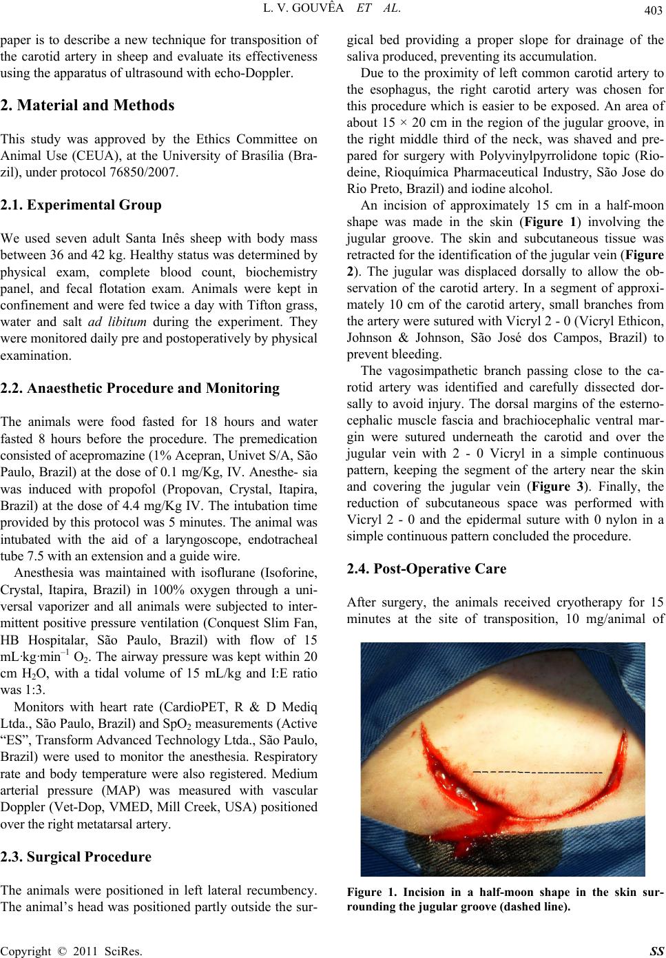
Surgical Science, 2011, 2, 402-405
doi:10.4236/ss.2011.27088 Published Online September 2011 (http://www.SciRP.org/journal/ss)
Copyright © 2011 SciRes. SS
Surgical Preparation of a Permanent Carotid
Transposition in Sheep
Liana V. Gouvêa, Ernane P. F. Novais, Ceci R. Leite, Fábio H. B. Ximenes,
Ricardo M. Almeida, R. B. Molás, J. G. C. Palermo, C. E. V. Silva,
Jose R. J. Borges, Eduardo M. M. Lima, Roberta Ferro Godoy
Large Animal Hospital, School of Veterinary Medicine, University of Brasilia, Brasilia, Brazil
E-mail: robertagodoy@unb.br
Received August 3, 2011; revised September 1, 2011; accepted September 10, 2011
Abstract
In large animal research, when frequent sampling of arterial blood is needed, the carotid artery transposition
is the most used technique. The objective of this paper is to describe a new technique for carotid artery
transposition in sheep and evaluate its effectiveness using the echo-Doppler ultrasound. The animals enrolled
in this study had their carotid surgically elevated to the subcutaneous level, by suturing underneath muscles
sternocephalic and brachiocephalic. None of the animals that underwent the procedure in this study had
trans- or postoperative complications. Six months after the suture removal, all animals presented patent ca-
rotids by echo-Doppler ultrasound. This is an easy and safe procedure for carrying out experiments that re-
quire the collection of arterial blood.
Keywords: Blood Gas Analysis, Eco-Doppler, Experimental Surgery
1. Introduction
Easy and rapid access to large-caliber arteries are fre-
quently needed in invasive studies about hemodynamics
and/or blood gas analysis in large animals, especially if
multiple samples are required. These arteries should be
accessible, but protected from trauma, and their patency
should be maintained for long periods [1]. Several de-
scriptions of displacement of the carotid artery to the
subcutaneous tissue of ruminants have been reported in
the literature [2-8]. Although this is a well studied tech-
nique, a new choice for this procedure allowing a patent
carotid artery for a prolonged time is presented, as well
as minimum complication is observed.
In the technique described in [4], an incision is made
parallel and ventral to the jugular vein, in the transition
between the midd le and d istal third of th e jugu lar groove.
The carotid artery is located and a 10 cm section is ex-
ternalized. The dorsal and the ventral margin of the
esternocephalic and brachiocephalic muscles are sutured
underneath the artery and above the jugular. The skin is
sutured over the carotid artery. The disadvantage of this
technique is that the carotid is not immobilized in the
subcutaneous tissue, hindering the collection of arterial
blood [1].
There is another described a technique [2] that differs
from the previous by suturing the skin under de carotid
artery, creating a skin tube containing the carotid artery.
This technique requires wrapping the animal’s neck to
prevent accidental puncture of the artery. Problems were
reported like skin necrosis and flow disturbances with
this technique, and the first two studies have reported
accidental carotid puncture [1,2,7].
A third technique described in [1], a modification of
the technique originally proposed in [3]. The incision
was made parallel and ventral to the jugular groove to
allow the identification of the jugular vein. The skin is
dissected, creating a flap which is folded dorsally. The
carotid artery is then isolated and displaced as in the
other techniques. The skin flap is returned to its original
position and the skin on top of the exposed carotid artery
is sutured around it. This technique has a lower risk of
accidental perforation of the carotid due to the way that
the artery is positioned.
A work [9] reported 94% success rate after surgery
relocation of carotid artery using the techniques from [2]
and [3]. However, it reported also complications includ-
ing skin necrosis and loss of arterial patency. The ideal
technique would be able to maintain the viability of the
carotid artery for a long period of time. The aim of this
L. V. GOUVÊA ET AL.
403
paper is to describe a new technique for transposition of
the carotid artery in sheep and evaluate its effectiveness
using the apparatus of ultrasound with echo-Doppler.
2. Material and Methods
This study was approved by the Ethics Committee on
Animal Use (CEUA), at the University of Brasília (Bra-
zil), under protocol 76850/2007.
2.1. Experimental Group
We used seven adult Santa Inês sheep with body mass
between 36 and 42 kg. Healthy status was determined by
physical exam, complete blood count, biochemistry
panel, and fecal flotation exam. Animals were kept in
confinement and were fed twice a day with Tifton grass,
water and salt ad libitum during the experiment. They
were monitored daily pre and postoperatively by physical
examination.
2.2. Anaesthetic Procedure and Monitoring
The animals were food fasted for 18 hours and water
fasted 8 hours before the procedure. The premedication
consisted of acepromazine (1% Acepran, Univet S/A, São
Paulo, Brazil) at the dose of 0.1 mg/Kg, IV. Anesthe- sia
was induced with propofol (Propovan, Crystal, Itapira,
Brazil) at the dose of 4.4 mg/Kg IV. The intubation time
provided by this protocol was 5 minutes. The animal was
intubated with the aid of a laryngoscope, endotracheal
tube 7.5 with an ext ensi on an d a g ui de wire.
Anesthesia was maintained with isoflurane (Isoforine,
Crystal, Itapira, Brazil) in 100% oxygen through a uni-
versal vaporizer and all animals were subjected to inter-
mittent positive pressure ventilation (Conquest Slim Fan,
HB Hospitalar, São Paulo, Brazil) with flow of 15
mL·kg·min–1 O2. The airway pressure was kept within 20
cm H2O, with a tidal volume of 15 mL/kg and I:E ratio
was 1:3.
Monitors with heart rate (CardioPET, R & D Mediq
Ltda., São Paulo, Brazil) and SpO2 measurements (Active
“ES”, Transform Advanced Technology Ltda., São Paulo,
Brazil) were used to monitor the anesthesia. Respiratory
rate and body temperature were also registered. Medium
arterial pressure (MAP) was measured with vascular
Doppler (Vet-Dop, VMED, Mill Creek, USA) positioned
over the right metatarsal artery.
2.3. Surgical Procedure
The animals were positioned in left lateral recumbency.
The animal’s head was positioned partly outside the sur-
gical bed providing a proper slope for drainage of the
saliva produced, preventing its accumulation.
Due to the proximity of left common carotid artery to
the esophagus, the right carotid artery was chosen for
this procedure which is easier to be exposed. An area of
about 15 × 20 cm in the region of the jugular groove, in
the right middle third of the neck, was shaved and pre-
pared for surgery with Polyvinylpyrrolidone topic (Rio-
deine, Rioquímica Pharmaceutical Industry, São Jose do
Rio Preto, Brazil) and iodine alcohol.
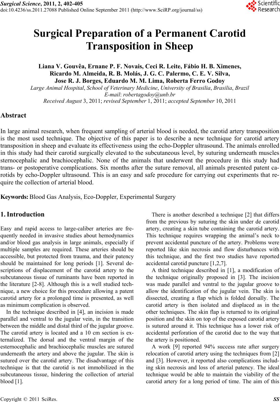
An incision of approximately 15 cm in a half-moon
shape was made in the skin (Figure 1) involving the
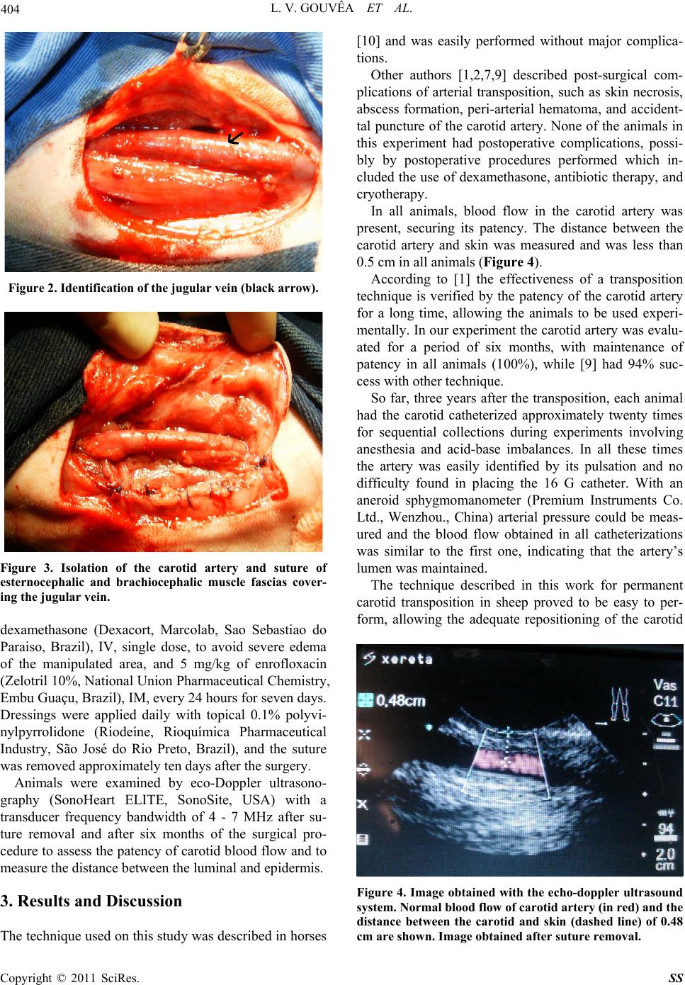
jugular groove. The skin and subcutaneous tissue was
retracted for the identification of th e jugular vein (Figure
2). The jugular was displaced dorsally to allow the ob-
servation of the carotid artery. In a segment of approxi-
mately 10 cm of the carotid artery, small branches from
the artery were sutured with Vicryl 2 - 0 (Vicryl Ethicon,
Johnson & Johnson, São José dos Campos, Brazil) to
prevent bleeding.
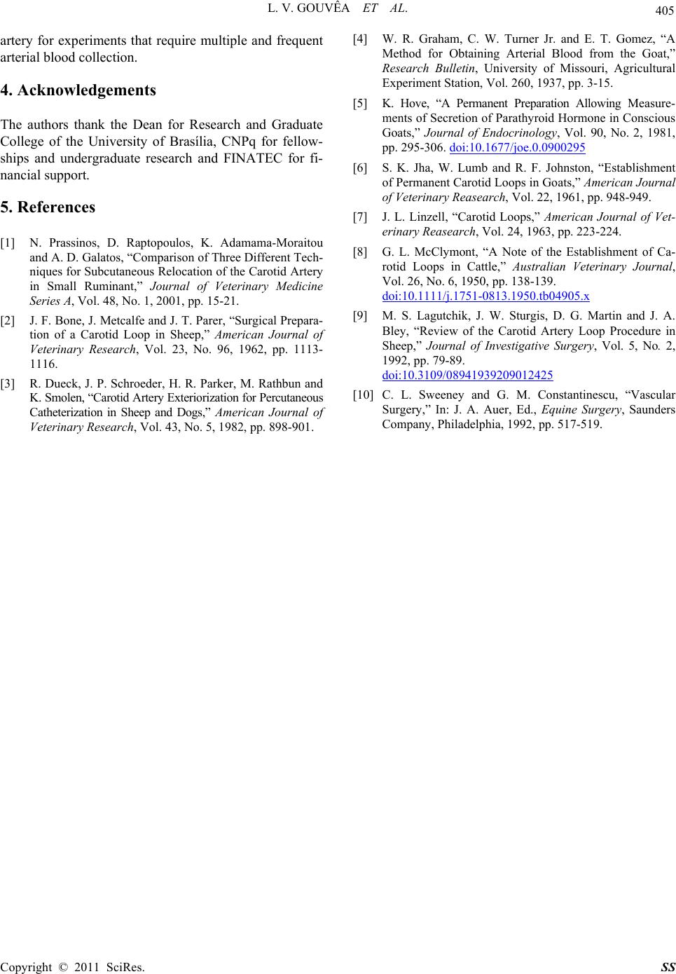
The vagosimpathetic branch passing close to the ca-
rotid artery was identified and carefully dissected dor-
sally to avoid injury. The dorsal margins of the esterno-
cephalic muscle fascia and brachiocephalic ventral mar-
gin were sutured underneath the carotid and over the
jugular vein with 2 - 0 Vicryl in a simple continuous
pattern, keeping the segment of the artery near the skin
and covering the jugular vein (Figure 3). Finally, the
reduction of subcutaneous space was performed with
Vicryl 2 - 0 and the epidermal suture with 0 nylon in a
simple continuous pattern concluded the procedure.
2.4. Post-Operative Care
After surgery, the animals received cryotherapy for 15
minutes at the site of transposition, 10 mg/animal of
Figure 1. Incision in a half-moon shape in the skin sur-
rounding the jugular groove (dashed line).
Copyright © 2011 SciRes. SS
404 L. V. GOUVÊA ET AL.
Figure 2. Identification of the jugular vein (black arrow).
Figure 3. Isolation of the carotid artery and suture of
esternocephalic and brachiocephalic muscle fascias cover-
ing the jugular vein.
dexamethasone (Dexacort, Marcolab, Sao Sebastiao do
Paraiso, Brazil), IV, single dose, to avoid severe edema
of the manipulated area, and 5 mg/kg of enrofloxacin
(Zelotril 10%, National Union Pharmaceutical Chemistry,
Embu Guaçu, Brazil), IM, every 24 hours for seven days.
Dressings were applied daily with topical 0.1% polyvi-
nylpyrrolidone (Riodeíne, Rioquímica Pharmaceutical
Industry, São José do Rio Preto, Brazil), and the suture
was removed approximately ten days after the surgery.
Animals were examined by eco-Doppler ultrasono-
graphy (SonoHeart ELITE, SonoSite, USA) with a
transducer frequency bandwidth of 4 - 7 MHz after su-
ture removal and after six months of the surgical pro-
cedure to assess the p atency of carotid blood flow and to
measure the distance between the luminal and epidermis.
3. Results and Discussion
The technique used on this study was described in horses
[10] and was easily performed without major complica-
tions.
Other authors [1,2,7,9] described post-surgical com-
plications of arterial transposition, such as skin necrosis,
abscess formation, peri-arterial hematoma, and accident-
tal puncture of the carotid artery. None of the animals in
this experiment had postoperative complications, possi-
bly by postoperative procedures performed which in-
cluded the use of dexamethasone, antibiotic therapy, and
cryotherapy.
In all animals, blood flow in the carotid artery was
present, securing its patency. The distance between the
carotid artery and skin was measured and was less than
0.5 cm in all animals (Figure 4).
According to [1] the effectiveness of a transposition
technique is verified by the patency of the carotid artery
for a long time, allowing the animals to be used experi-
mentally. In our experiment the carotid artery was evalu-
ated for a period of six months, with maintenance of
patency in all animals (100%), while [9] had 94% suc-
cess with other technique.
So far, three years after the transposition, each animal
had the carotid catheterized approximately twenty times
for sequential collections during experiments involving
anesthesia and acid-base imbalances. In all these times
the artery was easily identified by its pulsation and no
difficulty found in placing the 16 G catheter. With an
aneroid sphygmomanometer (Premium Instruments Co.
Ltd., Wenzhou., China) arterial pressure could be meas-
ured and the blood flow obtained in all catheterizations
was similar to the first one, indicating that the artery’s
lumen was maintained.
The technique described in this work for permanent
carotid transposition in sheep proved to be easy to per-
form, allowing the adequate repositioning of the carotid
Figure 4. Image obtained with the echo-doppler ultrasound
system. Normal blood flow of carotid artery (in red) and the
distance between the carotid and skin (dashed line) of 0.48
cm are shown. Image obtained after suture removal.
Copyright © 2011 SciRes. SS
L. V. GOUVÊA ET AL.
Copyright © 2011 SciRes. SS
405
artery for experiments that require multiple and frequent
arterial blood collection.
4. Acknowledgements
The authors thank the Dean for Research and Graduate
College of the University of Brasília, CNPq for fellow-
ships and undergraduate research and FINATEC for fi-
nancial support.
5. References
[1] N. Prassinos, D. Raptopoulos, K. Adamama-Moraitou
and A. D. Galatos, “Comparison of Three Different Tech-
niques for Subcutaneous Relocation of the Carotid Artery
in Small Ruminant,” Journal of Veterinary Medicine
Series A, Vol. 48, No. 1, 2001, pp. 15-21.
[2] J. F. Bone, J. Metcalfe and J. T. Parer, “Surgical Prepara-
tion of a Carotid Loop in Sheep,” American Journal of
Veterinary Research, Vol. 23, No. 96, 1962, pp. 1113-
1116.
[3] R. Dueck, J. P. Schroeder, H. R. Parker, M. Rathbun and
K. Smolen, “Carotid Artery Exteriorization for Percutaneous
Catheterization in Sheep and Dogs,” American Journal of
Veterinary Research, Vol. 43, No. 5, 1982, pp. 898-901.
[4] W. R. Graham, C. W. Turner Jr. and E. T. Gomez, “A
Method for Obtaining Arterial Blood from the Goat,”
Research Bulletin, University of Missouri, Agricultural
Experiment Station, Vol. 260, 1937, pp. 3-15.
[5] K. Hove, “A Permanent Preparation Allowing Measure-
ments of Secretion of Parathyroid Hormone in Conscious
Goats,” Journal of Endocrinology, Vol. 90, No. 2, 1981,
pp. 295-306. doi:10.1677/joe.0.0900295
[6] S. K. Jha, W. Lumb and R. F. Johnston, “Establishment
of Permanent Carotid Loops in Goats,” American Journal
of Veterinary Reasearch, Vol. 22, 1961, pp. 948-949.
[7] J. L. Linzell, “Carotid Loops,” American Journal of Vet-
erinary Reasearch, Vol. 24, 1963, pp. 223-224.
[8] G. L. McClymont, “A Note of the Establishment of Ca-
rotid Loops in Cattle,” Australian Veterinary Journal,
Vol. 26, No. 6, 1950, pp. 138-139.
doi:10.1111/j.1751-0813.1950.tb04905.x
[9] M. S. Lagutchik, J. W. Sturgis, D. G. Martin and J. A.
Bley, “Review of the Carotid Artery Loop Procedure in
Sheep,” Journal of Investigative Surgery, Vol. 5, No. 2,
1992, pp. 79-89.
doi:10.3109/08941939209012425
[10] C. L. Sweeney and G. M. Constantinescu, “Vascular
Surgery,” In: J. A. Auer, Ed., Equine Surgery, Saunders
Company, Philadelphia, 1992, pp. 517-519.