Open Journal of Pediatrics, 2011, 1, 30-33
doi:10.4236/ojped.2011.13008 Published Online September 2011 (http://www.SciRP.org/journal/ojped/ OJPed
).
Published Online September 2011 in SciR es. http://www.scirp.org/journal/OJPed
Chronological changes in Epstein-Barr virus genome and
subsets of peripheral mononuclear cells in a case of HLH
Yukayo Ukeba-Terashita1, Yoshinori Saita1, Yoshinori Ito2, Hirokazu Kanegane3, Hiroshi Kimura4,
Ichiro Kobayashi1,5*
1Department of Pediatrics, Kitami Red Cross Hospital, Kitami, Japan;
2Department of Pediatrics, Nagoya University Graduate School of Medicine, Nagoya, Japan;
3Department of Pediatrics, Graduate School of Medicine, University of T o yama, To yama, Japan;
4Department of Virology, Nagoya University Graduate School of Medicine, Nagoya, Japan;
5Department of Pediatrics, Hokkaido University Graduate School of Medicine, Sapporo, Japan.
Email: *ichikobaya@med.hokudai.ac.jp
Received 30 April 2011; revised 1 June 2011; accepted 15 July 2011.
ABSTRACT
Hemophagocytic lymphohistiocytosis (HLH) is a rare
disorder characterized by high grade fever, heap-
tosplenomegaly, lymphadenopathy and cytope- nia in
association with hypercytokinemia and mac- rophage
activation. Epstein-Barr virus (EBV) is a common
cause of HLH particularly in Asian countries. Al-
though EBV usually infects B cells and causes infec-
tious mononucleosis, it is detected predominantly in
CD8+ T cells in EBV-associated HLH. In the present
study we found the EBV genome in various lineages
of the lymphoid cells including T, B, and natural kil-
ler cells in a 14-months-old boy with mild EBV-asso-
ciated HLH. Furthermore, chronological changes in
the copy number of EBV-DNA in each lineage are
reported. Profound decrease in the number of pe-
ripheral natural killer cells at the early stage could be
involved in the development of HLH.
Keywords: Epstein-Barr Virus; Hemophagocytic Lympho-
histio Cytosis; Natural Killer Cell
1. INTRODUCTION
Hemophagocytic lymphohistiocytosis (HLH) is a rare
disorder characterized by high grade fever, hepatosple-
nomegaly, lymphadenopathy and cytopenia in associa-
tion with hypercytokinemia and macrophage activation
[1]. HLH is classified into two clinical entities; primary
HLH which develops in inherited diseases such as fa-
milial hemophagocytic lymphohistiocytosis (FHL) or pr-
imary immunodeficiency, and secondary HLH which
develops associated with other acquired disorders such
as infection, collagen vascular diseases and malignancy
[1]. Sixty percent of the secondary HLH in children is
associated with Epstein-Barr virus (EBV) in Japan [2].
Although most cases of EBV-HLH have been reported
from Asian countries, reports from other regions have
been increasing [3,4]. EBV is a ubiquitous human h erpes
virus which usually infects B cells and causes self-lim-
iting infectious mononucleosis (IM). On the other hand,
persistent or recurrent IM-like disease associated with
abnormally high levels of both anti-EBV antibodies and
EBV genome is known as chronic active EBV infection
(CAEBV) [5]. EBV-associated HLH (EBV-HLH) occurs
in association with either CAEBV or a primary infection
of the virus [1-5]. Recent reports have demonstrated that
EBV infects NK or CD4+ T cells in CAEBV, whereas it
dominantly infects CD8+ T cells in EBV-HLH [5,6]. We
report here a case of HLH associated with a primary
infection of EBV and the chronological changes in
EBV-DNA of each lymphoid cell subset.
2. CASE REPORT
A previously healthy 14-month-old boy was admitted to
our hospital because of fever up to 41˚C persisting for
four days. He was born to non-consanguineous healthy
parents and had no history suggesting immunodeficiency
or hypersensitivity to mosquito bite. On the third hospi-
tal day, cervical lymphadenopathy and hepatospleno-
megaly were noted. Laboratory examination showed:
white blood cell count 7.89 × 10⁹/l (neutrophils 17%,
lymphocytes 55%), hemoglobin 98 g/l, platelet count 45 ×
109/l, aspartate transaminase 543 IU/l, alanine transami-
nase 204 IU/l, total cho lesterol 0.81 g/l, triglyceride 2.24
g/l, C-reactive protein 115.3 mg/l, ferritin 2090 g/ l. Coa gu -
lation study showed: prothrombin time 17.8 seconds,
activated partial thromboplastin time 51.8 seconds, fi-
brinogen 2.66 g/l (reference range 2.00 g/l - 4.00 g/l) and
fibrin degradation products 93.9 g/ml (< 10 g/ml), sug-
Y. Ukeba-Terashita et al. / Open Journal of Pediatrics, 2011, 1, 30-33 31
gesting disseminated intravascular coagulation (DIC).
NK cell activity and a serum soluble CD25 was 14%
(normal; 18% - 45%) and 10,300 U/ml (normal < 520
U/ml), respectively. Although hemophagocytosis was
not evident in his bone marrow, seven of the eight items
of HLH criteria by Histiocyte Society were fulfilled [1].
Nafamostat mesilate was administered for DIC. Because
mild dilatation of coronary artery with a high echog enic-
ity of the arterial wall similar to that observed in Kawa-
saki disease was detected by echocardiography on the
7th day of hospitalization despite slight improvement of
both clinical and laboratory findings, high-dose intrave-
nous immunoglobulin (HD-IVIG) therapy (2 g/kg) was
started. Serological test for EBV before HD-IVIG ther-
apy showed IgG antibody to viral capsid antigen (VCA)
1:160, IgG antibody to early antigen 1:20, but negative
for IgG antibody to EBV nuclear antigen (EBNA) sug-
gesting a primary infection of EBV. Because his clinical
and laboratory findings rapidly improved, no further
therapy was performed.
Flow cytometric analysis of his peripheral blood at the
onset of HLH demonstrated a high level of CD8+HLA-
DR+ cells, wher eas the number of CD19+, CD20+, CD5 6+,
CD16+ , and CD57+ cells was low (Ta b l e 1 ). The num-
ber of CD8+HLA-DR+ cells declined in 20 days which
was associated with recovery of CD16+ or CD57+ cells.
The ratio of each subset has returned to normal ranges 3
months after the onset.
The copy number of EBV-DNA was analyzed by a
real-time polymerase chain reaction after separation of
his peripheral blood mononuclear cells (PBMC) into
CD19+, CD3, CD4+, CD8+, CD16+ and 56+ cells by an
immunobeads method (DynaBeads; Dynal A/S, Oslo,
Norway) as previously reported [7]. DNA was extracted
Table 1. Flow cytometric analyses of the peripheral mononu-
clear cells .
Date Apr 5 Apr 25 Jul 11 Nov 6
Absolute number
of PBMC (/mm3) 3666 3846 3872 5728
CD4 19.2% 41.6% 44.1% 36.4%
CD8 70.7% 40.6% 38.7% 34.0%
CD8×HLA-DR 56.7% 17.7% 11.2% 8.9%
CD16 2.3% 17.2% 12.2% 20.0%
CD56 1.4% ND 6.5% 14.9%
CD57 4.4% 7.6% 6.6% 12.2%
CD19 0.9% 0.7% 12.7% 18.2%
CD20 1.2% 0.6% 9.2% 12.9%
Abbreviation: PBMC, peripheral mononuclear cells; ND, not done.
from 2 × 10⁶ PBMC using a QIAmp Blood Kit (Qiagen,
Hilden, Germany). A real-time quantitative PCR assay
with a fluorogenic probe was performed using TaqMan
PCR kit (PE Applied Biosystems, Foster City, CA). The
amount of EBV-DNA was calculated as the number of
virus copies per microgram PBMC DNA. The copy
number of EBV-DNA was extremely high at the onset in
all of CD19+, CD3, CD4+, CD8+, CD16+ and 56+ cells. A
high level infection with CD16+ or 56+ cells persisted at
least for five months, although the copy number of the
viral DNA declined with the clinical improvement (Ta-
ble 2). One year after the onset EBV-DNA was detect-
able only in CD19+ cells at a latent infection level com-
parable to normal individuals with a past infection of the
virus.
After the recovery from HLH, he has not shown any
IM-like symptoms or abnormal laboratory findings dur-
ing 2 years of follow up. IgG antibody to EBV early
antigen disappeared soon, whereas serum levels of IgG
antibodies to VCA and EBNA gradually elevated to
1:1280 and 1:40 in 5 months, respectively. NK activity
restored to 45%. No mutation was detected in either
SH2D1A or XIAP gene.
3. DISCUSSION
In the present study, we analyzed chronological changes
of EBV-DNA in each subset of PBMC in a case of
EBV-HLH. From his clinical course and the profile of
anti-EBV antibodies, HLH was likely associated with a
primary infection of EBV rather than CAEBV. Although
coronary lesion was transiently observed, the diagnostic
criteria for Kawasaki disease was not fulfilled [8]. In
addition to vasculitis such as Kawasaki disease, coronary
lesions develop in association with infection of the virus
to the endothelial cells in CAEBV or with systemic in-
flammation as observed in systemic-onset juvenile idio-
pathic arthritis [9,10].
Our case showed increased number of peripheral
CD8+HLA-DR+ cells and a decrease in both the number
and the activity of NK cells at the diagnosis, which is
consistent with the previous reports of EBV-HLH [11].
Table 2. Chronological changes of EBV-DNA (copies/g DNA) in
each subset.
DateApr 5 Apr 25 Jun 7 Sep5 Jan 9 Apr 16, 2008
PBMC110 × 1044.77 × 104269 858 54 86
CD317.9 × 1043.46 × 10420 823 ND ND
CD41.22 × 1040.74 × 10417 249 ND ND
CD840.2 × 1043.55 × 1040 527 ND ND
CD16ND 5.14 × 1041483 311 99 ND
CD5697.8 × 1045.13 × 1041295 1769 ND ND
CD1913.9 × 1042.16 × 104119 932 173 508
Abbreviation: PBMC, peripheral mononuclear cell s ; ND, not done.
C
opyright © 2011 SciRes. OJPed
Y. Ukeba-Terashita et al. / Open Journal of Pediatrics, 2011, 1, 30-33
32
The dominant population of EBV-infected cells is re-
ported to be CD8+ T cells in EBV-HLH, whereas the
ma jor target s of EBV in CAEBV are CD4+ T cells or NK
cells [5-7]. EBV-infected CD8+Tcells are activated and
produce cytokines such as IFN-γ, TNF-α and inter-
leukin-6, which activate macrophages and endothelial
cells resulting in hemophagocytosis, DIC, and multi-
organ failure [1,5,12]. NK cells, as well as EBV-specific
cytotoxic T cells (CTL), play a critical role in the elimi-
nation of EBV-infected cells [1]. This is supported by the
fact that the functional defects in cytotoxic activity of
CTL and NK cells such as X-linked lymphoproliferative
syndrome and FHL predispose for EBV-HLH [1]. In
acute IM the number of NK cells, although it usually
increases, is inversely correlated with the severity of the
disease [13]. Thus, the numerical defect of NK cells as
observed in our case could cause the development of
EBV-HLH or severe IM. Our case showed subsequent
recovery of both NK activity and the number of CD16+ ,
56+ or 57+cells as well as decline in CD8+HLA-DR+
cells, which was associated with clinical improvement.
Furthermore, no mutation was detected in causative
genes of XLP such as SH2DA or XIAP gene. Thus, al-
though mutations of other FHL-related genes have not
been tested, the numerical and functional abnormality
was unlikely to be intrinsic to his NK cells. Of note was
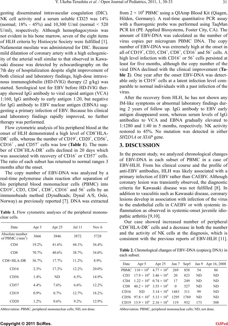
the finding that high lev els of EBV-DNA was detected in
CD16+ and 56+ cells, in addition to T and B cells, despite
the decreased number of the subsets in our case. Al-
though other methods such as in situ hybridization and
immunohistochemistry were not performed, a high level
of infection in CD 16+ and 56+ cells was confirmed by
serial examination for several months. The copy number
of EBV-DNA in PBMC of our case was comparable to
that in previously reported cases of EBV-HLH, CAEBV
and post-transplantational lymphoproliferative disorder
[14]. Although EBV-HLH has a tendency to have larger
viral burdens than acute phase of IM, it is difficult to
differentiate between these two diseases simply by viral
load in whole PBMC [15]. The copy number of EBV-
DNA gradually declined in all of the subsets and was
finally detectable in B cells only at a latency level one
year after the onset of the disease. These findings are in
contrast to NK cell-type CAEBV or NK cell lymphoma
which shows a clonal expansion of EBV-infected NK
cells [5]. It is known that EBV is able to infect NK cells
at an early stage of IM, although the precise mechanisms
of infection remain unclear [16]. As well, EBV genome
is also detected in bo th NK and B cells under some con-
ditions such as EBV-HLH but usually at a lower level
than CD8+ T cells [6]. Isobe et al have reported that in
vitro infection of EBV induces apoptosis of NK cells
[17]. Thus, it is possible that the infection by EBV in-
duced apoptosis of NK cells in vivo and allowed un-
regulated activation of EBV-infected CD8+T cells. De-
creased number of peripheral B cells in association with
HLH as observed in our case has also been reported [18].
Imashuku et al have demonstrated depletion of B cells in
the spleen of the patients with HLH and suggested in-
volvement of cytokines produced by activated T or NK
cells and/or Fas-FasL-mediated apoptosis [18].
Although our case showed both clinical and labora-
tory improvement before the commencement of HD-
IVIG therapy suggesting self-limiting nature, it is possi-
ble that the therapy accelerated the improvement by its
anti-inflammatory mechanism [19]. Further studies are
required to clarify the mechanisms of the decrease in the
number of peripheral NK cells in EBV-HLH without un-
derlying primary immunodeficiencies or FHL.
In conclusion, EBV infected various subsets of PBMC
including NK cells in a case of HLH associated with a
primary infection of EBV. EBV genome gradually de-
clined in association with his clinical improvement and
was finally detectable in only B cells at a latency level.
Transient decrease in NK cells could be involved in the
development of EBV-HLH in our case.
REFERENCES
[1] Henter, J.-I., Horne, A.C., Aricó, M., et al. (2007) HLH-
2004: Diagnostic and therapeutic guidelines for hemo-
phagocytic lymphohistiocytosis. Pediatric Blood & Can-
cer, 48, 124-131. doi:10.1002/pbc.21039
[2] Ishii, E., Ohga, S., Imashuku, S., et al. (2007) Nation-
wide survey of hemophagocytic lymphohistiocytosis in
Japan. International Journal of Hematology, 86, 58-65.
doi:10.1532/IJH97.07012
[3] Beutel, K., Gross-Wieltsch, U., Wiesel, T., Zur Stadt, U.,
Janka, G. and Wagner, H.-J. (2009) Infection of T lym-
phocytes in Epstein-Barr virus-associated hemophago-
cytic lymphohistiocytosis in children of non-Asian origin.
Pediatric Blood & Cancer, 53, 184-190.
[4] Sonke, G.S., Ludwig, I., Van Oosten, H., et al. (2008)
Poor utcomes of chronic active Epstein-Barr virus infec-
tion and hemophagocytic lymphohistiocytosis in non-Ja-
panese adult patients. Clinical Infectious Diseases, 47,
105-108. doi:10.1086/588790
[5] Kimura, H. (2006) Pathogenesis of chronic active Ep-
stein-Barr virus infection: Is this an infectious disease,
lymphoproliferative disorder, or immunodeficiency? Re-
views in Medical Virology, 16, 251-261.
doi:10.1002/rmv.505
[6] Kasahara, Y., Yachie, A., Takei, K., et al. (2001) Differ-
ential cellular target of Epstein-Barr virus (EBV) infec-
tion between acute EBV-associated hemophagocytic lym-
phohistiocytosis and chronic active EBV infection. Blood,
98, 1882-1888. doi:10.1182/blood.V98.6.1882
[7] Kimura, H., Hoshino, Y., Kanegane, H., et al. (2001) Cli-
nical and virological characteristics of chronic active Ep-
stein-Barr virus infection. Blood, 98, 280-286.
doi:10.1182/blood.V98.2.280
C
opyright © 2011 SciRes. OJPed
Y. Ukeba-Terashita et al. / Open Journal of Pediatrics, 2011, 1, 30-33
Copyright © 2011 SciRes.
33
OJPed
[8] Ayusawa, M., Sonobe, T., Ue mura, S., et al., (2005) Revi-
sion of diagnostic guidelines for Kawasaki di sease (the 5th
revised edition). Pediatrics International, 47, 232-234.
|d oi:10.1111/j.1442-200x.2005.02033.x
[9] Kikuta, H., Sakiyama, Y., Matsumoto. S., et al. (1993)
Detection of Epstein-Barr virus DNA in cardiac and aor-
tic tissues from chronic, active Epstein-Barr virus infec-
tion associated with Kawasaki disease-like coronary ar-
tery aneurysms. Journal of Pediatrics, 123, 90-92.
doi:10.1016/S0022-3476(05)81546-X
[10] Binstadt, B.A., Levine, J.C., Nigrovic, P.A., et al. (2005)
Coronary artery dilation among patients presenting with
systemic-onset juvenile idiopathic arthritis. Pediatrics,
116, e89-e93. doi:10.1542/peds.2004-2190
[11] Kogawa, K., Lee, S.M., Vilanueva, J., et al. (2002) Per-
forin expression in cytotoxic lymphocytes from patients
with hemophagocytic lymphohistiocytosis and their fam-
ily member s. Blood, 99, 61-66.
doi:10.1182/blood.V99.1.61
[12] Chuang, H.-C., Hsie, W.-C., Wang, H.-C., et al. (2005)
Epstein-Barr virus LMP1 inhibits the expression of SAP
gene and upregulates Th1 cytokines in the pathogenesis
of hemophagocytic syndrome. Blood, 106, 3090-3096.
doi:10.1182/blood-2005-04-1406
[13] Williams, H., McAulay, K., Macsween, K.F., et al. (2005)
The immune response to primary EBV infection: A role
for natural killer cells. British Journal of Haematology,
129, 266-274. doi:10.1111/j.1365-2141.2005.05452.x
[14] Kimura, H., Morita, M., Yabuta, Y., et al. (1999) Quanti-
tative analysis of Epstein-Barr virus load by using real-
time PCR assay. Journal of Clinical Microbiology, 37,
132.
[15] Kimura, H., Hoshino, Y., Hara, S., et al. (2002) Viral lo ad
in Epstein-Barr virus-associated hemophagocytic syn-
drome. Microbiology and Immunology, 46, 579-582.
[16] Trempat, P., Tabiasco, J., Andre, P., et al. (2002) Evi-
dence for early infection of nonneoplastic natural killer
cells by Epstein-Barr virus. Journal of Virology, 76,
11139-11142. doi:10.1128/JVI.76.21.11139-11142.2002
[17] Isobe, Y., Sugimoto, K., Yang, L, et al. (2004) Epstein-
Barr virus infection of human natural killer cell lines and
peripheral blood natural killer cells. Cancer Research, 64,
2167-2174.
[18] Imashuku, S., Obayashi, M., Hosoi, G., et al. (2000)
Splenectomy in haemophagocytosis: Report of histopa-
thological changes with CD19; B-cell depletion and
therapeutic results. British Journal of Haematology, 108,
505-510. doi:10.1046/j.1365-2141.2000.01904.x
[19] Nimmerjahn, F. and Ravetch, J.V. (2008) Anti-inflam-
matory actions of intravenous immunoglobulin. Annual
Review of Immunology, 26, 513-533.
doi:10.1146/annurev.immunol.26.021607.090232