Open Access Library Journal
Vol.03 No.12(2016), Article ID:72595,6 pages
10.4236/oalib.1103189
Prenatal Sonographic Diagnosis of Talipes Equinovarus (Clubfoot) in a Non-Selected Population of Northwest of Iran
Mostafa Ghavami1, Ramin Abedinzadeh2, Mohammad-Hossein Biglu3*
1Radiology Department, Paramedical Faculty, Tabriz University of Medical Sciences, Tabriz, Iran
2Day Medical Imaging Center, Tabriz, Iran
3Basic Sciences Department, Paramedical Faculty, Tabriz University of Medical Sciences, Tabriz, Iran

Copyright © 2016 by authors and Open Access Library Inc.
This work is licensed under the Creative Commons Attribution International License (CC BY 4.0).
http://creativecommons.org/licenses/by/4.0/


Received: September 1, 2016; Accepted: December 4, 2016; Published: December 7, 2016
ABSTRACT
Background: Clubfoot is one of the most common malformations of children, in which the infant’s foot is turned into inner side. The objective of current study is to determine the infants with Clubfoot and associated anomalies in the second and third trimester of gestation by transabdominal sonography. Methods: We analyzed retrospectively the ultrasound examination of 7300 women who referred to our Medical Imaging Center during March 2005-March 2015. All women went under routine anomaly scanning at the second or third trimester of gestation. Results: A total number of 128 cases with Clubfoot were diagnosed during the last decade with an incidence of 2.4:1000, which 67 (52%) of cases were determined with isolated clubfoot and 61 cases (48%) had associated anomaly. 82 (64%) cases were male and 46 (36%) of cases were female. The gestational age at diagnosis period ranged from 14 to 34 weeks. Conclusion: When the clubfoot is identified in sonography, it is vital to determine whether it is bilateral or unilateral, complex or isolated. Amniocentesis should be offered in complex cases and considered in bilaterally isolated cases. Serial sonography in cases with Clubfoot has demonstrated that there is reclassification to the complex group during pregnancy which is important for counseling given the difference in prognosis.
Subject Areas:
Computational Biology
Keywords:
Clubfoot Sonography, Prenatal, Anomaly

1. Introduction
Clubfoot is one of the most common malformations of children, in which the infant’s foot is turned into inner side. The clubfoot appears sometimes so severely that the bottom of the foot turns even upward. It is estimated that about one infant in every 1000 live births may have clubfoot; therefore, it is one of the high prevalence congenital foot malformations [1] .
According to the estimation of previous studies, the prevalence of clubfoot among male children is two times higher than female children [2] . The malformation affects both ankle and subtalar joints.
Although the clubfoot is not reported to be painful during infancy, if the affected children are not treated, their foot will remain deformed, and consequently the affected person will not be able to walk normally in the future.
Clubfoot may cause the affected person to suffer from further complications in the future, like reduced movement and/or foot sores, so that she/he would not be able to do his/her daily activities. Clubfoot influences both physically and psychologically on affected persons. The physical influences are including: arthritis, lack of walking adjustments, and inability of walking normally. Poor self-imagination seems to be the most common psychological influence of clubfoot.
Appropriate treatment should begin shortly after birth, which will help a great number of children to enjoy the normal physical activities, in spite of little sign of deformity in their foot. Though many clubfoot cases are well corrected with nonsurgical ways, almost 50 percent of treatments need orthopedic intervention [3] . According to the results of studies, the cause of clubfoot remains still unclear. The accepted theory about the cause of clubfoot is the combination of genetic and environmental factors, however, there is an increased risk in families with a history of clubfoot. “Clubfoot can be unilateral or bilateral, isolated, familial, or seen in association with skeletal dysplasia, syndromes, aneuploidy, and musculoskeletal and neurologic conditions such as arthrogryposes and spinal bifida” [4] . Despite the high incidence of clubfoot, few studies designated the involved genes. The study of Alvarado, David M., et al. identified a mutation in the bicoid-related homeodomain transcription factor gene PITX1 (MIM 602- 149), in a multigenerational family with mainly isolated clubfoot [5] .
2. Patient and Methods
This current study is a retrospective analysis of Ultrasound screening in second and third trimester of pregnancy for women who referred to the Day Medical Imaging Center (DMIC) from March 2005 to March 2015.
All fetuses diagnosed with talipes equionovarus (Clubfoot) went under analysis.
The ultrasound examinations were performed in “Day medical imaging center” by using the LOGICS 500 MACHINES (General electric USA).
Approximately a total number of 7300 patients refer to the DMIC yearly. We took only the patients, those who were diagnosed as clubfoot under consideration. The suspicious patients, an also those who were missed to be diagnosed as having clubfoot, were excluded from the study.
The gestational age at diagnosis of Clubfoot, other anomalies associated with clubfoot, and the fetal sex were taken into consideration and reported in the study.
We classified the diagnosed club-foot in two categories (I. Isolated clubfoot; II. Complex Clubfoot).
In addition, the patients were classified in 5 different types as: Unilateral or Bilateral Clubfoot; Isolated or complex Clubfoot; Amniotic fluid; Volume; Gestational age of diagnosis.
3. Results
Analysis of data showed that from all patients’ women who referred to our center throughout the period of study, only 128 of them had fetuses with clubfoot.
The gestational age at diagnosis of Clubfoot ranged from 14 to 34 weeks. The sonographic diagnosis indicated that 88 (69%) of fetuses had bilateral-clubfoot and 40 (31%) of them had unilateral club-foot. 61 (48 %) of the 128 affected fetuses diagnosed with associated anomalies. 67 (52%) fetuses diagnosed with Isolated Clubfoot.
The study furthermore revealed that associated anomalies were often multiple and most commonly involved the musculoskeletal, and central nervous system.
Ultrasound examinations diagnosed that complex Clubfoot was associated with conditions such as: 31 limb, 15 CNS and spine anomalies, 6 urinary system, 6 IUCR, 4 cardiac 2 Diaphragmatic hernia, 2 cleft lip 5 oligohydramnios, 5 polyhydramnios.
Figure 1 shows the clubfoot images in ultrasound for fetus that diagnosed as clubfoot. Figure 2 shows the photo of a newborn with bilateral clubfoot.

Figure 1. The Ultrasound images of fetus diagnosed as clubfoot.
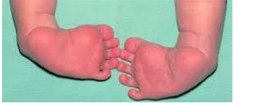
Figure 2. Photo of newborn diagnosed as bilateral clubfoot.
4. Discussion
Congenital talipes equinovarus or club-foot is a common developmental disorder of the lower limb. In talipes equinovarus the foot deviates medially at the ankle and remains fixed at a right angle to the distal tibia and fibula. This causes the long axis of the foot to reside in the same longitudinal plane of section as the tibia and fibula. Normally in this plane of section the ankle and foot are seen in short axis. The foot growth is influenced by many factors, such as neuromuscular conditions, genetics, syndromes, aneuploidy, amniotic fluid volume, multiple gestations and hereditary factors [6] . There is also risk reported with the exposure of teratogens during pregnancy, and this teratogenic influence must occur before 7 - 8 menstrual weeks for there to be any profound effect on musculoskeletal development, because the extremities have differentiated into adult form structures by the end of the embryonic period (the 10th menstrual week). Unfortunately, most teratogenic influence occurs within this period as often the patient is unaware that they are pregnant. There is also debate about the significance of prenatal teratogenic exposure. i.e.: perhaps the male should be made more aware that his actions before conception could also have profound effects on the fetus [7] . The birth prevalence varies among different ethnic groups, and the highest rates are seen in individuals of Polynesian (7 per 100 live births) and the lowest in Asian populations (0.57 per 1000 live births). The prevalence of clubfoot is 1 to 3 per 1000 live births in Caucasians [1] [2] .
Sonography may provide important information about fetal anomalies. In the complex forms depending on the severity of the associated anomalies, future parents have the opportunity to choose between different management options such as termination of pregnancy or continuation of pregnancy with non-intervention or standard management.
The diagnosis of clubfoot can be made as early as 13 weeks, gestation by transvaginal sonography and at 16 weeks by transabdominal ultrasound [8] .
The present study was performed in a large, non-selected population. We found that 128 cases of TEV among 52,100 pregnant women an incidence of 1:1000, that is similar to the 107:103,228 found in a large unselected study by Bakalis et al. [9] . Our data showed that 88 cases (69%) had bilateral, and 40 cases (31%) had unilateral clubfoot. The distribution of TEV in male to female (2:1) was similar to that in published reports [8] . In our study 67 cases (52%) had isolated TEV and 61 cases (48%) had associated anomalies.
Anomalies of the musculoskeletal, central nervous system were the most common associated abnormalities. In our prenatal complex TEV group, there were 31 cases (51%) with a musculoskeletal, 15 cases (24%) with CNS and spine abnormalities, 6 cases (10%) had urinary system anomalies, 5 (8%) cases with polyhydramnios, 5 (8%) with oligohydramnios, 5 (8%) with IUGR, 4 (6.5%) cases with cardiac anomaly, 2 (3%) with cleft lip and 2 (3%) diaphragmatic hernia.
A study by Shipp and Benacerraf retrospectively reviewed their database of sonographic reports over an 18-year period for the sonographic finding of a clubfoot abnormality. Fetuses with associated anomalies were excluded from the study. Their final analysis was of those in which follow-up was obtained from an initial group of 87 fetuses with isolated clubfoot abnormality. 68 had follow-up. 38 fetuses had bilateral clubfoot verified after birth with correct prenatal sonographic identification. 8 fetuses were thought to have clubfoot abnormality but were normal at birth. 22 cases had unilateral clubfoot [10] . In other study by Rijhsinghani et al., the authors retrospectively reviewed their sonographic database of 23,863 pregnant women for cases of clubfoot abnormality. Their analysis revealed clubfoot abnormality in 35 cases from 17 to 37 weeks’ gestation. 18 cases were unilateral and 17 were bilateral. 7 cases were categorized as isolated, additional abnormalities were found in 28 of 35 patients (neural tube, hydrocephalus, ventriculomegaly cardiac defects, and diaphragmatic hernia [11] .
E. Bar-On et al. diagnosed club-foot in 51 fetuses between 1996 and 2003 at a mean gestation age of 22 weeks. Of the 51 fetuses 31 cases had isolated clubfoot, 20 fetuses had complex clubfoot. Ultrasound diagnosed both complex clubfoot and the associated conditions was: five arthrogryposes, four multiple anomalies, two anencephalies, one macrocephaly, one ventricolomegaly, one myelomeningocele, one scoliosis, and growth retardation, one leg-length discrepancy, and neck widening, one hydronephrosis, and small penis one congenital knee dislocation, one polyhydramnios and one rubinstein- Taybi syndrome [12] .
5. Conclusion
We should inform the pregnant women complicated by clubfoot, about the risk of combination of clubfoot with other structural anomalies.
The sonographic scanning of all parts of fetuses, for finding the possible anomalies is an essential procedure.
Acknowledgements
The authors acknowledge all people who helped us during the period of study.
Ethical Issues
The study was approved by research affairs of Tabriz University of Medical Sciences. All participants were asked to sign an informed written consent before the start of data collection.
Conflict of Interests
Authors declare that there is no conflict of interests.
Financial Support
Researchers received no financial support.
Cite this paper
Ghavami, M., Abedinzadeh, R. and Biglu, M.-H. (2016) Prenatal Sonographic Diagnosis of Talipes Equinovarus (Clubfoot) in a Non-Selected Population of Northwest of Iran. Open Access Library Journal, 3: e3189. http://dx.doi.org/10.4236/oalib.1103189
References
- 1. http://orthoinfo.aaos.org/topic.cfm?topic=a00255
- 2. http://radiopaedia.org/articles/congenital-talipes-equinovarus
- 3. http://orthoinfo.aaos.org/topic.cfm?topic=a00255
- 4. Kinnander, C. (2015) Ultrasound: The Requisites. Acta Radiologica, 56, NP54.
https://doi.org/10.1177/0284185115612970 - 5. Alvarado, D.M., Aferol, H., McCall, K., Huang, J.B., Techy, M., Buchan, J., Cady, J., Gonzales, P.R., Dobbs, M.B. and Gurnett, C.A. (2010) Familial Isolated Clubfoot Is Associated with Recurrent Chromosome 17q23. 1q23. 2 Microduplications Containing TBX4. The American Journal of Human Genetics, 87, 154-160.
https://doi.org/10.1016/j.ajhg.2010.06.010 - 6. Moore, K.L. and Persoud, T.V.N. (1998) Before We Are Born-Essentials of Embryology and Birth Defects. 5th Edition, W. B. Saunders Company, Sydney, 420-423.
- 7. Canto, M.J., Cano, S., Palau, J. and Ojeda, F. (2008) Prenatal Diagnosis of Clubfoot in Low-Risk Population: Associated Anomalies and Long-Term Outcome. Prenatal Diagnosis, 28, 343-346.
https://doi.org/10.1002/pd.1984 - 8. Bakalis, S., Sairam, S., Homfray, T., Harrington, K., Nicolaidess, K. and Thilaganatjan, B. (2002) Outcome of Antenatally Diagnosed Talipes Equinovarus in an Undelected Obstetric Population. Ultrasound in Obstetrics & Gynecology, 20, 226-229.
https://doi.org/10.1046/j.1469-0705.2002.00780.x - 9. Offerdal, K., Jebens, N., Blaas, H.G.K. and Eik-Nes, S.H. (2007) Prenatal Ultrasound Detection of Talipes Equinovarus in a Non-Selected Population of 49 314 Deliveries in Norway. Ultrasound in Obstetrics & Gynecology, 30, 838-844.
https://doi.org/10.1002/uog.4079 - 10. Shipp, T.D. and Benacerraf, B.R. (1998) The Significance of Prenatally Identified Isolated Clubfoot: Is Amniocentesis Indicated? American Journal of Obstetrics & Gynecology, 178, 600-602.
https://doi.org/10.1016/S0002-9378(98)70445-4 - 11. Rijhsinghani, A., Yankowitz, J., Kanis, A.B., Mueller, G.M., Yankowitz, D.K. and Williamson, R.A. (1998) Antenatal Sonographic Diagnosis of Clubfoot with Particular Attention to the Implications and Outcomes of Isolated Clubfoot. Ultrasound in Obstetrics & Gynecology, 12, 103-106.
- 12. Bar-On, E., Mashiach, R., Inbar, O., Weigl, D., Katz, K. and Meizner, I. (2005) Prenatal Ultrasound Diagnosis of Clubfoot. The Bone & Joint Journal, 87, 990-993.