Materials Sciences and Applicatio ns, 2011, 2, 1298-1301
doi:10.4236/msa.2011.29175 Published Online September 2011 (http://www.SciRP.org/journal/msa)
Copyright © 2011 SciRes. MSA
A Top-Down Approach of Making Sn-3.5Ag
Nanosolder Alloy by Swirl Method
Hsin Jen Pan1, Chao Yang Lin1*, Jung Hua Chou1, Udit Surya Mohanty2*, Yuan Gee Lee3
1Department of Engineering Science, National Cheng Kung University, Tainan, Taiwan (China); 2Department of Materials Science
and Engineering, National Cheng Kung University, Taiwan (China); 3Department of Automation Engineering and Institute of
Mechatronoptic Systems, Chien-Kuo Institute of Technology, Changhua, Taiwan (China).
Email: *n9895121@mail.ncku.edu.tw, *suryaudit@yahoo.com
Received January 18th, 2011; revised February 9th, 2011; accepted March 20th, 2011.
ABSTRACT
The nanoparticles of tin-silver sold er, Sn-3.5Ag, of necklace geometry were made in a swirl batch. It was found that the
addition of the element, Ag, did not vary the microstru cture of the solder matrix, but Ag simp ly diluted into th e Sn ma-
trix randomly. The swirl flow facilitated th e formation of particles with different sizes. It was found that the size distri-
bution of the nanoparticles was strongly related to the height in the swirl batch. In addition, the aggregation of the
nanoparticles was explored and the dispersion of the nanoparticles was achieved by adjusting the pH value of the solu-
tion near the neu tral value.
Keywords: Swirl Flow, Particle Size Distribution, Nanoparticle Aggregation
1. Introduction
Due to the advances in semiconductor manufacturing
technology, the feature size of the complementary
metal-oxide semiconductor (CMOS) structure could ap-
proache nano-scale length. Metal tin is always a major
component of the solder matrix which serves as an inter-
connection between the device and substrate. However, it
cannot withstand the mechanical requirements at ele-
vated temperatures. Traditional lead alloys are being
phased out [1]. Many researchers have been looking for
alternatives to replace lead [2-4]. Eutectic tin-silver,
Sn-3.5Ag, is a popular replacement since the soldering
temperature and surface tension near to that of Sn-37Pb
solder [5]. Metal nanoparticles have recently received
great attention. The nano-metal is not only employed in
photonics, but it also has potential applications in opto-
electronics [6]. Moreover, tin particles also follow Bar-
bier-type allyation reactions and also serve as an abrasive
to antiwear under loading, resulting in reduction of the
particle size to the nanometer scale [7]. Based on these
specific advantages, the size of the Sn-3.5Ag alloy is
scaled down to nanometer scale using the swirl method; a
technique to sieve solid p article size in fluidic bed facili-
ties [8].
2. Experimental Procedure
Metals Sn (99.8%), Ag (99.9%) and paraffin oil (99.8%)
were commercial chemicals. 9.65 grams of Sn powder
was blended with 0.35 grams of Ag powder in a sealed
vacuum glass tube. The tube was heated to 260˚C in an
oven for 2 hours with intermittent shaking. This alloy
was then checked for uniformity and then immersed in
the heating solvent swirl batch of paraffin oil, at 320˚C
for 10 hours. Aliquots were removed and quenched into
liquid nitrogen to prevent aggregation. The viscous solu-
tion, composed of nanoparticles and paraffin, was
blended with chloroform to dilute th e paraffin. The solu-
tion was centrifuged and then was diluted in turn three
times. The nanoparticles formed as black sediments at
the bottom of the tube. The nanoparticles were washed
with water and acetone several times until pH = 7 then
vacuum dried at 40˚C for 12hrs. The upper zone, chloro-
form, was dried out and then the black sediment was
sampled to identify its phase with X-ray diffraction (Ri-
kagu). To prevent the nanoparticles from aggr egation, the
rest of the sediment was dissolved in ammonium hy-
droxide (NH4OH) and dispersed using an ultrasonic
shaker. The alkaline, NH4OH solution was dispensed
onto a glass substrate. After drying, the morphology of
the particle was investigated by a scanning electron mi-
A Top-Down Approach of Making Sn-3.5Ag Nanosolder Alloy by Swirl Method1299
croscope (SEM 2500, Hitachi) and the diffraction pattern
was obtained by a high resolution TEM (JEOL 2100).
3. Results and Discussion
3.1. Morphology Investigation and Phase
Identification
Uniform Sn-3.5Ag nanoparticles were prepared and dis-
persed successfully by the swirl method. However, the
nanoparticles did not disperse individually but joined
together with necklace morphology. The nanoparticles
thus formed showed active surfaces [9].
The trace element, Ag, was resolved in the tin matrix
homogeneously. Figure 1 shows the HRTEM diffraction
pattern of Sn-3.5Ag nanoparticles. This pattern was iden-
tified to be a space group of I41/amd with lattice parame-
ters, a, 5.819 Å, and b, 3.175 Å, respectively. This pat-
tern is consistent with the tetragonal system [10], as that
of the synthesized tin. Thus, the addition of the silver did
not vary the microstructure of the tin matrix.
3.2. Kinetics of the Formation of Sn-3.5Ag
Nanoparticles
The size of the nanoparticles was determined by Oswald
ripening and the rotational speed of the swirl. Figure 2
showed the particle size variation for subsequent sam-
pling time with a constant sampling height. It is noted
that the particle size decreased initially, then reached the
minimum value and finally recoiled appreciably. This
indicated a specific formation procedure as proposed in
the following section .
Figure 1. HRTEM diffraction pattern of Sn-3.5Ag nanosol-
der.
Figure 2. Particle size variation with respect to sampling
time.
Initially, the swirl flow quickly reached steady state.
The bulk homogeneous Sn-Ag alloy was heated and
showed a viscous fluid with a coherence force to main-
tain its spherical-like shape. Because the Sn-Ag alloy did
not resolve in the paraffin solvent, the coherence force
prevailed. When the viscous Sn-Ag alloy was deposited
on the floor of the rotational bath, the immiscible phase,
paraffin and also the Sn-3.5Ag were subjected to fluid
shear stresses. Because the shear force was larger than
the coherence force, the bulk viscous fluid in the bottom
layer was dissevered. This dissevered debris climbed
peripherally up and was dissevered again. Meanwhile the
quantum effect emerged due to the small size of the
Sn-Ag alloy while approaching the nano-scale [11]. Fi-
nally, the shear force could not eclipse the incoherent
force and the alloy vo lume was maintained. The shape of
the nanoparticles could not maintain its sph erical features
when the quantum effect balanced the incoherent force
[12]. The swirl flow facilitated a particle size distribution
with respect to different sampling heights of the swirl
batch as shown in Figure 3. The size of the nanoparticles
decreased with increasing sampling height. A force bal-
ance model was constructed to analyze the particles in
the swirl batch. In the swirl flow, the nanoparticles were
balanced by the flow force and rotated with the same
velocity as that of the fluid. The velocity was a function
of both the radical distance and height of the batch. Ex-
cept at the center of the batch, the nanoparticles experi-
enced a net force perpendicular to the surface of the swirl
flow layer.
3.3. Dispersion of the Aggregated Nanoparticles
The aggregated nanoparticles were dispersed freely by
Copyright © 2011 SciRes. MSA
A Top-Down Approach of Making Sn-3.5Ag Nanosolder Alloy by Swirl Method
1300
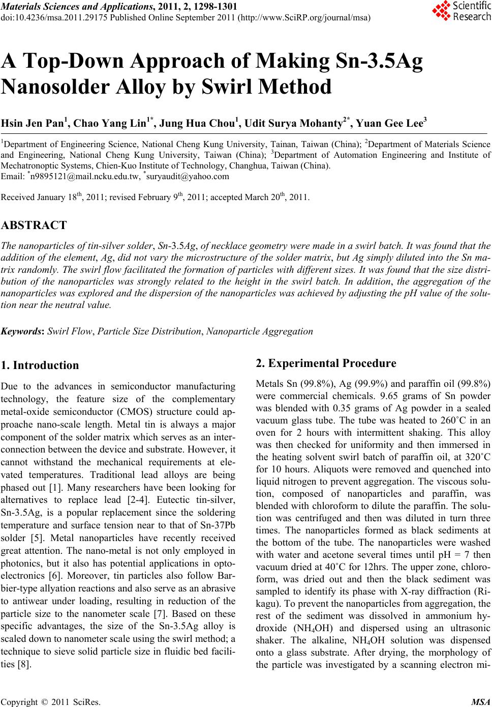
Figure 3. The average particle size dipped at different
heights.
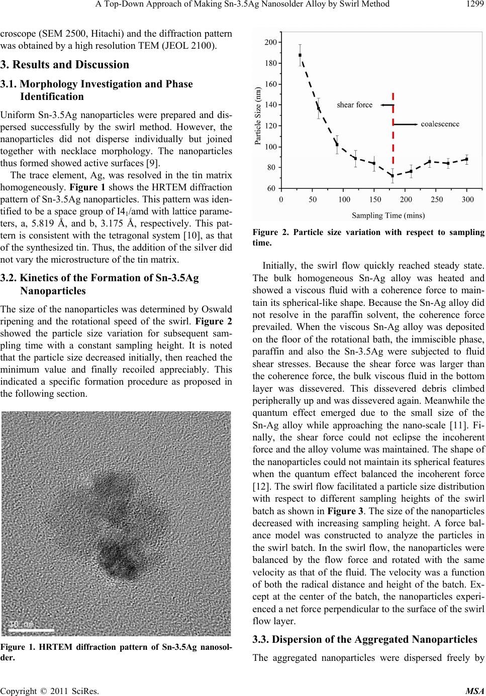
adjusting the pH value of the solution. Particle size de-
creased with increasing pH values (Figure 4) up to 8.54,
but then increased appreciably up to pH value of 11.2.
However, the nanoparticles aggregated again, as the so-
lution changed from neutral to alkaline. It has been re-
ported that the metallic nanoparticles possess negative
electrical carriers [13]. In acidic solutions the hydrogen
ions, neutralizes the nanoparticle’s surface. The zeta po-
tential of the nanoaparticles thus approaches zero. An
increase in pH changed the charge of the nanoparticles
from positive to negative. The repulsive force also in-
creased to counteract the attractive force. However, the
nanoparticles became unstable as the negative surface
charge became immoderate. The systematic energy thus
had to be reduced due to the flocculation of the particles
[14]. The particle size was no more spherical but
tetragonal. The non-spherical shape of the particle indi-
cated that some force dominated the lattice assembly.
The shape hence showed a distinct morphology; namely
polyhedral rather than the traditiona l spherical shape.
Figure 4. Particle size distribution v.s pH values in the dis-
persive solution.
4. Conclusions
Tin-silver nanoparticles were prepared successfully by a
swirl method with the trace element, Ag, resolved in the
tin matrix homogeneously. Because the particle size
could approach a steady state value, the kinetics was in-
vestigated in this study. Th e size of the nanoparticles was
determined by the rotational velocity of the swirl and
height off the bottom. To avoid the aggregation of the
nanoparticles, the pH value of the solution was adjusted
and the aggregated particles were dispersed successfully.
5. Acknowledgements
We are grateful for the financial support provided by the
Chien-kuo Techno logy University (C TU-95-RP-AE-007-
007-A).
REFERENCES
[1] Y. Li and C. P. Wong, “Recent Advances of Conductive
Adhesives as a Lead-Free Alternative in Electronic Pack-
aging: Materials, Processing, Reliability and Applica-
tions,” Materials Science and Engineering, R: Reports,
Vol. 51, No. 1-3, 2006, pp. 1-35.
doi:10.1016/j.mser.2006.01.001
[2] M. Abtew and G. Selvaduray, “Lead-Free Solders in
Microelectronics,” Material Science Engineering, R:
Reports, Vol. 27, No. 5-6, 2000, pp. 95-141.
[3] M. McCormack, S. Jin, G. W. Kammlott and H. S. Chen,
“New Lead-Free, Sn-Ag-Zn-Cu Solder Alloy with Im-
proved Mechanical Properties,” Applied Physics Letters,
Vol. 63, No. 1, 1993, pp. 15-17.
[4] I.E. Anderson, J. C. Foley, B.A. Cook, J. Harringa, R.L.
Terpstra and O. Unal, “Alloying Effects in Near-Eutectic
Sn-Ag-Cu Solder Alloys for Improved Microstructural
Stability,” Journal of Electronic Materials, Vol. 30, No. 9,
2001, pp. 1050-1059. doi:10.1007/s11664-001-0129-5
[5] P.T. Vianco and A.C. Claghorn, “Effect of Substrate Pre-
heating on Solderability Performance as a Guideline for
Assembly Process Development Part 1: Baseline Analy-
sis,” Soldering & Surface Mount Technology, Vol. 8, No.
3, 1996, pp. 12-18. doi:10.1108/09540919610777708
[6] S. A. Maier, M. L. Brongersma, P. G. Kik, S. Meltzer, A.
A. G. Requicha and H. A. Atwater, “Plasmonics a Route
to Nanoscale Optical Devices,” Advanced Materials, Vol.
13, No. 19, 2001, pp. 1501-1505.
doi:10.1002/1521-4095(200110)13:19<1501::AID-ADM
A1501>3.0.CO;2-Z
[7] Z. Zha, S. Qiao, J. Jiang, Y. Wang, Q. Miao and Z. Wang,
“Barbier-Type Reaction Mediated with Tin Nano-Parti-
cles in Water,” Tetrahedron, Vol. 61, No. 9, 2005, pp.
2521-2527. doi:10.1016/j.tet.2004.12.063
[8] M. Athar, U. C. Kothyari and R. J. Garde, “Distribution
of Sediment Concentration in the Vortex Chamber Type
Sediment Extractor,” International Journal of Hydraulic
Research, Vol. 41, No. 4, 2003, pp. 427-438.
Copyright © 2011 SciRes. MSA
A Top-Down Approach of Making Sn-3.5Ag Nanosolder Alloy by Swirl Method
Copyright © 2011 SciRes. MSA
1301
doi:10.1080/00221680309499987
[9] K. S. Le Corre, E. Valsami-Jones, P. Hobbs, B. Jefferson
and S. A. Parsons, “Agglomeration of struvite crystals,”
Water Research, Vol. 41, No. 2, 2007, pp. 419-425.
doi:10.1016/j.watres.2006.10.025
[10] Y. Zhao, Z. Zhang and H. Dang, “Preparation of Tin
Nanoparticles by Solution Dispersion,” Material Science
and Engineering A, Vol. 359, No. 1-2, 2003, pp. 405-407.
doi:10.1016/S0921-5093(03)00395-2
[11] A. Bhardwaj, P. Srivastava and H. K. Sehgal, “Chemi-
cally Deposited PbSe Nanoparticle Films Variation in
Shape and Size,” Journal of Electrochemical Society, Vol.
154, No. 10, 2007, pp. 83-86. doi:10.1149/1.2763963
[12] E. Pan, Y. Zhang, P. W. Chung and M. Denda, “Strain
Energy on the Surface of an Anisotropic Half-Space Sub-
strate: Effect of Quantum-Dot Shape and Depth,” CMES
—Computer Modeling in Engineering & Sciences, Vol.
24, No. 2-3, 2008, pp. 157-167.
[13] J. Duan and J. Gregory, “Coagulation by Hydrolysing
Metal Salts,” Advances in Colloid Interface Science and
Technology, Vol. 10, 2003, pp. 100-102.
[14] J. Gregory, “Particles in Water: Properties and Proc-
esses,” IWA Publishing, London, 2006.