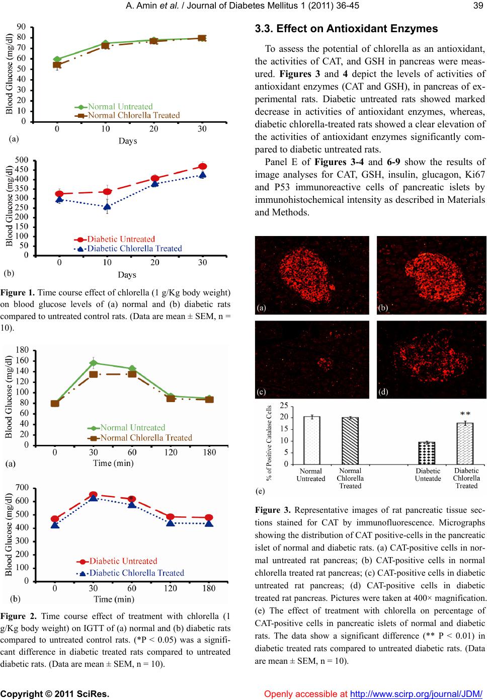
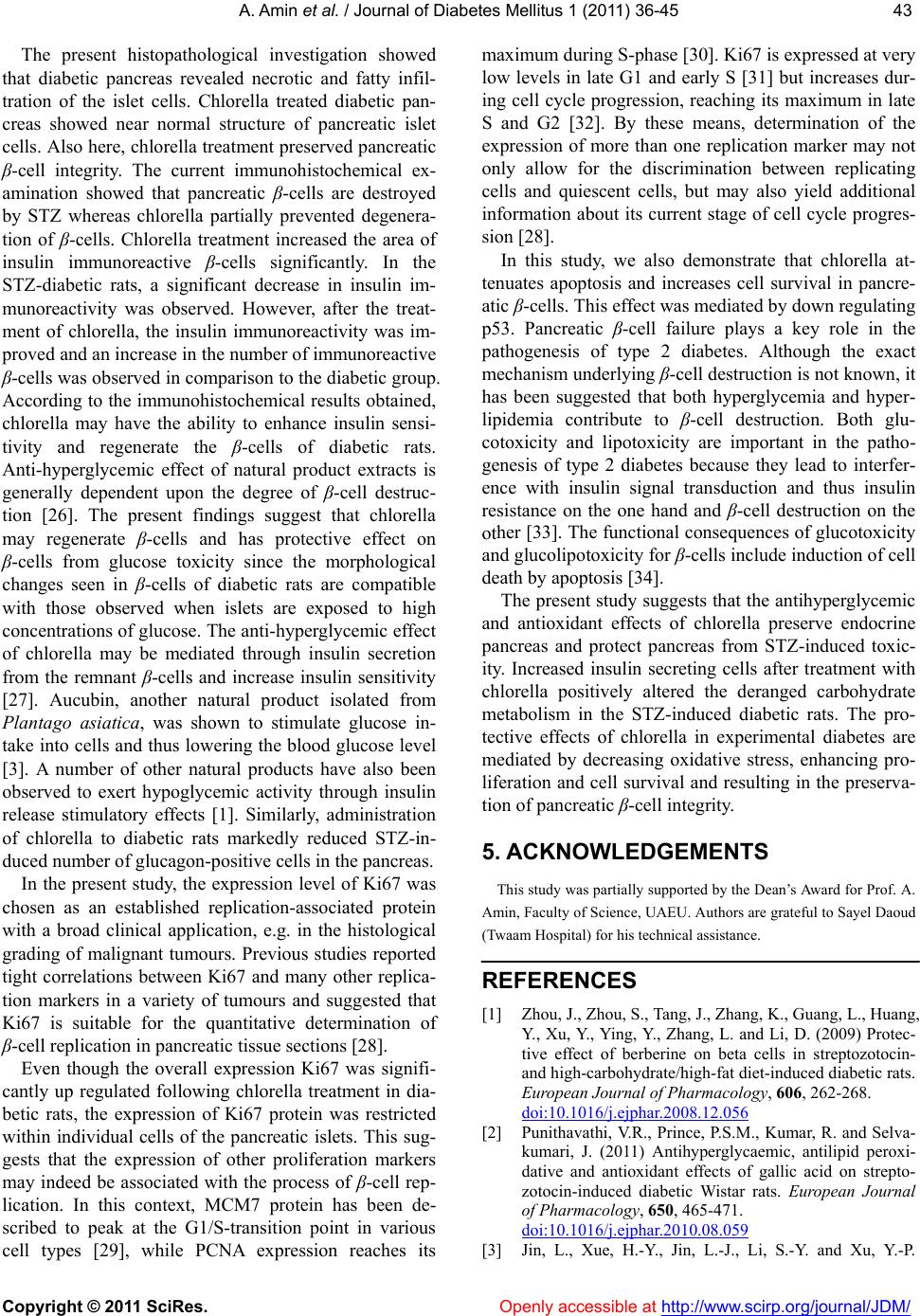
A. Amin et al. / Journal of Diabetes Mellitus 1 (2011) 36-45
Copyright © 2011 SciRes. Openl y accessible at http:// www.scirp.org/journal/JDM/
44
(2008) Antioxidant and pancreas-protective effect of au-
cubin on rats with streptozotocin-induced diabetes. Euro-
pean Journal of Pharmacology, 582, 162-167.
doi:10.1016/j.ejphar.2007.12.011
[4] Frode, T.S. and Medeiros, Y.S. (2008) Animal models to
test drugs with potential antidiabetic activity. Journal of
Ethnopharmacology, 115, 173-183.
doi:10.1016/j.jep.2007.10.038
[5] Baynes, J.W. and Thorpe, S.R. (1996) The role of oxida-
tive stress in diabetic complications. Current Opinion of
Endocrinology, 3, 277-284.
doi:10.1097/00060793-199608000-00001
[6] Ihara, Y., Toyokuni, S., Uchida, K., Odaka, H., Tanaka, T.,
Ikeda, H., Hiai, H., Seino, Y. and Yamada, Y. (1999) Hy-
perglycemia causes oxidative stress in pancreatic beta-
cells of GK rats a model of type 2 diabetes. Diabetes, 48,
927-932. doi:10.2337/diabetes.48.4.927
[7] Saxena, A.K., Srivastava, P., Kale, R.K. and Baquer, N.Z.
(1993) Impaired antioxidant status in diabetic rat liver.
Effect of vanadate. Biochemical Pharmacology, 45, 539-
542. doi:10.1016/0006-2952(93)90124-F
[8] Maritim, A.C., Sanders, R.A. and Watkins, J.B. (2003)
Effect of alpha lipoic acid on biomarkers of oxidative
stress in streptozotoc in-induced diabetic rats. The Jour-
nal of Nutritional Biochemistry, 14, 288-294.
doi:10.1016/S0955-2863(03)00036-6
[9] Ames, B.N. (1989) Endogenous oxidative DNA damage,
aging, and cancer. Free Radical Research Communica-
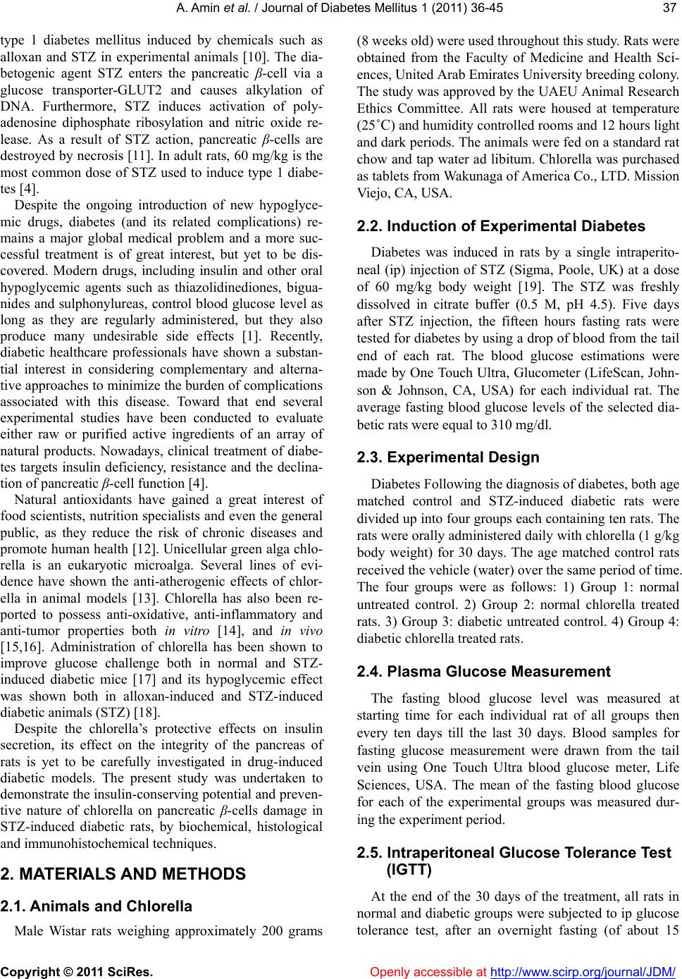
tion, 7, 121-128. doi:10.3109/10715768909087933
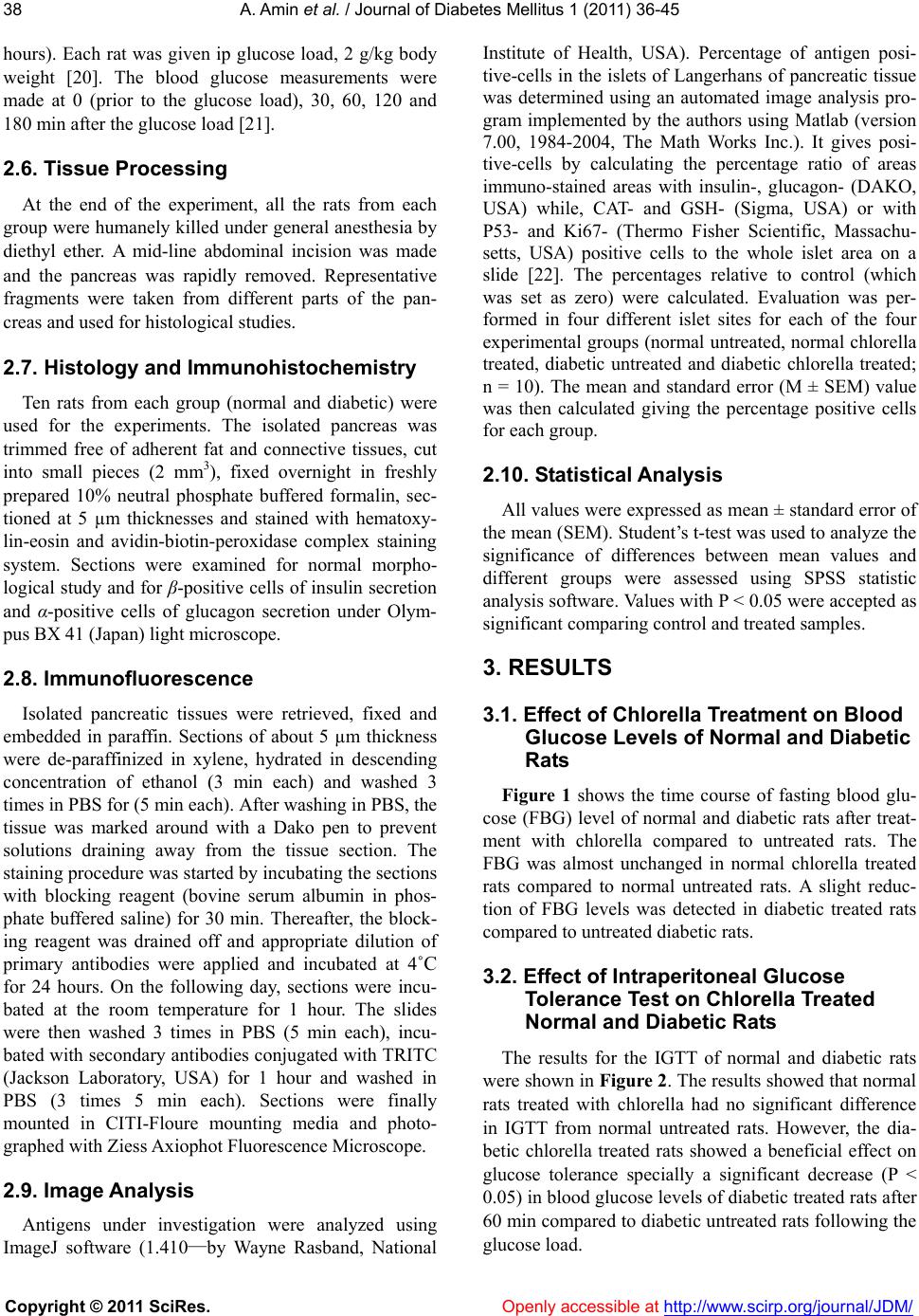
[10] Ku, Y.P., Jin, M., Kim, K.H., Ahn, Y.J., Yoon, S.P., You,
H.J. and Chang, I.Y. (2009) Immunolocalization of
8-OHdG and OGG1 in pancreatic islets of streptozoto-
cin-induced diabetic rats. Acta Histochemica, 111, 138-144.
doi:10.1016/j.acthis.2008.05.008
[11] Mythili, M.D., Vyas, R., Akila, G. and Gunasekaran, S.
(2004). Effect of streptozotocin on the ultrastructure of
rat pancreatic islets. Microscopy Research and Technique,
63, 274-281. doi:10.1002/jemt.20039
[12] Jagtap, U.B. and Bapat, V.A. (2010) Artocarpus: A re-
view of its traditional uses, phytochemistry and pharma-
cology. Journal of Ethnopharmacology, 129, 142-166.
doi:10.1016/j.jep.2010.03.031
[13] Lee, H.S., Choi, C.Y., Cho, C. and Song, Y. (2003) At-
tenuating effect of Chlorella supplement on oxidative
stress and NF-kB activation in peritoneal macrophage
and liver of C57BL/6 mice fed on an atherogenic diet. Bio-
science biotechnology and biochemistry, 67, 2083-2090.
doi:10.1271/bbb.67.2083
[14] Guzman, S., Gato, A. and Calleja, J.M. (2001) Anti- im-
flammatory activities of the marine microalgae. Chlorella
Stigmatophora and Phaeodactylum tricornutum. Phyto-
therapy Research, 15, 224-230. doi:10.1002/ptr.715
[15] Amin, A. (2008) Chemopreventive effect of chlorella on
the antioxidant system in DMBA-induced oxidative stress
in liver. International Journal of Pharmacology, 4, 169-
176. doi:10.3923/ijp.2008.169.176
[16] Amin, A. (2009) Protective effect of green algae against
7,12-dimethylbenzanthrancene (DMBA)-induced breast
cancer in rats. International Journal of Cancer Research,
5, 12-24. doi:10.3923/ijcr.2009.12.24
[17] Cherng, J.-Y. and Shih, M.-F. (2005) Potential hypogly-
cemic effects of Chlorella in streptozotocin-induced dia-
betic mice. Life Sciences, 77, 980-990.
doi:10.1016/j.lfs.2004.12.036
[18] Cherng, J.-Y. and Shih, M.-F. (2006) Improving glyco-
genesis in Streptozocin (STZ) diabetic mice after ad-
ministration of green algae Chlorella. Life Sciences, 78,
1181-1186. doi:10.1016/j.lfs.2005.06.050
[19] Adeghate, E. (1999) Effect of subcutaneous pancreatic
tissue transplants on streptozotocin-induced diabetes in
rats. I. Morphological studies on normal, diabetic and
transplanted pancreatic tissues. Tissue and Cell, 31, 66-
72. doi:10.1054/tice.1999.0008
[20] Caluwaerts, S., Lambin, S., van Bree, R., Peeters, H.,
Vergote, I. and Verhaeghe, J. (2007) Diet-induced obesity
in gravid rats engenders early hyperadiposity in the off-
spring. Metabolism, 56, 1431-1438.
doi:10.1016/j.metabol.2007.06.007
[21] Sharma, B., Balomajumder, C. and Roy, P. (2008) Hypo-
glycemic and hypolipidemic effects of flavonoid rich ex-
tract from Eugenia jambolana seeds on streptozotocin
induced diabetic rats. Food and Chemical Toxicology, 46,
2376-2383. doi:10.1016/j.fct.2008.03.020
[22] Salazar-Montes, A., Ruiz-Corro, L., López-Reyes, A.,
Castrejón-Gómez, E. and Armendáriz-Borunda, J. (2008)
Potent antioxidant role of pirfenidone in experimental cir-
rhosis. European Journal of Pharmacology, 595, 69-77.
doi:10.1016/j.ejphar.2008.06.110
[23] Upadhyay, O.P., Singh, R.M. and Dutta, K. (1996) Stud-
ies on antidiabetic medicinal plants used in Indian
folk-lore. Aryavaidyan, 9, 159-167.
[24] Reuter, S., Gupta, S.C., Chaturvedi, M.M. and Aggarwal,
B.B. (2010) Oxidative stress, inflammation, and cancer:
How are they linker? Free Radical Biology and Medicine,
49, 1603-1616. doi:10.1016/j.freeradbiomed.2010.09.006
[25] Picton, S.F., Flatt, P.R. and Mcclenghan, N.H. (2001)
Differential acute and long term actions of succinic acid
monomethyl ester exposure on insulin secreting BRAIN-
BD 11 cells. International Journal of Experimental Dia-
betes Research, 2, 19-27. doi:10.1155/EDR.2001.19
[26] Grover, J.K., Vats, V. and Rathi, S.S. (2000) Anti-hy-
perglycemic effect of Eugenia jambolana and Tinospora
cordifolia in experimental diabetes and their effects on
key metabolic enzymes involved in carbohydrate me-
tabolism. Journal of Ethnopharmacology, 73, 461-470.
doi:10.1016/S0378-8741(00)00319-6
[27] Leng, S.H., Lu, F.E. and Xu, L.J. (2004) Therapeutic
effects of berberine in impaired glucose tolerance rats
and its influence on insulin secretion. Acta Pharma-
cologica Sinica, 25, 496-502.
[28] Köhler, C.U., Kreuter, A., Rozynkowski, M.C., Rahmel,
T., Uhl, W., Tannapfel, A., Schmidt, W.E. and Meier, J.J.
(2010) Validation of different replication markers for the
detection of beta-cell proliferation in human pancreatic
tissue. Regulatory Peptides, 162, 115-121.
doi:10.1016/j.regpep.2009.12.021
[29] Shohet, J.M., Hicks, M.J., Plon, S.E., Burlingame, S.M.,
Stuart, S., Chen, S.Y., Brenner, M.K. and Nuchtern, J.G.
(2002) Minichromosome maintenance protein MCM7 is
a direct target of the MYCN transcription factor in
neuroblastoma. Cancer Research, 62, 1123-1128.
[30] Jaskulski, D., deRiel, J.K., Mercer, W.E., Calabretta, B.
and Baserga, R. (1988) Inhibition of cellular proliferation
by antisense oligodeoxynucleotides to PCNA cyclin.