Open Journal of Urology, 2011, 1, 56-59
doi:10.4236/oju.2011.13012 Published Online August 2011 (http://www.SciRP.org/journal/oju)
Copyright © 2011 SciRes. OJU
A Case of Incidentally Found Bilateral Retroperitoneal
Cavernous Hemangiomas
Shunsuke Yui1, Masayoshi Zaitsu1, Koji Mikami1, Y ut a Take shi ma1, Akiko Tonooka2,
Toshimasa Uekusa2, Takumi Takeuchi1
1Department of Ur ol o gy, Kanto Rosai Hospital, Kawasaki, Japan
2Pathology, Kanto Rosai Hospital, Kawasaki, Japan
E-mail: takeuchit@abelia.ocn.ne.jp
Received July 5, 2011; revised July 31, 2011; accepted August 9, 2011
Abstract
A 59-year-old male was admitted to our hospital because of incidentally found right retroperitoneal tumor.
He had undergone removal of a hemangioma in the left oral cavity four years before. An abdominal CT scan
performed in our hospital revealed poorly enhanced bilateral retroperitoneal tumors adjacent to kidneys.
Those tumors were of low signal intensity on T1-weighted images and high on T2-weighted images by
magnetic resonance imaging. The right retroperitoneal tumor of 2.5 cm in size was surgically removed and
histopathological examination indicated cavernous hemangioma. The smaller left retroperitoneal tumor of
1.1 cm in size was left untouched to be followed up, as we supposed that it has the same benign pathology.
There have been no previous cases of retroperitoneal cavernous hemangioma as a presentation of multiple
hemangiomas.
Keywords: Hemangioma, Bilateral, Retroperitoneum
1. Introduction
Primary retroperitoneal tumors just account for 0.2% of
all tumors in a old article publish ed in 1954 [1] and there
are no available reports definitely describing the recent
prevalence of retroperitoneal tumors. Braasch et al. col-
lected 101 cases of retroperitoneal tumor from the litera-
ture and reported in 1967 that more than 80% of cases
were malignant and symptomatic [2]. Nowadays benign,
smaller, and less symptomatic retroperitoneal tumors
must be diagnosed with the improvement and prevalence
of advanced imaging modalities as CT scan, magnetic
resonance imaging, and ultrasonography. Nakajima et al.
reported in 1997 that 10 (40%) out of 25 retroperitoneal
tumors were malignant [3], thus the percentage of ma-
lignancy among all diagnosed retroperitoneal tumors
seems decreased than before.
Retroperitoneal hemangioma is very uncommon,
while the true incidence of that disorder among retrop-
eritoneal tumors is basically unknown, as most of them
may be asymptomatic and remain undiagnosed. That
there have been only few reports of retroperitoneal he-
mangioma diagnosed only with radiological imagings [4]
may indicate that surgical procedure has been chosen for
both diagnostic and therapeutic purposes. Here we show
a case of bilateral retroperitoneal cavernous hemangio-
mas which was difficult to be diagnosed before surgical
extirpation.
2. Case Report
A 59-year-old male was admitted to our hospital because
of incidentally found right retroperitoneal tumor. Tumor
markers as well as regular laboratory data were unre-
markable. Urine cytology was negative. An abdominal
CT scan performed in our hospital revealed a right re-
troperitoneal tumor of 2.5 cm in size and a left retroperi-
toneal tumor of 1.1 cm in size adjacent to either kidney.
Those tumors with speckled calcification were not en-
hanced so much both in early phase and late phase (Fig-
ure 1). By an abdominal magnetic resonance imaging,
those tumors were of low signal intensity on T1-
weighted images and high on T2-weighted images (Fig-
ure 2). We have decided to extirpate the right retroperi-
toneal tumor using transperitoneal approach, because
those tumors could not be determined to be of benign
pathology by imaging studies. We did not adopt lararo-
scopic and retroperitoneoscopic surgery because of un-
S. YUI ET AL.
57
known pathology.
At operation, there was little adhesion between the
tumor and the surrounding structures. The tumor was
supposed to be originated from the vascular sheath of the
right renal artery and there were small feeders from the
sheath. The tumor was irregular on surface and reddish
like a raspberry in color (Figure 3). We utilized renal
arterial clump for a short time in order to reduce gradual
bleeding from the tumor surface which occurred when
the preparation of the tumor was initiated. The resected
tumor was 3.0 × 1.5 × 1.5 cm in size. The cut section
showed sponge-like appearance containing solid com-
ponent (Figure 4).
Histology of the tumor (Figure 5) was diagnosed as
cavernous hemangioma composed of elaborately intera-
nastomosing vascular spaces of large dilated blood-filled
vessels. Vascular channels with fibrous walls were lined
by flattened endothelial cells which have no atypism.
The smaller left retroperitoneal tumor of 1.1 cm in size
was left untouched to be followed up, as we supposed
that it has the same benign pathology.
3. Discussion
We have experienced a relatively rare case of bilateral
retroperitoneal cavernous hemangiomas occurred in an
elder man. Hemangioma is often found in infants, espe-
cially in the skin [5]. Hemangioma is histologically de-
fined as a benign tumor or malformation made up of
mature well-formed vessels usually lined by a single
layer of endothelium and is classified into six categories,
i.e. capillary hemangioma, cavernous hemangioma, ve-
nous hemangioma, epithelioid hemangioma, pyogenic
Figure 1. Example of a figure caption A CT scan demon-
strating bilateral retroperitoneal tumors. Arrows indicate
tumors.
Figure 2. An MR imaging demonstrating bilateral retro-
peritoneal tumors. Arrows indic a te tumors.
V
A
T
K
Figure 3. A photograph during operation. T: right retro-
peritoneal tumor, A: taped right renal artery, V: right renal
vein, K: right kidney.
Figure 4. A macroscopic cut section of the right retro-
pritoneal tumor.
granuloma, and acquired tirfted hemangioma [6]. He-
mangioma is also known to be found in the oral region
[7] as was observed in the present case.
In the present case, hemangiomas were multiple in bi-
lateral retroperitoneal spaces as well as in the mouth.
Multiple hemangiomas are often a part of established
syndromes such as Sturge-Weber syndrome, Maffucci
syndrome, Kasabach-Merritt syndrome, and von Hippel-
Copyright © 2011 SciRes. OJU
58 S. YUI ET AL.
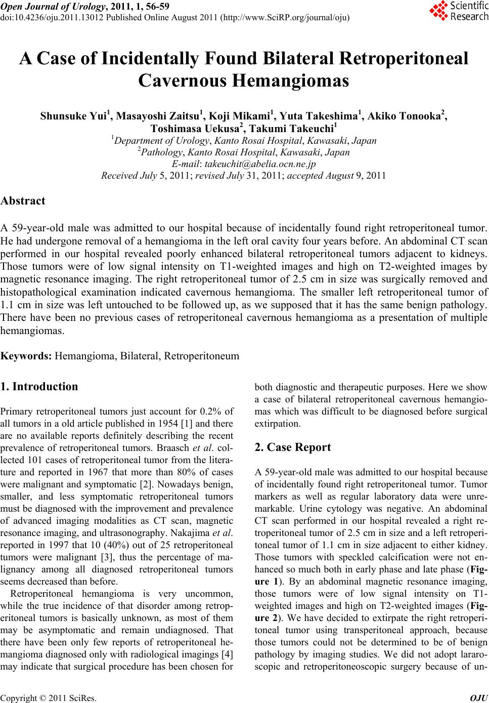
Figure 5. Hematoxylin and eosin staining of the right re-
troperitoneal tumor showing cavernous hemangioma.
Lindau disease, but this case did not seem to apply to
them as it lacks in the characteristics of those disorders.
There have been no previous cases of retroperitoneal
cavernous hemangioma as a presentation of multiple
hemangiomas.
Surgical removal of retroperitoneal tumors is indicated
as long as the possibility of poten tial malignan cy remains.
Maximum diameter of malignant retroperitoneal tumors
is reported to be larger than that of benign ones (11.45 ±
1.90 cm vs 5.31 ± 0.43 cm) [3]. The tumor size of the
present case was relatively small and the probability of
malignancy was not very high in the view points of tu-
mor size, then we might have followed up the interval
change in the tumor size before indicating surgery. Addi-
tionally, retroperitoneal hemangiomas have a risk of
rupture and bleeding [8] even they are histologically be-
nign, leading to a rational of choosing surgical extirpa-
tion especially when they are large. Although we adopted
open surgical approach, laparoscopic resection of retrop-
eritoneal cavernous hemangioma has been reported
[9,10]. It may be appropriate to remove retroperitoneal
hemangiomas laparoscopically or retroperitoneoscopi-
cally if they are probably benign .
Besides surgical treatments, there are some options as
corticosteroid therapy and radiotherapy in the treatment
of hemangioma. While corticosteroid therapy is mainly a
choice of treatments of capillary hemangioma [11], there
is a report of unresectable 8 × 5 cm retroperitoneal cav-
ernous hemangioma diagnosed in a female neonate 20
days after birth and showing anemia and thrombocyto-
penia, which disappeared following oral corticosteroid
therapy [12]. Radiotherapy is also given to treat symp-
tomatic hemangiomas [13].
In the present case, the smaller left retroperitoneal tu-
mor of 1.1 cm in size was left untouched to be followed
up, as we supposed that it has the same ben ign pathology.
When the left retroperiton eal tumor grows larger enough ,
we may need to re-consider if he needs some treatment
including surgery.
4. References
[1] G. T. Pack and E. J. Tabah, “Collective Review: Primary
Retroperitoneal Tumors; a Study of 120 Cases,” Surgery,
Gynecology & Obstetrics, Vol. 99, No. 3, 1954, pp. 209-
231.
[2] J. W. Braasch and A. B. Mon, “Primary Retroperitoneal
Tumors,” Surgical Clinics of North America, Vol. 47, No.
3, 1967, pp. 663-678.
[3] J. Nakashima, M. Ueno, K. Nakamura, M. Tachibana, S.
Baba, N. Deguchi, H. Tazaki and M. Murai, “Differential
Diagnosis of Primary Benign and Malignant Retroperito-
neal Tumors,” International Journal of Urology, Vol. 4,
No. 5, 1997, pp. 441-446.
doi:10.1111/j.1442-2042.1997.tb00282.x
[4] N. Takaha, M. Hosomi, K. Se kii, S. Nakamori, K. Itoh, S.
Sagawa, T. Kido, M. Satani, S. Kawamoto and K. Sato,
“Retroperitoneal Cavernous Hemangioma: A Case Re-
port,” Hinyokika Kiyo, Vol. 37, No. 7, 1991, pp. 725-728.
[5] B. A. Drolet, N. B. Esterly and I. J. Frieden, “Heman-
giomas in Children,” New England Journal of Medicine,
Vol. 341, No. 3, 1999, pp. 173-181.
doi:10.1056/NEJM199907153410307
[6] S. W. Weiss, “Histological Typing of Soft Tissue Tu-
mours,” Vol. 2, Springer-Verlag, New York, 1994.
[7] N. Kawachi, “A Comparative Histopathological and Im-
munohistochemically Study of Capillary Hemangioma,
Pyogenic Granuloma and Cavernous Hemangioma in the
Oral Region: With Special Reference to Vascular Prolif-
eration Factors,” International Journal of Oral-Medical
Sciences, Vol. 9, No. 3, 2011, pp. 241-251.
doi:10.5466/ijoms.9.241
[8] T. L. Forbes, “Retroperitoneal Hemorrhage Secondary to
a Ruptured Cavernous Hemangioma,” Canadian Journal
of Surgery, Vol. 48, No. 1, 2005, pp. 78-79.
[9] Y. S. Choi and H. K. Oh, “Laparoscopic Resection of a
Retroperitoneal Hemangioma Arising from Ovarian Ves-
sels,” Journal of Minimally Invasive Gynecology, Vol. 16,
No. 6, 2009, pp. 778-780.
doi:10.1016/j.jmig.2009.07.017
[10] R. N. P. Martín, J. E. Zarranz, C. V. F. Mdel, C. C. Re-
dondo, J. A. Sesmero and J. Martinez-Sagarra, “Laparo-
scopic Resection of Retroperitoneal Venous Heman-
gioma,” Journal of Urology, Vol. 171, No. 1, 2004, p.
336.
[11] M. T. Edgerton, “The Treatment of Hemangiomas: With
Special Reference to the Role of Steroid Therapy,” An-
nals of Surgery, Vol. 183, No. 5, 1976, pp. 517-532.
doi:10.1097/00000658-197605000-00009
[12] Y. Kasubuchi, T. Sawada and T. Nakamura, “Successful
Treatment of Neonatal Retroperitoneal Hemangioma with
Corticosteroid,” Journal of Pediatric Surgery, Vol. 8, No.
1, 1973, pp. 59-62.
Copyright © 2011 SciRes. OJU
S. YUI ET AL.
Copyright © 2011 SciRes. OJU
59
doi:10.1016/0022-3468(73)90296-0
[13] R. K. Aich, A. R. Deb, A. Banerjee, R. Karim and P.
Gupta, “Symptomatic Vertebral Hemangioma: Treatment
with Radiotherapy,” Journal of Cancer Research and
Therapeutics, Vol. 6, No. 2, 2010, pp. 199-203.
doi:10.4103/0973-1482.65248