J. Biomedical Science and Engineering, 2009, 2, 287-293
doi: 10.4236/jbise.2009.25043 Published Online September 2009 (http://www.SciRP.org/journal/jbise/
JBiSE
).
Published Online September 2009 in SciRes.http://www.scirp.org/journal/jbise
A new size and shape controlling method for producing
calcium alginate beads with immobilized proteins
Yan Zhou1, Shin’ichiro Kajiyama1, Hiroshi Masuhara2*, Yoichiro Hosokawa2*, Takahiro Kaji2*,
Kiichi Fukui1**
1Department of Biotechnology, Grad. School of Engineering, Osaka University, 2-1 Yamadaoka, Suita, 565-0871, Osaka, Japan;
2Department of Applied Physics, Grad. School of Engineering, Osaka University, 2-1 Yamadaoka, Suita, 565-0871, Osaka, Japan;
*Present address: Nara Institute for Science and Technology, 8916-5, Takayama, Ikoma 630-0192, Nara, Japan.
Email: kfukui@bio.eng.osaka-u.ac.jp
Received 29 April 2009; revised 10 May 2009; accepted 15 May 2009.
ABSTRACT
A method for producing size- and shape-con-
trolled calcium alginate beads with immobilized
proteins was developed. Unlike previous cal-
cium alginate bead production methods, pro-
tein-immobilized alginate beads with uniform
shape and sizes less then 20 micrometers in
diameter could successfully be produced by
using sonic vibration. BSA and FITC-conjugated
anti-BSA antibodies were used to confirm pro-
tein immobilization in the alginate beads. Pro-
tein diffusion from the beads could be reduced
to less than 10% by cross-linking the proteins to
the alginate with 1-ethyl-3-(3-dimethylamino-
propyl)carbodiimide (EDC) and N-hydroxysul-
fosuccinimide (NHSS). The calcium alginate
beads could also be arranged freely on a slide
glass by using a femtosecond laser.
Keywords: Calcium Alginate Beads; Size Controlla-
ble Production Method; Protein Immobilized Beads;
Femtosecond Laser; Laser Manipulation
1. INTRODUCTION
Calcium alginate beads have been widely used for
immobilizing DNA [1,2,3,4], proteins [5,6], and cells
[7] for applications in a variety of fields. In our labo-
ratory, alginate beads have successfully been used for
DNA transfection into microorganisms [1], plants [2,
3], and [4] animal cells. Another important application
of calcium alginate beads is protein-immobilized
alginate beads. Protein-immobilized alginate beads
can be used for oral drug delivery [8], protein charac-
terization [9], etc.
The size of the beads is an important factor for appli-
cations of calcium alginate beads, since it have been
reported that smaller beads are more biocompatible than
larger beads [10] and that lower shear forces due to re-
duced size may increase their long-time stability [11].
Several methods for producing protein-immobilized
calcium alginate beads have been reported in previous
studies, such as dropping an alginate solution into a gen-
tly stirred calcium chloride solution [12], adding an
alginate solution and a calcium chloride solution into a
gently stirred oil phase [13], and dropping an alginate
solution into a calcium chloride solution containing a
surfactant using a high voltage electrostatic generator
[14]. However, while some of those methods produce
calcium alginate beads less than 200 m in diameter [14],
it is difficult to produce beads under 50 m with a uni-
form size. Moreover, protein-retention capacity seriously
affects the future applications of protein-immobilized
alginate beads.
In this study, we produced protein-immobilized cal-
cium alginate beads with uniform shape smaller than 20
m in size by using a vibration method. The small beads
made by this method are easy to arrange by optical
tweezers or laser manipulation. This should open the
door to new applications of protein-immobilized calcium
alginate beads, such as the development of protein arrays
using such alginate particles. To enhance the pro-
tein-retention capacity of the bio-beads, the analyte pro-
teins were cross-linked to the alginate carboxyl groups
with 1-ethyl-3-(3-dimethylaminopropyl)carbodiimide (E
DC) and N-hydroxysulfosuccinimide (NHSS). EDC is
commonly used for the covalent linking of proteins to
other molecules [15], and catalyzes the formation of
amide bonds between the carboxylic groups of alginate
and the amine groups of proteins. The cross-linking re-
action is promoted by NHSS [16]. The beneficial effec-
tiveness of cross-linking on protein retention is demon-
strated. In addition, femtosecond laser irradiation of the
target calcium alginate beads and laser arrangement of
the calcium alginate beads into alphabetical patterns was
performed.
288 Y. Zhou et al. / J. Biomedical Science and Engineering 2 (2009) 287-293
SciRes Copyright © 2009 JBiSE
2. MATERIALS AND METHODS
Chemical materials Sodium alginate with a viscosity of
100~150 cP, isoamyl alcohol, isopropyl alcohol, 1-ethyl
-3-(3-dimethylaminopropyl)carbodiimide (EDC) and N-
hydroxysulfosuccinimide (NHSS) were purchased from
Wako Co. (Osaka, Japan). Bovine serum albumin (BSA)
was purchased from Nakalai Tesque Co. (Kyoto, Japan).
Bovine serum albumin labeled with fluorescein isothio-
cyanate (FITC) and anti-bovine serum albumin antibody
were purchased from Sigma Co. (St. Louis, MO, USA).
EZ-Label™ FITC Protein Labeling Kit was purchased
from Takara Co. (Shiga, Japan).
Calcium alginate beads production A solution
containing isoamyl alcohol, isopropyl alcohol, and aq.
CaCl2 (2:1:1) was added into a 1.5 ml test tube. So-
dium alginate solution (alginate concentration: 1 %
w/w) containing protein (25 g/ml FITC-labeled BSA)
was forced from a 100 l syringe (1710RN 100 l GL
Sciences, Tokyo, Japan) by a syringe pump (MSP-RT
As One, Osaka, Japan) through a fused silica capillary
(30-75 m) (GL Sciences) at a constant flow rate (0.1-2
l/min) (Table 1), and dropped into the mixture while
vibrating with a loudspeaker (FR-8, 4 , Visaton,
Germany) which was connected to a sine wave sound
generator (AG-203D Kenwood, Tokyo, Japan) to pro-
duce calcium alginate beads (Figure 1). The frequency
of the sine wave sound generator was set to 200 Hz. To
harvest the calcium alginate beads produced, the test
tube was centrifuged at 5,000 rpm for 3 min. The up-
per isoamyl alcohol phase was discarded, taking care
not to remove the calcium alginate beads. After adding
100 mM CaCl2, the suspension was mixed using a mi-
cro-tube mixer (CST-040; Asahi Technoglass, Tokyo,
Japan) until the precipitated calcium alginate beads
were completely re-suspended. Centrifugation was
conducted at 5,000 rpm for 3 min. This washing step
was repeated at least 3 times, and the final volume was
adjusted to 50 l.
Calcium alginate beads size measurement Calcium
alginate beads were produced under 7 different condi-
tions (Table 1). Adequate amounts of calcium alginate
beads were re-suspended in a fresh 100 mM CaCl2 solu-
tion on a glass slide and digital images of calcium algi-
nate beads were captured through an inverted fluorescent
Figure 1. Apparatus for producing calcium alginate beads by the vibration method, com-
prising a syringe pump for forcing sodium alginate solution from a syringe, a loudspeaker,
and a sine wave sound generator.
Y. Zhou et al. / J. Biomedical Science and Engineering 2 (2009) 287-293 289
SciRes Copyright © 2009 JBiSE
Table 1. Conditions for calcium alginate beads production.
Conditions 1 2 3 4 5 6 7
Capillary (m) 75 75 75 75 75 30 30
Flow rate (l/ min) 2 1 0.8 0.5 0.4 0.2 0.1
Diameter of beads 14.09±1.90 12.96±2.35 11.87±1.919.61±1.24 8.77±1.04 8.72±0.62 6.36±1.34
Number of beads measured 52 53 53 53 53 53 54
microscope (IX-70 Olympus, Tokyo, Japan) equipped
with an RGB color CCD video camera. The original
images of the calcium alginate beads were introduced
into a personal computer and the area of each bead in the
images was measured with ImageJ
R image analysis
software. The calcium alginate beads were assumed to
be spherical, and their diameters were determined from
the projection area. For each condition, at least 100
beads were collected, and of these, 371 isolated beads in
total were measured.
BSA and anti-BSA antibody reaction in calcium
alginate beads Anti-BSA antibody was labeled with
FITC by an EZ-Label™ FITC Protein Labeling Kit ac-
cording to manufacturer’s instructions. BSA (50 g/ml)
protein was immobilized in calcium alginate beads. Cal-
cium alginate beads without protein and calcium alginate
beads with non-specific protein Glutathione S-trans-
ferase (GST 50 g/ml) were used as negative controls.
After washing 3 times, the beads were collected into
three 1.5 ml tubes. Aqueous 5% skim milk was prepared
as a blocking solution; since the skim milk was difficult
to dissolve, it was centrifuged (4°C, 1,500 rpm, 10 min),
and the supernatant was used.
The beads were incubated with 0.5 ml blocking solu-
tion for 1 hour. After blocking, the beads were washed 3
times with aq. CaCl2 (100 mM). FITC-antiBSA antibody
was diluted 5,000-fold with aq. CaCl2 (100 mM). Into
each of the 3 tubes was added 200 l aq. FITC-antiBSA,
followed by incubation for another hour. After washing 3
times, the beads were investigated by using the CCD
video camera-equipped fluorescence microscope.
Protein-retention capacity observation EDC and
NHSS were added to a sodium alginate solution (1%
w/w) to give a final concentration of 2.5 g/ml EDC and
0.8 g/ml NHSS. The protein solution (FITC-BSA 25
g/ml) was mixed with this cross-linker-containing algi-
nate solution (1:2 v/v), and stood at room temperature
for 15 minutes.
The solution containing isoamyl alcohol, isopropyl
alcohol, and aq. CaCl2 (2:1:1) was added into the test
tube to generate a CaCl2 concentration gradient. The
protein (25 g/ml FITC-BSA) and aq. alginate (100 l),
with or without cross-linker, was forced from a syringe
through a silica capillary by the bead-production instru-
ment (capillary 75 l, flow rate 2 l/min), and dropped
into the mixture solution.
The protein-retention capacity was evaluated by ana-
lyzing the intensity of fluorescence of each bead’s sur-
face. Adequate amounts of calcium alginate beads were
re-suspended in a fresh 100 mM CaCl2 solution and
placed on a glass slide and digital images of the calcium
alginate beads were captured through an inverted fluo-
rescent microscope equipped with the CCD video cam-
era. The original images of the calcium alginate beads
were introduced into the personal computer and the in-
tensity value of each bead was analyzed with MAT-
LAB
R software. Images of beads were taken at the 3rd
day, the 6th day, and the 14th day after bead production.
From each sample, the fluorescence intensities of 30~50
beads were measured.
Calcium alginate beads arrangement Sample cal-
cium alginate beads produced by the vibration method
were deposited on a 2% 3-aminopropyltrimethoxysilane
(APS, Tokyo Chemical Industry Co. Tokyo, Japan)-
coated cover glass by a Cytospin centrifuge (Shanpon
Cytospin
R 4, Thermo Scientific, Cheshire, UK) at 2,000
rpm for 5 min and placed above a target slide glass. A
water layer of 100 m was maintained between the two
glasses by a silicone rubber spacer. The source and target
substrates were set on an inverted microscope (Olym-
pus), equipped with a 100× objective lens (PLN100XO,
NA 1.25, WD 0.15, Olympus). The laser beam from a
regeneratively amplified Ti:sapphire laser (Spectra
Physics, Hurricane, 800 nm, 120 fs) was introduced to
the inverted microscope. The beam diameter was ad-
justed with collimator lenses to be about 5 mm to match
the size of the back aperture of the 100× objective lens,
and the laser beam was focused on the image plane of
the microscope. The protein-beads were patterned by
scanning a motorized microscope stage (BIOS-102T,
Sigma Koki, Tokyo, Japan) with a linear velocity of 90
µm/s, while irradiating a focused femtosecond laser
pulse train with a repetition rate of 1 kHz. The laser
pulse energy was 63 nJ/pulse (Figure 2).
Figure 2. Experimental setup for micro-patterning calcium
alginate beads by focused femtosecond laser.
290 Y. Zhou et al. / J. Biomedical Science and Engineering 2 (2009) 287-293
SciRes Copyright © 2009 JBiSE
3. RESULTS
Calcium alginate beads production Protein-immobilized
calcium alginate beads with uniform size were success-
fully produced using the bead-production equipment
(Figure 3). When the bead-production conditions were set
as capillary , 75 m, and flow rate, 2 l/min, the average
diameter of the calcium alginate beads was approxi-
mately 14 m. At a flow rate of 0.8 l/min, the size
decreased to approximately 12 m. When the flow rate
was further reduced to 0.4 l/min, the bead size did not
change. To get smaller beads, the capillary was
changed to 30 m, and the diameter of most of the
beads could be controlled to approximately 5 m (Fig-
ure 4, Table 1).
Figure 3. Images of protein-immobilized calcium alginate beads made by the vibration method. Im-
ages were photographed under a fluorescent microscope by cooled CCD camera. Bars: 20 m. (a) mi-
croscope image of FITC-BSA-immobilized beads. (b) fluorescence image of the same beads.
Figure 4. (a) Mean values of the sizes of at least 50 beads for each of 7 different conditions for cal-
cium alginate beads production. Condition 1: capillary m, flow rate 2 l/min. Condition 2:
capillary m, flow rate 1 l/min. Condition 3: capillary m, flow rate 0.8 l/min. Condi-
tion 4: capillary m, flow rate 0.5 l/min. Condition 5: capillary m, flow rate 0.4 l/min.
Condition 6: capillary m, flow rate 0.2 l/min. Condition 7: capillary m, flow rate 0.1
l/min. (b) Calcium alginate beads made under the 1st condition. (c) Calcium alginate beads made
nder the 7th condition. Bars: 20 m. u
Y. Zhou et al. / J. Biomedical Science and Engineering 2 (2009) 287-293 291
SciRes Copyright © 2009 JBiSE
BSA and anti-BSA antibody reaction in calcium
alginate beads To confirm that the protein was immobi-
lized in the alginate beads, antigen-antibody reaction in
the alginate beads was performed by using BSA and
FITC-labeled anti-BSA. Alginate beads without any en-
capsulated proteins and beads with encapsulated non-
specific protein (GST) were used as negative controls.
BSA-encapsulated beads were clearly observed with
FITC-labeled anti-BSA antibody under a fluorescence
microscope. Almost no fluorescence was detected from
GST protein-immobilized calcium alginate beads (Fig-
ure 5(b)). Weak signals were observed from non-protein
calcium alginate beads (Figure 5(a)). However the in-
tensity was barely more than a third that of
BSA-immobilized calcium alginate beads (Figure 5(c)).
These results suggest that the protein-immobilized cal-
cium alginate beads would be useful for detecting anti-
gen-antibody reactions.
Protein-retention capacity observation The protein-
Figure 5. Calcium alginate beads produced by the vibra-
tion method. The images were taken under a fluores-
cence microscope by cooled CCD camera. Bars: 20m. (a)
Negative control, calcium alginate beads without any
immobilized protein. (b) Negative control, calcium algi-
nate beads with nonspecific protein (GST). (c) calcium
alginate beads with immobilized BSA.
retention capacity was observed by using 2 types of cal-
cium alginate beads: protein-immobilized alginate beads
produced by the vibration method either with or without
cross-linking. One group of calcium alginate beads had
FITC-BSA cross-linked to the alginate carboxyl groups by
EDC and NHSS, whereas the standard beads had no
FITC-BSA cross-linking. After analyzing the captured
images of the samples, the fluorescence data showed that
the small alginate beads made by this vibration method
showed a good protein-retention capacity. Two weeks
after production of the beads, the image intensity of the
standard beads had decreased only 22%, while the inten-
sity reduction of the cross-linked beads was less then 10%
(Figure 6) and the cross-linked beads could hold more
protein than the standard beads. These results suggest that
both of the standard beads and protein-cross-linked beads
have excellent ability for protein-retention.
Calcium alginate beads arrangement Calcium algi-
nate beads produced by using the vibration method were
deposited on an APS (2%)-coated cover glass by cen-
trifugation. The cover glass was placed above another
glass slide where the calcium alginate beads would be
arranged. A water layer of 100m was maintained be-
tween the two glasses by a silicone rubber spacer. The
source and target slides were set on an inverted micro-
scope equipped with a 100× objective lens. Laser scan-
ning arranged the beads on the target slide into the pat-
tern “F U K U I” (Figure 7). This result suggests that a
a
b
c
Figure 6. Intensity changes for cross-linked beads and stan-
dard beads. Squares, cross-linked beads. Triangles, standard
beads.
Figure 7. Microsopic image of target slide after laser irradiation with a 63 nJ/pulse energy. Bars: 200 m.
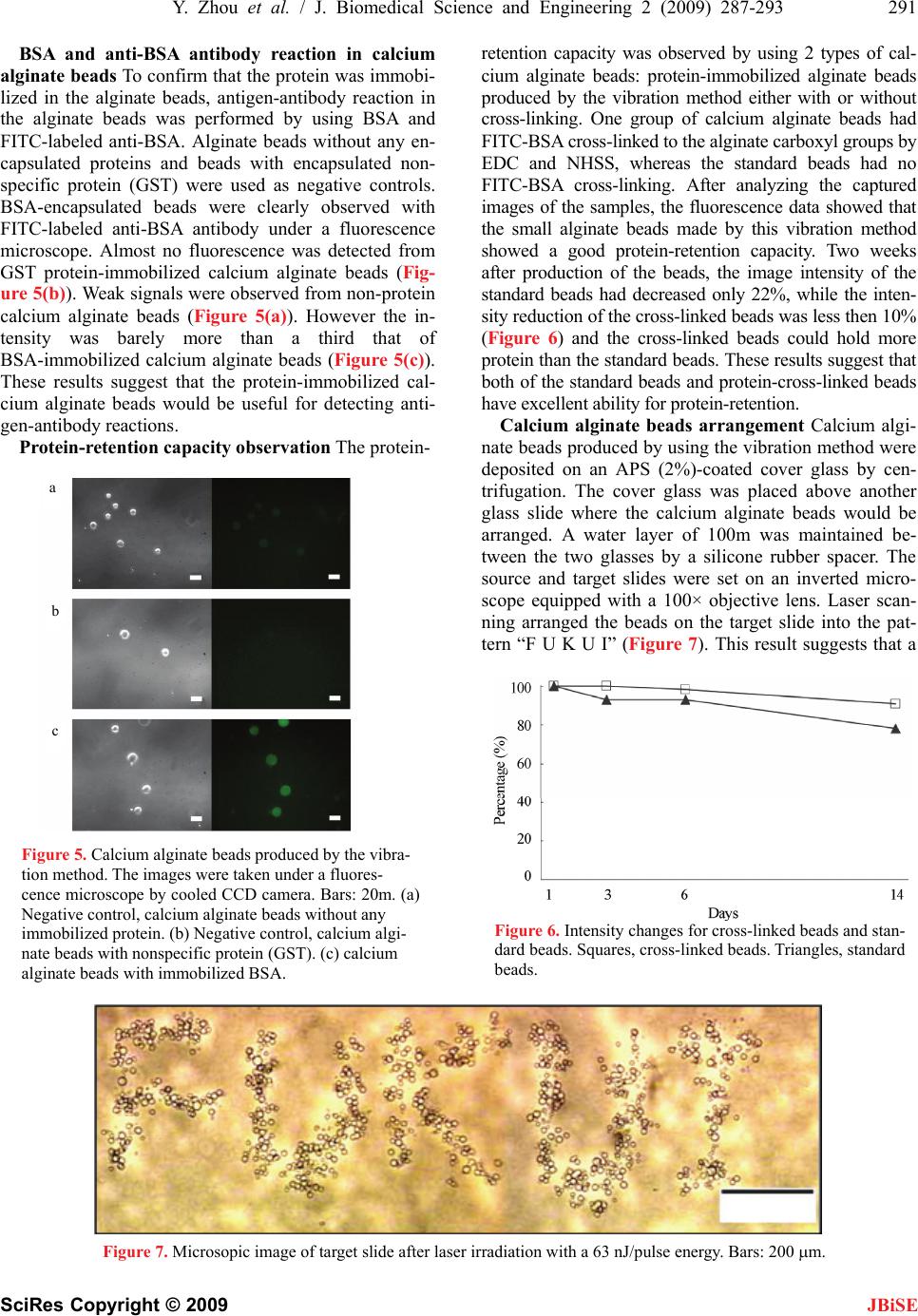
292 Y. Zhou et al. / J. Biomedical Science and Engineering 2 (2009) 287-293
SciRes Copyright © 2009 JBiSE
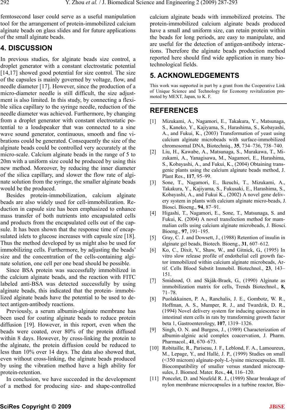
femtosecond laser could serve as a useful manipulation
tool for the arrangement of protein-immobilized calcium
alginate beads on glass slides and for future applications
of the small alginate beads.
4. DISCUSSION
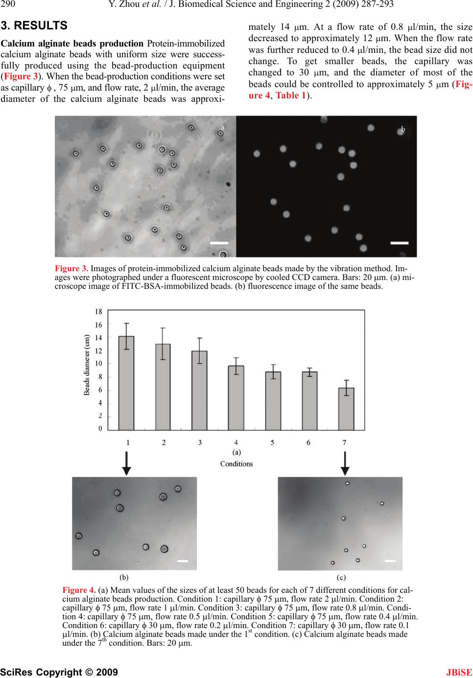
In previous studies, for alginate beads size control, a
droplet generator with a constant electrostatic potential
[14,17] showed good potential for size control. The size
of the capsules is mainly governed by voltage, flow, and
needle diameter [17]. However, since the production of a
micro-diameter needle is still difficult, the size adjust-
ment is also limited. In this study, by connecting a flexi-
ble silica capillary to the syringe needle, reduction of the
needle diameter was achieved. Furthermore, by changing
from a droplet generator with constant electrostatic po-
tential to a loudspeaker that was connected to a sine
wave sound generator, continuous, smooth and fine vi-
brations could be generated. Consequently the size of the
alginate beads could be controlled very accurately at the
micro-scale. Calcium alginate beads in the range of 5 to
20m with a uniform size could be produced by using this
new method. Moreover, by reducing the inner diameter
of the silica capillary, and slower the flow rate of algi-
nate solution from the syringe, the smaller alginate beads
would be the produced.
Besides protein-immobilization, calcium alginate
beads are also widely used for cell-immobilization. Re-
duction in capsule size has been emphasized to enhance
mass transfer of both nutrients into encapsulated cells
and products from the encapsulated cells out of the cap-
sule. It has been shown that the response time of encap-
sulated islets to glucose increases with capsule size [18].
Thus the method developed by us might also be used for
immobilizing cells. Furthermore, by adjusting the beads’
size and the concentration of the cells-containing algi-
nate solution, one cell per one bead should be possible.
Since BSA protein was successfully immobilized in
the calcium alginate beads, and the reaction with FITC
labeled anti-BSA was detected successfully by using
alginate beads, this indicated that the protein- immobi-
lized alginate beads have the potential to be used to de-
tect antigen-antibody reactions.
Previously, a serum albumin-alginate membrane has
been used for coating alginate beads to reduce protein
diffusion [19]. However, in this report, even when the
beads were coated, over 80% of the protein diffused
within 8 days. However, by cross-linking the protein to
the alginate, the protein diffusion could be reduced to
less than 10% over 14 days. The data also showed that,
even without cross-linking, the alginate beads produced
by using the vibration method have a high ability for
protein-retention.
In conclusion, we have succeeded in the development
of a method for producing size- and shape-controlled
calcium alginate beads with immobilized proteins. The
protein-immobilized calcium alginate beads produced
have a small and uniform size, can retain protein within
the beads for long periods, are easy to manipulate, and
are useful for the detection of antigen-antibody interac-
tions. Therefore the alginate beads production method
reported here should find wide application in many bio-
technological fields.
5. ACKNOWLEDGEMENTS
This work was supported in part by a grant from the Cooperative Link
of Unique Science and Technology for Economy revitalization pro-
moted by MEXT, Japan, to K. F.
REFERENCES
[1] Mizukami, A., Nagamori, E., Takakura, Y., Matsunaga,
S., Kaneko, Y., Kajiyama, S., Harashima, S., Kobayashi,
A., and Fukui, K., (2003) Transformation of yeast using
calcium alginate microbeads with surface-immobilized
chromosomal DNA, Biotechniq., 35, 734–736, 738–740.
[2] Liu, H., Kawabe, A., Matsunaga, S., Murakawa, T., Mi-
zukami, A., Yanagisawa, M., Nagamori, E., Harashima,
S., Kobayashi, A., and Fukui, K., (2004) Obtaining trans-
genic plants using the calcium alginate beads method, J.
Plant Res., 117, 95–99.
[3] Sone, T., Nagamori, E., Ikeuchi, T., Mizukami, A.,
Takakura, Y., Kajiyama, S., Fukusaki, E., Harashima, S.,
Kobayashi, A., and Fukui K., (2002) A novel gene deliv-
ery system in plants with calcium alginate micro-beads, J.
Biosci. Bioeng., 94, 87–91.
[4] Higashi, T., Nagamori, E., Sone, T., Matsunaga, S. and
Fukui, K. (2004) A novel transfection method for mam-
malian cells using calcium alginate microbeads, J. Biosci.
Bioeng., 97, 191–195.
[5] Gray, C. J. and Dowsett, J., (1988) Retention of insulin in
alginate gel beads, Biotech. Bioeng., 31, 607–612.
[6] Ko, C., Dixit, V., Shaw, W., and Gitnick, G., (1995) In
vitro slow release profile of endothelial cell growth fac-
tor immobilized within calcium alginate microbeads, Ar-
tif. Cells Blood Substit Immobil. Biotechnol., 23, 143–
151.
[7] Smidsrød, O. and Skjåk-Braek, G., (1990) Alginate as
immobilization matrix for cells, Trends Biotechnol., 8,
71–78.
[8] Puolakkainen, P. A., Ranchalis, J. E., Gombotz, W. R.,
Hoffman, A. S., Mumper, R. J., and Twardzik, D. R.,
(1994) Novel delivery system for inducing quiescence in
intestinal stem cells in rats by transforming growth factor
beta 1, Gastroenterology, 107, 1319–1326.
[9] Singh, O. N. and Burgess, J., (1989) Characterization of
albumin-alginic acid complex coacervation, J. Pharm.
Pharmacol., 41, 670–673.
[10] Robitaille, R., Pariseau, J. F., Leblond, F. A., Lamoureux,
M., Lepage, Y., and Hallé, J. P., (1999) Studies on small
(<350 microm) alginate-poly-L-lysine microcapsules. III.
Biocompatibility of smaller versus standard microcap-
sules, J. Biomed. Mater. Res., 44, 116–120.
[11] Poncelet, D. and Neufeld R. J., (1989) Shear breakage of
nylon membrane microcapsules in a turbine reactor, Bio-
Y. Zhou et al. / J. Biomedical Science and Engineering 2 (2009) 287-293 293
SciRes Copyright © 2009 JBiSE
technol. Bioeng., 5, 95–103.
[12] Sakai, S., Ono, T., Ijima, H., and Kawakami, K., (2000)
Synthesis and transport characterization of alginate/
aminopropyl-silicate/alginate microcapsule: application
to bioartificial pancreas, Biomater., 22, 2827–2834.
[13] Srivastava, R. and McShane, M. J., (2005) Application of
self-assembled ultra-thin film coatings to stabilize mac-
romolecule encapsulation in alginate microspheres, J.
Microencapsul., 22, 397–411.
[14] Wang, S. B., Chen, A. Z., Weng, L. J., Chen, M. Y., and
Xie, X. L., (2004) Effect of drug-loading methods on
drug load, encapsulation efficiency and release properties
of alginate/poly-L-arginine/chitosan ternary complex
microcapsules, Macromol. Biosci., 4, 27–30.
[15] Timkovich, R., (1997) Detection of the stable addition of
carbodiimide to proteins, Anal. Biochem., 179, 135–143.
[16] Grabarek, Z. and Gergely, J., (1990) Zero-length cross-
linking procedure with the use of active esters, Anal.
Biochem, 185, 131–135.
[17] Strand, B. L., Gåserød, O., Kulseng, B., Espevik, T., and
Skjåk-Baek, G., (2002). Alginate-polylysine-alginate
microcapsules: effect of size reduction on capsule prop-
erties, J. Microencapsul., 19, 615–30.
[18] Chicheportiche, D. and Reach, G., (1988) In vitro kinet-
ics of insulin release by microencapsulated rat islets: ef-
fect of the size of the microcapsules, Diabetologia., 31,
54–57.
[19] Hurteaux, R., Edwards-Lévy, F., Laurent-Maquin, D.,
and Lévy, M. C., (2005) Coating alginate microspheres
with a serum albumin-alginate membrane: application to
the encapsulation of a peptide, Eur. J. Pharm. Sci., 24,
187–197.