 Journal of Biosciences and Medicines, 2016, 4, 33-37 Published Online March 2016 in SciRes. http://www.scirp.org/journal/jbm http://dx.doi.org/10.4236/jbm.2016.43006 How to cite this paper: Nagayama, K., Kurihara, T., Amano, Y. and Tanahashi, M. (2016) Particle Simulation of Skin Basal Layer Formation. Journal of Biosciences and Medicines, 4, 33-37. http://dx.doi.org/10.4236/jbm.2016.43006 Particle Simulation of Skin Basal Layer Formation Katsuya Nagayama1, Takeshi Kurihara1, Yasuko Amano2, Masanori Tanahashi2 1Kyushu Institute of Technology, Fukuoka, Japan 2Kao Corporation, Tokyo, Japan Received 2 October 2015; accepted 10 March 2016; published 17 March 2016 Abstract There has been increasing concern regarding the cosmetic aspects of skin in recent years. Compu- tational simulation can be useful in understanding the mechanism underlying skin formation. The bottom of the epidermis is called the basal layer and is very undulation. In this study, we focus on the basal layer formation. We created a particle model, which forms an undulation basal layer and regenerates the basal layer formation by numerical simulation. At first, two-dimensional basal layer formation without epidermal turnover was simulated. The results showed film shape changes and the stability, as a layer in the process of long-ti me with an increase and decrease of basal cells. Next, the model was applied to three-dimensional basal layer formation with epidermal turnover. As the structure of the basal layer was deformed, the upper structure of the epidermis comprising the cells divided from the basal layer also became irregular. The simulation results accurately represented and reproduced the three-dimensional basal layer formation and epidermis turnover process. Keywords Numerical Simulation, Skin Formation, Particle Model, Basal Layer 1. Introduction Skin is the largest organ of the human body. We can diagnose epidermal conditions and provide appropriate care particularl y because the epidermis is the most external part of the skin. In recent years, there has been an in- creasing concern regarding the cosmetic aspects of skin care in both men and women, prompting research stu- dies on anti-ageing therapy and cosmetics. The epidermis consists of four differentiated layers. In particular, the basal layer, which is at the bottom of epidermis, is undulationly formed. This undulationness is considered to be associated with spots and ageing. Therefore, it is important to elucidate the mechanism of the formation of an undulation basal layer. However, there are many unexplained phenomena in skin metabo lis ma nd the formation of undulation layers because the in situ observation of basal layer formation is difficult. Computational simulation can be useful in further understanding the mechanisms of skin development, and 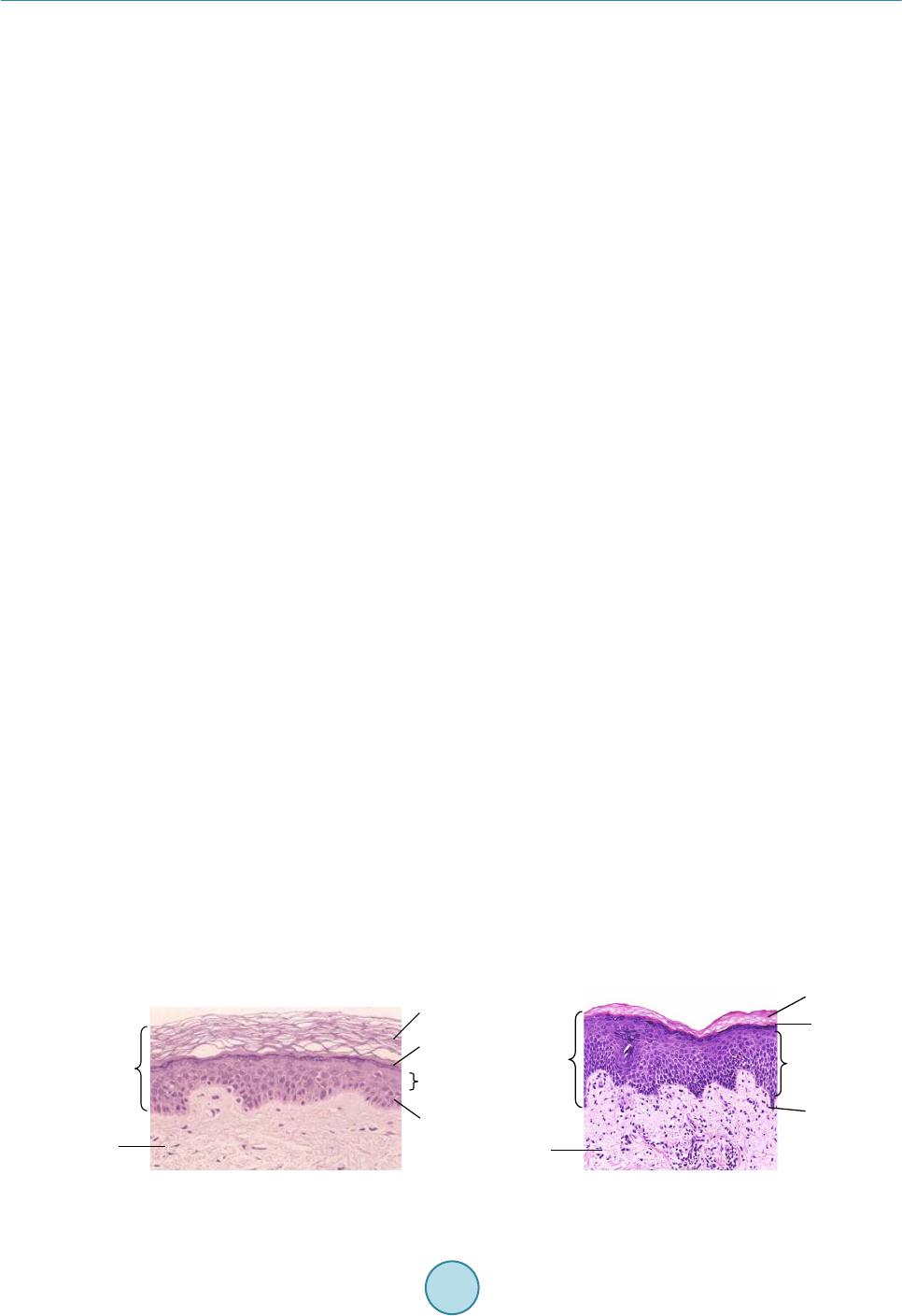 K. Nagayama et al. several models have been proposed [1]-[4]. However, the analysis of the epidermis requires an ability to not on- ly increase and decrease epidermal cells but also to assess the transition of cell shape and physical properties. For this reason, previous methods are not good at analyzing all the four layers of the epidermis with their re- spective characters, simultaneously. Thus, in this study, we propose a particle model that can handle complex biological phenomena, including cell interactions such ascell division, motion, deformation, and transition [5]-[7]. Furthermore, we believe that it is a suitable method for simulating skin formation. We developed an analytical method for studying the formation and turnover process of the skin using the particle model [8]-[10]. This study conducted the numerical simulation of cutification, which includes basal layer undulation ness, us- ing the particle method. The particle method can simulate three-dimensional skin formation with cornification and by changing physical properties, whereas also being able to increase and decrease basal cell production. Our aim was to elucidate the phenomena of epidermis formation using this model to contribute to medical skin treatment and the development of cosmetics. 2. Analysis Object and Model Description 2.1. Analysis Object Figure 1 depicts a cross-section of the skin [11], and the roles of each cell layer are described. The epidermis is the outermost layer of the skin and is primarily composed of cells called keratinocytes. The epidermis consists of four layers. A basal layer, which is the lowest layer of the epidermis, provides new cells by dividing each day. The dividing cells are called the prickle layer, which are pushed and moved up toward the skin surface, trans- forming into the granular layer and stratum corneum, which finally detaches from the skin surface. Skin cells change not only in their shape but also in their physical properties during this process. This process is referred to as turnover and occurs at approximately 4-week intervals. Dermis is located under the epidermis, and is divided by the basal layer. Furthermore, capillaries in the dermis supply nutrition and oxygen for basal cells. 2.2. Model Description The particle model [5]-[7] is introduced to simulate the epidermis formation process [8]-[10]. The model con- siders the interaction between the particles and pursues motions of the particles in a Lagrangian way. This me- thod is suitable for analysis with large deformations, or when the numbers of calculation points are changing. The cellular particles move in response to inter-particle forces, such as volume conservation force and spring force. The volume conservation force works to keep the distance between the particles. Because of the repulsive force, particles eventually move to a stable distance. The spring force works to make the continuum of the cel- lular particles structural. Spring force has already introduced cornification into our model [8]-[1 0]. In this paper, we describe the application of spring force to the basal layer. The actual basal layer maintains a layer with film shape, which includes undulationness. To introduce this film shape to our model, we use spring force to produce intervals in basal cells. Moreover, the distance which spring force works to is variable because it can fill the gaps generated by the basal layer. By summing up these forces from the surrounding particles, the particles gradually move to the position of the force balance. In addition, each basal cell is a stem cell and can divide into two daughter cells. This division has three pat- terns as shown in Figure 2 [12] [13]. Pattern.1 is where both cells become basal cells, and Pattern.2 is where one cell remains as a basal cell and another cell becomes a prickle cell, while in Pattern.3, both cells change into prickle cells. Each basal cell follows a pattern among these three patterns at random. We modeled the basal layer Figure 1. Cross-section of the skin [11]. Corneum Granular layer Prickle layer Basal layer Epidermis Dermis Corneum Granular layer Prickle laye r Basal laye r Epidermis Dermi s 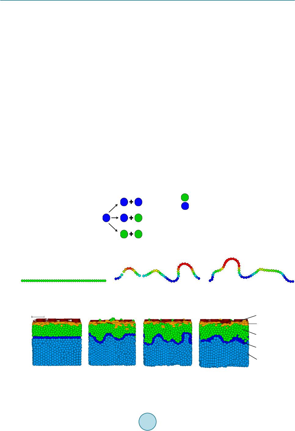 K. Nagayama et al. using function of film shape and stem cells having abilities to follow three patterns. When basal cells increase, the film shape suffers a change, because the increase in basal cells presses them against other cells. Besides, when basal cells decrease, surrounding basal cells fill the gap and keep the film shape. 3. Results and Discussion Two cases of simulation were performed. One is two-dimensional basal layer formation without epidermal turn- over to check the basal layer shape. Another is three-dimensional basal layer formation with epidermal turnover to simulate close to real phenomena. 3.1. Two-Dimensional Basal Layer Formation Figure 3 presents the simulation results of two-dimensional basal layer formation without epidermal turnover. Figures 3(a)-(c) are taken after 45, 100, and 1000 days of analysis time, respectively. Red colour indicates that the particle is moving upward, light green indicates it is stationary, a nd blue indicates it is moving downward. The results show film shape changes and the stability, as a layer in the long-time process with an increase and decrease of basal cells. 3.2. Three-Dimensional Basal Layer Formation with Epidermal Turnover Figure 4 and Figure 5 present the simulation results of three-dimensional basal layer formation with epidermal turnover. Figures 4(a)-(d) are taken after 45, 100, 300, and 1000 days of analysis time, respectively. From the basal layer (blue), prickle cells (green) split and move up, which then develop into the granular layer (yellow). Finally, the corneum (red) peels off. The cells in the course of the spinous layer transform within the stratum corneum, from spherical-shaped into elliptical-shaped cells, by extending in the horizontal direction to form the skin. Figure 4 shows formation of natural undulationness of the basal layer and the stability of a film shape as a Figure 2. Cell division of basal layer cell [12] [13] . (a) (b) (c ) Figure 3. Results of two-dimensional basal layer formation simulation. (a) 45 days; (b) 100 days; (c) 1000 days. (a) (b) (c) (d) Figure 4. Results of three-dimensional skin formation simulation (Side view). (a) 45 days; (b) 100 days; (c) 300 days; (d) 1000 days. Basal cell Prickle cell 8% 84% 8% Probability Pattern.1 Pattern.2 Pattern.3 50[μm]Corneum Granular layer Dermi s Prickle layer Basal layer 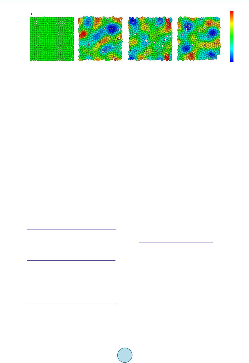 K. Nagayama et al. (a) (b) (c) (d) Fig ure 5. Results of three-dimensional skin formation simulation (Top-down view of basal layer). (a) 45 days; (b) 100 days; (c) 300 days; (d) 1000 days. layer in the process of long-time epidermal formation with an increase and decrease of basal cells. Moreover, the basal layer plays a role in the film function because particles do not get mixed up between the prickle layer and dermis. Figure 5 only displays the basal layer of the same model as Figure 4. However, it should be noted that this result is the top-down view of the analysis domain. Although there are partial biases by increasing and de- creasing the basal cell layer, particles are placed uniformly in the whole of the basal layer. In addition, undula- tionness is formed all over the field. 4. Conclusion In this study, the basal layer formation and the epidermis turnover were modeled using the particle model. We created a particle model which forms an undulation basal layer and regenerates the basal layer formation by nu- merical simulation. At first, two-dimensional basal layer formation without epidermal turnover was simulated. The results showed film shape changes and the stability, as a layer in the long-time process with an increase and decrease of basal cells. Next, the model was applied to three-dimensional basal layer formation with epidermal turnover. As the structure of the basal layer was deformed, the upper structure of the epidermis comprising cells divided from the basal layer also became irregular. The simulation results accurately represented and reproduced the three-dimensional basal layer formation and epidermis turnover process. References [1] Querleux, B. (2014 ) Computational Biophysics of the Skin. Pan Stanford Publishing. [2] Sütterlin, T., Huber, S., Dickhaus, H. and Grabe, N. (2009) Modeling Multi-Cellular Behavior in Epidermal Tissue Homeostasis via Finite State Machines in Multi-Agent Systems. Bioinformatics, 25, 2057-20 63. http://dx.doi.org/10.1093/bioinformatics/btp361 [3] Hirashima, T., Hosokawa, Y., Iino, T. and Nagayama, M. (2013) On Fundamental Cellular Processes for Emergence of Collective Epithelial Movement. Biology Open, 2, 600-666. http://dx.doi.org/10.1242/bio.20134523 [4] Adra, S., Sun, T., MacNeil, S., Holcombe, M. and Smallwood, R. (2010) Development of a Three Dimensional Mul- tiscale Computational Model of the Human Epidermis. Open Journal PLoS ONE, 5, e8511. http://dx.doi.org/10.1371/journal.pone.0008511 [5] Nagayama, K., Nitta, J. and Miura, I. (2009) Numerical Analysis on Angiogenesis in Cancer Using a Particle Model. Theoretical and Applied Mechanics Japan, 58, 321-32 4. [6] Hashiguchi, S. and Nagayama, K. (2011) Construction of a Hair Formation Analysis Method Using the Particle Model, 2011 Bioengineering Conference, 23th Japan Society of Mechanical Engineers, Japan, 355-356. [7] Naga ya ma, K., Matsuoka, S., Morisaki, N. and Taguchi, H. (2015) 3D Numerical Simulation of Hair Formation Process Using a Particle Model. Open Journal of Regenerative Medicine. http://file.scirp.org/Html/1-2390044_53849.htm [8] Uehara, T., Nagayama, K., Amano, Y. and Tanahashi, M. (2013) Numerical Simulation of Epidermis Formation Using Particle Model. The Second BMIRC International Symposium on Frontiers in Computational Systems Biology and Bioeng ineeri ng, Japan, 30 January 2013. [9] Amano, Y., Uehara, T., Tanahashi, M. and Nagayama, K. (2012) An Epidermal Turnover Model Predicts Undulation  K. Nagayama et al. Skin Tone with Aging. SY01.01ICB S (World Congress on the International Society for Biophysics and Imaging of the Skin), Copenhagen, 29 November 2012. [10] Nagayama, K., Uehara, T., Amano, Y. and Tanahashi, M. (2015) 3D Numerical Simulation of Epidermis Turnover Process Using a Particle Model. Journal of Biosciences and Medicines. http://dx.doi.org/10.4236/jbm.2015.33007 [11] Shimizu, H.S. (2011) Textbook of Modern Dermatology. Nakayamashoten Co. 38 , Japan. [12] Clayton, E., Doupe, P.D., Klein, M.A., Winton, J.D., Simons, D.B. and Jones, H.P. (2007) A Single Type of Progenitor Cell Maintains Normal Epidermis. Nature, 446, 18 5 -189. http://dx.doi.org/10.1038/nature05574 [13] Jones, H.P., Simons, D.B. and Watt, M.F. (2007) Sic Transit Gloria: Farewell to the Epidermal Transit Amplifying Cell? Cell Stem Cell Review, 1, 371-381. http://dx.doi.org/10.1016/j.stem.2007.09.014
|