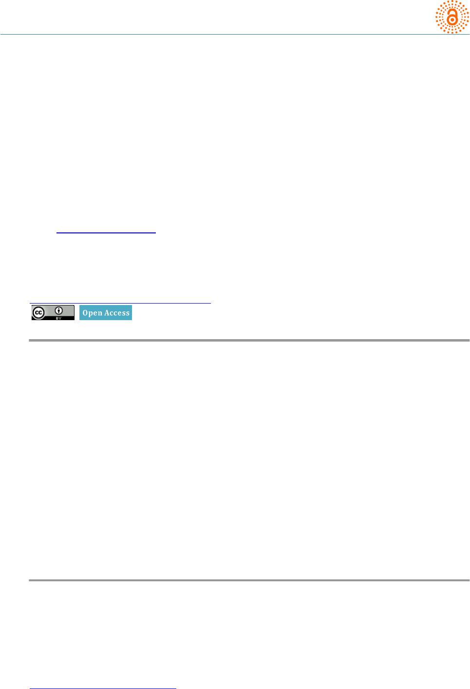
Open Access Library Journal
How to cite this paper: Latha, S., Venkata Sai, P.M. and Bagadi, R.K. (2014) Ultrasound Guided Saline Hydrostatic Reduction:
A Non-Surgical Procedure for the Management of Intussusception in Children. Open Access Library Journal, 1: e644.
http://dx.doi.org/10.4236/oalib.1100644
Ultrasound Guide d Saline Hydrostatic
Reduction: A Non-Surgical Procedure for
the Management of Int us s us cepti on in
Children
Suvarna Latha1, P. M. Venkata Sai1, R. K. Bagadi2
1Depart men t of Radiology, Sri Ramachandra University, Porur, Chennai, India
2Pediatric Surgery, Sri Ramachandra University, Poru r, Chennai, India
Email: kiranlathap@yahoo.co.in
Received 3 April 2014; revised 13 May 2014; accepted 3 July 2014
Copyright © 2014 by authors and OALib.
This wor k is licens ed under the Creati ve Commons Attribution International License (CC BY).
http://creativ ecommon s.org/l icens es/by/4.0/
Abstract
Introductio n: Intus suscep tion is the most com mon cau se of intestinal ob s tr u ction in infant &
toddler age group. Intussusceptio n is the telesc oping of one segment of bow el into the conti gu ou s
distal segment. Peak age i ncid ence is be tween 2 mo nth s to 3 yea rs of l ife. Obje ctive of the study: To
prove the efficacy of Ultrasound guided saline hydrostati c reductio n, a non surgical, non radiati o n
method in the manageme nt of intussu scept ion in children. Material s and meth ods : Fifty -three
children in th e a ge group of 2 mon th s to 3 yea rs with cli nical diag nosis of intussusception on
whom the Ultras o n og r am (USG ) guided hydros ta tic redu ctio n with norm al saline were attemp te d .
500 - 1000 ml of no rmal s aline w as used for reducti on. High r esol uti on USG (7 .5 MHz) w as used to
assess the p rog ress of r edu ction during the p roce dure. Results: Our initial ex p erience in the l as t
six years from April 2007 to Marc h 2013 sh owed succ essful r ed uc ti on in 45 chil dren ou t of 53 at-
tempted red u cti o ns .
Keywords
Intussuscep tion, H ydro st atic Redu ct ion, Ultrasonography (USG)
Subject Areas: Clinical Trials, P edia trics
1. Introduction
Intussusception is one of the common causes of intestinal obstruction in children with peak incidence between 2
mont hs to 3 years of age [1]. Intussusception is in vagi natio n of a segment of proximal bowel into the c onti guous
S. Latha et al.
OALibJ | DOI:10.4236/oalib.1100644 2 July 2014 | Volume 1 |
e644
lume n of distal bowel loop. The proxi mal telescoping loop is called as intussusceptum and the d istal l oop is
called as intu s suscipiens [1]. When this happens, the mesentery and blood vessels, which have telescoped along
with the intussusceptum, will get compressed in the outer loop resulting in ische mia and edema of the bowel
wall pre senting wi th severe pai n.
If intussusception is not diagnosed and not relieved, it may result in complica tions like bowel wall necrosis
and perforation.
Fifty percent are affected in the first year of life [1] and 24% presentin g in the second year. Approximately
2/3 of patients are boys. Most common cause of intussusception in this age group is idiopathic.
If intussusception occurs outside the above menti oned age group, p ossibility of pathologic lead point is to be
suspected as a cause of int uss usc epti ons (Figure 1).
Symptoms
In our study, 35 children presented with abdominal pain and palpable abdominal mass. Eigh t children presented
with features of intestinal obstruction, anot he r ten children presented with a combination of abdominal pain and
bloody diarrhea.
2. Materials and Met hod s
The study incl udes the patients for a period of 6 years, from April 2007 to March 2013.
A total number of 53 cases clinically diagnosed as intussusception and confir med by ultrasonographic ap-
pearance using a linear array transducer of 7.5 - 10 MHz using ALOKA SSD 5500 Ultrasound machine.
Patients were evaluated furt her for viabilit y of the bowel by demonstrating the va sc ul arity using color Dopp-
ler.
The following criteria were used to decide the suitabili ty of the patient for the Ultrasound guided sali ne hy-
drostatic reduct i on.
2.1. Inclusi on Criteria
1) Patients presenting within 48 hours of onset of symptoms.
2) Presence of palpable mass for more than 6 hours .
3) Absence of signs of perforation and perito nitis, including no radiographic evidence of free intraperitoneal
air (especially in whom lar ge amount of free fluid was seen in the peritoneum).
2.2. Exclusion Criteria
Absolute cont ra indications are perforation, peritonitis, sever e dehydration and hypovolemic shock.
Other contra indications include:
Figure 1. Intussusception is telesco ping of one loop of bowel
in to the other. Herniating loop is called intussusceptum and
the distal loop is called Intussuscipiens.
S. Latha et al.
OALibJ | DOI:10.4236/oalib.1100644 3 July 2014 | Volume 1 |
e644
Absence of vascul arity on color Doppler imaging;
Appearance of “Dissection sign”;
Presence of large amount of free fluid in the peritoneal cavity.
2.3. Procedure
The patie nt’s parents were explained about the procedure in detail and informed consent was obtained.
The pediatric surgi cal tea m was also present in ultras ound room throu ghout the proced ure.
The child was well hydrated before the procedure, for the best resul t s [1].
A 16 F Foley’s catheter introduced into the rectum with the child put on left lateral position, after lubric ating
tip of the catheter with 2% Lignocaine.
The Foley’s bul b was the n inflated with about 10 - 15 ml of normal sali ne. The but t ocks taped t ogethe r tightly
to provide a seal. The other end of the catheter was connected to a bottle of normal saline (at normal body tem-
perature) thro ugh an intr avenous l ine a nd the saline bag was suspended at more than 100 cm above the bed level
and normal saline wa s allowed to flow into the rectum under gent l e hydrostatic pressure.
Distending colonic loops and grad ual passa ge of saline into the ileal loops monitor ed under real time Ultra-
sonography [1]-[3]. Complete red uction was assumed onc e t he passage of saline through the ileo-cecal valve in-
to the terminal ileum was seen (Figure 3). Saline flow was allowed into several loops of ileu m. 500 - 1000 ml of
saline was used depending up on the size of the patient. The amount of saline required was recorded in each
case.
Once red uction was found to be achieved, the saline was drained by lowering t he saline b ag below the level of
the bed. Following this, Foley’s bulb was deflated and the catheter was removed. Clinical condition of the pa-
tient was closely and carefully monitored thr oughout t he procedure (Figure 2 and Figure 3).
If the intussusception is not reduced after a 3 minutes of sustained p ress ure, the sa l ine pressure has to be lo-
wered and child should be rested for 3 min . Thr ee such attempts can be made before consi der ing the intussus -
ception irred ucib le and sending the patient for surgi cal management.
Procedure should be abandoned if the saline d issects between the la ye rs of intuss uscepti on. This sign is de-
scribed as “dissection sign” . Which indicates that thereduction is less likely” to happen by nonsurgical method.
[4].
Afte r the procedure the patie nt was sh ifted to observation room under pediatric ia n ’s super vi si on. A co urse of
antibiotic therapy was started. In each case follo w-up, High Resolution Ultraso und was done after 24 hours to
rule o ut recurrence [5].
3. Results
Fort y-five cases out of fifty-three were succ essfully reduced using sal ine hydrostatic re d uction under ultras ono -
graphy guidance. In seven children in whom, the Ultrasonogr am (USG) showed an appearance of ileoileoco lic
Figure 2. Intussusception: Ultrasound image of the colon during the
procedure. Colonic loop distending with gradual passage of saline.
S. Latha et al.
OALibJ | DOI:10.4236/oalib.1100644 4 July 2014 | Volume 1 |
e644
Figure 3. Intussusception: Ultrasound image during the procedure.
Saline is distending the ileo-caecal region and outlining the ileo-
caecal valve. Complete reduction was assumed once the passage of
saline through the ileo-caecal val ve into the terminal ileum was
seen.
intussuscep tion (double loop intuss usceptio n), ultrasound guided reduction was attempted in view of positive
incl us i on crite r ia . Real time monitoring showed accumulation of Saline between the loops of intussusceptum
and intussuscip iens. No saline entered into the ileal loops even after introducing around 500 ml, at 3 safe suc-
cessive attempts of 3 minutes each suggest i ng the need for surgi c al ma nagement. Pr oced ure abandoned and the
reduction was achieved surgically.
Bowel perforation occurred in one patient, which was eventually managed surgic a l ly.
4. Discussion
The re are several types of intuss uscep tion described depending upon the anatomical parts of the bowel i nvolve d,
such as (a) Ileo Colic, (b) Ileo Ileal, (c) Colo Colic, (d) Ileo Ileo Colic, (e) multiple and (f) retrograde.
The Ileo Colic t ypes of Intussusceptions are the commonest type constituting about 90% of cases [1].
4.1. Incidence
Majo r ity of Int ussus c epti ons are idiopathic. 50% occur in first year and two-thirds of the patients are boys.
Outside this age gr oup there is an increased incid ence of lead points e.g. polyps, meckel’s diver ticulum, non-
Hodgkin’s lymp homa, duplication cyst .
4.2. Clinical Presentation
1) Intermittent colicky abdominal pain is the most common presentation;
2) Palpable abdominal ma s s;
3) Intestinal obstruction;
4) Vomiting;
5) Diarrhea, sometimes with blood in the stool;
6) Some patients present with dehydration, hypovolemic shock and lethargy intermittent.
4.3. Imaging Evaluation
Plain X-ray abdomen may show a soft tissue mass a nd signs of intestinal obstruction, sometimes free air.
Presence of free air suggests perforation which is a contra indication for hydrostatic re duction.
Ultrasound on trans ve rse scan may show, a “doughnut sign”, central echogenic mucos a with sur r ounding
edematous wall of the i ntussusceptum or a “target sign”, co ncentric ri ngs of alternating sonolucency and echo-
genicit y. On longitudinal scan a “pseudo kidney sign” is evident.
S. Latha et al.
OALibJ | DOI:10.4236/oalib.1100644 5 July 2014 | Volume 1 |
e644
The s e characteristic signs are easily recognized at the leading edge of int ussus c epti ons (Figur e 4 and Figure
5).
Presence of large amount of free fluid in the peritoneal cavity is a contraindic a tion where as small amount is
common in intussusception and does not contraindicate the pr oced ure.
Color Doppler can detect the viability of involved bowel by sho wing presence or absence of flow.
Absence of blood flow in color Doppler stud y in the intuss us c e ptio n suggests bowel isc hemia and co rrelates
significantly with irre ducibility.
4.4. Treatment Options Available Are
Operative reduction
Non operative red uctio n using procedures like:
a) USG guided reduction using n ormal saline;
b) Fluoroscopy guided air enema reductio n.
USG guided reduction usi ng no rmal sali ne is the rece ntly adopted technique. This method uses about 500 -
1000 ml of normal sali ne at a heig ht of 100 - 130 cm, maint a i ning a constant hydrostatic pressure [1]-[7].
Figure 4. Intussusception: Doughn ut s i gn or Target sign. Ul-
trasound appearance of concentric layers of alternating echo-
genic and hypoechogenic bands.
Figure 5. Intussusception: Pseudo kidney sign. Ultrasound ap-
pearance of oblique longitudinal section of the intussuscepted
bowel. Echogenic mesentery herniated along th e Intussusci-
piens appears like hilum of the kidney.
S. Latha et al.
OALibJ | DOI:10.4236/oalib.1100644 6 July 2014 | Volume 1 |
e644
4.5. Non Operative Reduction Is Not Rec ommended in the Following
Absence of vascula r i ty on color Doppler imaging;
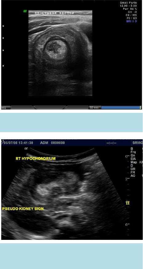
Appearance of “dissec tion sign”;
Presence of large a mount of free fluid in the peritoneal cavity;
No signs of reduction even after three attempts of three minutes each with an interva l r est period of 3 mi-
nutes [7].
However our experience showed that in the presence of pediatr ic surge ry team readily available in the proce-
dure room to deal with any complicatio ns if they arise and if the patie nt is able to tolerate the procedure, the
number of attempts can be increased up to 5 and the duration of each attempt can be pro l onge d up to 5 minutes.
4.6. Advantages
1) The procedure is simple, easily available, effective, and economical;
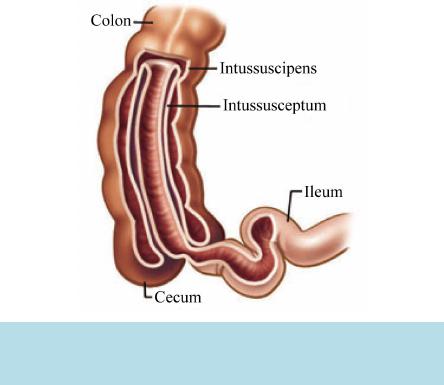
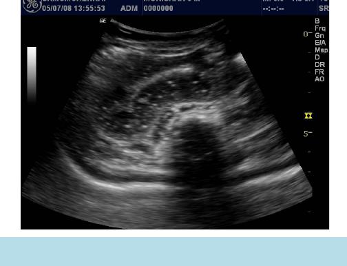
2) Facilitates Real time monito r i ng. This hel ps to observe and confirm complete reduction.
Also allows immediate identifica tion of perforation in case it happ ens.
3) Pre-procedural ultrasou nd and color Doppler assessment hel ps impro ving the percentage of positive out-
come of non surgica l manageme nt;
4) No radiation hazard;
5) Less morbidity;
6) Real ti me observation under ultra sound avoids the pitfall of pseudo-reduction as seen in fluoroscopic
guided procedures;
7) Disadva nt ages of air insuffla tion like fluct ua t i on of intra colonic pressure can be avoided.
Disadva nt ages of barium enema (messy procedure & Barium peritoni tis, if perforation occurs) are overcome
by saline reducti on.
8) Recurrence can also be treated by the same method;
9) Can be readily repeated in case of recurrent intuss usceptio n.
4.7. Limitations
1) The person performing the procedure needs to have expertise to perform real time ultrasound scan on a pe-
diatric patient;
2) The child usua lly struggl es d uring the sal i ne infusion due to discomfort, making t he real time ultra sound
more difficult;
3) Risk of perforation is high if the Foley’s balloon is inflated ;
4) Saline leaks though the anal canal if the Foley’s balloon is not inflated and adequate pressure will not be
built to reduce the intussuscepti ons.
5. Conclusions
No mor e complicatio ns! No more radiation hazard! No more mess!
USG-guided hydrostatic reduction of intussusceptio n in chil dre n using normal s a l ine is a simp le and cost ef-
fective technique wh ich requires minimal hospital stay.
USG guidance equipped with color Doppler helps proper patient selection which improves the success rate of
this non sur gi cal ma nagemen.
References
[1] Applegate, K.E. Intussusception, Chapter 108, Caffey’s Pediatric Diagnostic Imaging. 12th Edition.
[2] Bolia, A. A. (1985) Diagnosis and Hydrostatic Reduction off an Intussusception under Ultrasound Guidance—Case
Report. Clinical Radiology, 36, 655-657. http://d x.doi .o rg/10.1016/ S000 9-9260(85)80269-5
[3] Peh, W.C., Khon g, P.L. an d Chan, K.L. (1996 ) Sonographically Guided Hydrostatic Reduction of Childhood Intussus-
ception Using Hartmann’s Solution. American Journal of Roentgenology, 167, 1237-1241.
http://dx.doi.org/10.2214/ajr.167.5.8911188
[4] Fishman, M.C., Borden, S. and Cooper, A. (1984) The Dissection Sign of Nonreducible Ileocolic Intussusception.
S. Latha et al.
OALibJ | DOI:10.4236/oalib.1100644 7 July 2014 | Volume 1 |
e644
American Journal of Roentgenology (AJR), 143, 5-8. http://www.ncbi.nlm.nih.gov/pubmed/6610330
[5] Kumar, K., Hameed, S. and Umamaheswari (2006) Ultrasound Guided Hydrostatic Reduction in the Management of
Intussusception. The Indian Journal of Paediatrics, 73, 217-220.
[6] Khong, P.L., Peh, W.C. and Lam, C.H. (2000) Ultrasound -Guided Hydrostatic Reduction of Childhood Intussusception:
Technique and Demonstration. Radiographics, 20, 1818.
[7] Man esh, Y., Glover-Ad d y, H . and Twi re, V.E. (2011) Ultrasound Guided Hydrostatic Reduction of Intussusception in
Children at Korle Bu Teaching Hospital, an Initial Experience. Ghana Medical Journal, 45, 128-131.