Advances in Bioscience and Biotechnology
Vol.1 No.4(2010), Article ID:2857,5 pages DOI:10.4236/abb.2010.14039
Cellular effects of an aqueous solution of Losartan(R) on the survival of Escherichia coli AB1157 in the presence and absence of SnCl2, and on the physiological property (osmotic fragility) of the erytrocyte
![]()
1Centro de Ciências da Saúde, Faculdade de Medicina, Departamento de Biofísica e Fisiologia, Universidade Severino Sombra, Vassouras, Brazil;
2Departamento de Biofísica e Biometria, Instituto de Biologia Roberto Alcantara Gomes, Universidade do Estado do Rio de Janeiro, Rio de Janeiro, Brazil.
Email: thais.zaidan@gmail.com
Received 12 July 2010; revised 19 July 2010; accepted 25 July 2010.
Keywords: Losartan®; Surveillance; Escherichia Coli AB1157; Osmotic fragility; Erythrocyte
ABSTRACT
The angiotensin receptors type 1 (AT1) have affinity by Losartan®, low affinity to non-peptides antagonists and similar effect as Angiotensin-convert-enzyme inhibitors. It have been reported that natural and synthetic products might reduce the genotoxic and cytotoxic effects related to stannous chloride (SnCl2). SnCl2 is used in nuclear medicine as a reducing agent to obtain technetium-99 m-radiopharmaceuticals. The aim of this work was to evaluate the cellular effects produced by a solution of Losartan® (25 mg/ml) on the survival of Escherichia coli AB1157 in the presence and absence of SnCl2, and on the osmotic fragility of erythrocytes of the blood of Wistar rats. Briefly, blood sample was withdrawn by Wistar rats with heparinized syringe and incubated with Losartan® solution. Saline (NaCL 0.9%) was used as a control. The samples were gently mixed with hypotonic solutions of NaCl. After that it was centrifuged and the supernadant isolated for optical determination of the hemoglobin present. E. coli AB1157 cultures (exponential growth phase) were collected by centrifugation, washed and resuspended in 0.9% NaCl. Samples were incubated in water bath shaker with: (a) SnCl2 (25 μg/ml), (b) Losartan® (25 mg/ml) and (c) SnCl2 (25 μg/ml) + Losartan® (25 mg/ml). Incubation with 0.9% NaCl was also carried out (control). At 60 min intervals, aliquots were withdrawn, diluted, spread onto Petri dishes with solid LB medium and incubated overnight. The colonies formed were counted and the survival fractions calculated. Statistical analysis was performed. The results showed that there was a significantly increase (P < 0.05) in the osmotic fragility of the blood cells treated with Losartan®. Moreover, Losartan® was also able to protect the E. coli cultures against the lesive action of SnCl2. Although, in erythrocyte the osmotic fragility was increased by the presence of Losartan® that could 1) alter the physical properties of this cell, or 2) had a direct or indirect effect on the intracellular sodium concentration or 3) had acted on the cardiovascular system. It suggested that the Losartan® did interfere strongly with cellular metabolism and did alter the survival fractions of E. coli AB1157.
1. INTRODUCTION
Losartan® is an angiotensin II receptor antagonist drug used mainly to treat high blood pressure due to cardiovascular diseases. As with all angiotensin II type 1 receptor (AT1) antagonists, Losartan® is indicated for the treatment of hypertension [1]. Losartan® may also delay progression of diabetic nephropathy and is also utilized for the reduction of renal disease progression in patients with type-2 diabetes or hypertension or microalbuminuria or proteinuria [2]. Losartan® has been found to downregulate the expression of transforming growth factor beta (TGF-β) types I and II receptors in the kidney of diabetic rats, which may partially account for its nephroprotective effects [3]. Alterations on TGF-β expression may also account for its potential efficacy in Marfan syndrome and Duchenne muscular dystrophy (DMD)–Losartan® has been shown to prevent aortic aneurysm and certain pulmonary complications in a mouse model of the disease [4].
Blood contains many types of cells with very different functions, ranging from the transport of oxygen to the production of antibodies. The sodium-potassium pump has a direct role in regulating red blood cell (RBC) volume: It controls the solute concentration inside the cell, thereby regulating the osmotic forces that can make a cell swell or shrink [5]. The capability of RBC to resist hemolysis characterizes what is called the osmotic fragility (OF) of the membrane. The OF is classically used as a general screening procedure [6]. The “fragility curve” reflects the structural and geometrical changes in RBC. Hemolytic results from a structural perturbation of the RBC and its cytoskeleton caused by its high partition in the membrane [7,8].
Some deleterious effects of stannous chloride (SnCl2) have been described in humans, it has been reported that it is highly irritant to the mucous membrane and skin, although it presents low systemic toxicity [9]. In animals, it can produce stimulation or depression of the central nervous system [9]. As for bacterial assays, as experiments with Escherichia coli survival, SnCl2 appears to be capable of inducing and/or producing injuries in deoxyribonucleic acid (DNA), being considered as a potential genotoxic agent. These effects may be, at least in part, attributed to free radicals (FR), generated during SnCl2 treatment [10-13].
The aim of this work was to evaluate the cellular effects produced by a solution of Losartan® (25 mg/ml) on the survival of Escherichia coli AB1157 in the presence and absence of SnCl2, and on osmotic fragility of erythrocytes of the blood withdrawn from Wistar rats.
2. MATERIAL AND METHODS
2.1. Losartan® Preparation
The solution of Losartan® was prepared by a dilution of 250 mg of Losartan® dust in 10 ml of saline. The final concentration of Losartan® was considered 25 mg/ml.
2.2. Osmotic Fragility
Blood was withdrawn from rat Wistar (2.5 ml) with a heparinized syringe. The osmotic fragility evaluations of the RBC were performed (once) with from rat Wistar (2.5 ml) blood samples incubated with Losartan® (0.5 ml; 25 mg/ml) or with 0.15 M NaCl as a control, for 60 minutes at room temperature. The blood samples (100 µl) after treatment were gently mixed with 5ml of hypotonic NaCl solutions with concentrations from 0.02 to 0.12 M. After 30 min, these tubes were centrifuged at 3500 rpm/ 5 min and the supernatants were isolated to determine the optical density (OD) of the hemoglobin in a spectrophotometer (540 nm). It was performed 9 (triplicate) experiments total [8]. The results were compared with the control samples and statistical analysis was performed by independent t-test [8].
2.3. Bacterial Cultures
E. coli AB1157, a wild-type strain, proficient to repair damage in the DNA, was used in this work. From stock (in glycerol 50% v/v), a sample (50 µl) of the culture was grown on liquid LB medium (5 ml, Luria and Burrous, 1957) at 37°C overnight on a shaking water bath (reciprocal water bath shaker (Quimis Equipamentos Industriais Ltda, model 215.1, São Paulo, BR) up to the stationary growth phase.
2.4. Bacteria Inactivation
A sample (200 μl) was taken from this culture and further incubated (20 ml; liquid LB medium) under the same conditions to exponential growth (108 cells/ml). The cells were collected by centrifugation, washed twice in 10 ml of saline and suspended again in the same solution until they reached 108 cells/ml. Samples (1.0 ml) of these washed cultures (108 cells/ml) were incubated on the shaking water bath with 1) 0.5 ml of SnCl2 (75 μg/ml) and 0.1 ml of saline, or 2) 0.1 ml of Losartan® solution (25 mg/ml) and 0.5ml of saline, or 3) 0.1 ml of Losartan® solution (25 mg/ml) and 0.5ml of SnCl2 (75 μg/ml), or 4) 0.6 ml of saline as a control, on initial time and after 60 min, at 37°C. During the assay, at 0 and 60 min, aliquots (100 μl) were diluted with saline and spread onto Petri dishes containing solidified LB medium (1.5% agar). Colonies units formed, after overnight incubation at 37°C, were determined. The survival fraction was calculated dividing the number of viable cells obtained per ml in each time of the treatment (N) by the number of viable cells obtained per ml in zero time (N0).
2.5. Statistical Analysis
The ANOVA test following by Boferroni test was performed and the significance is accepted if P < 0.05. This statistical test was realized according the methodology showed by Cavalcanti et al., 2003 for the osmotic fragility test.
3. RESULTS
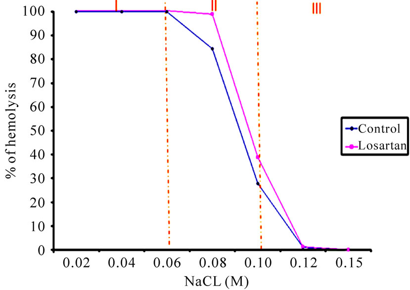
Figure 1 shows the osmotic fragility of the rats erythrocyte incubated with Losartan® solution when reacted with different NaCl hypotonic solutions. The curve tendency shows that in small concentrations of the salt, the hemolysis increased.
The results showed that there is a significantly increase (p < 0.05) in the osmotic fragility of the Losartan® treated cells on the curve interval between the 0.06 and 0.10 M

Figure 1. Osmotic fragility tendency of red blood cell, treated with Losartan®. A blood sample was withdrawn Wistar rats (2, 5 mL), rats with heparinized syringe and incubated with Losartan® (0.5 mL; 25 mg/mL) or with solution of sodium chloride (NaCl 0.15 M) a control, for 60 minutes at room temperature. Blood sample (25 mL), treated or not were gently mixed with 5 mL of hypotonic NaCl solutions with concentrations from 0.02 to 0.12 M after treatment or not, were gently mixed with hipotonic of NaCl (from 0.02 to 0.15 M), were centrifuged 3500 rpm/5 min and the supernatants were isolated to determine the optical density (OD) of the hemoglobin in a spectrophotometer (540 nm).
concentrations of NaCl, that is a hypotonic interval of the osmotic curve. The isotonic interval between 0.10 and 0.15 M of NaCl, the osmotic fragility also increased significantly (p < 0.05) in the presence of Losartan®.
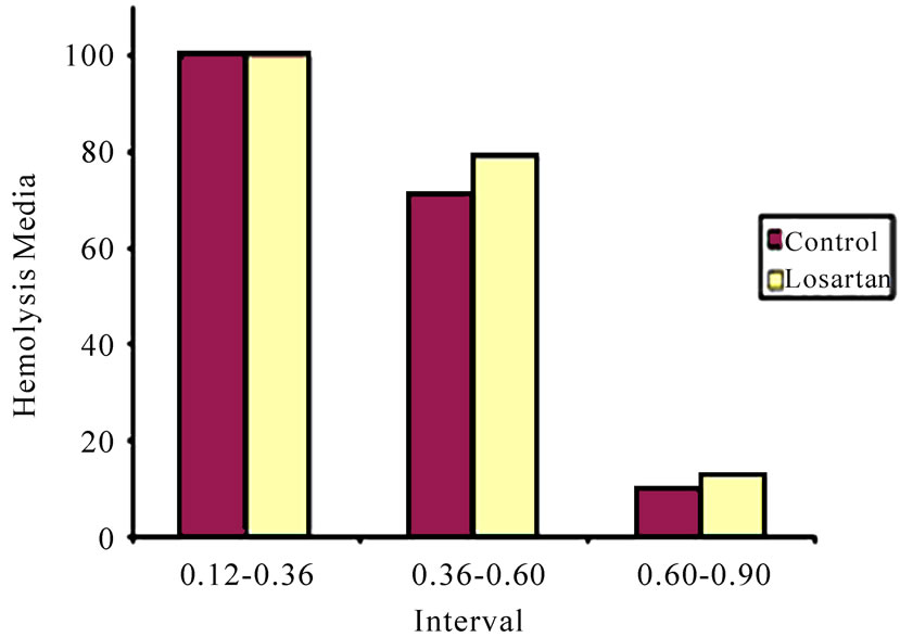
Figure 2 shows the mean of the osmotic fragility after analysis of the three NaCl concentrations intervals obtained of the osmotic curve of the Figure 1. The analysis of the results showed a significant statistical increase (p < 0.05) on osmotic fragility of erythrocyte incubated with Losartan® solution in the intervals 2 (0.06 until 0.10 M) and 3 (0.10 until 0.15 M).
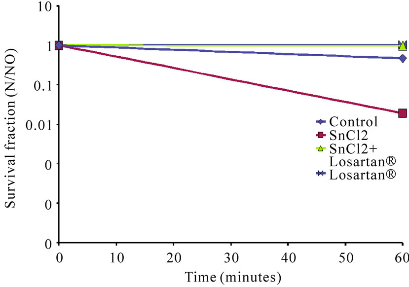
Figure 3 shows that Losartan® was able to protect the E. coli cultures against the lesive action of SnCl2. The Losartan® also did not interfere with the survival of the cultures. It suggested that the Losartan® did interfere strongly with cellular metabolism and did alter the survival fractions of E. coli AB1157
4. DISCUSSION
A large number of drugs that cause alterations on the shape and physiology of the red cells have been cited by some authors [14-17].
The results obtained with the quality comparison of the shape of the RBC (non treated and treated with natural extracts) under optical microscopy could justify the modifications in the uptake of 99 mTc for the red blood cells in the presence of Mentha crispa extract, similar to that observed with the extract of Maytenus iliciofolia

Figure 2. The osmotic fragility of red blood cells treated with Losartan®. The ANOVA test following by Boferroni test was performed and the significance is accepted if P < 0.05. This statistical test was realized according the methodology showed by Cavalcanti et al., 2003 for the osmotic fragility test.

Figure 3. Losartan®’s solution effect on the survival of E.coli AB1157 culture treated or not with SnCl2. Samples of E. coli cultures with 108 cells/ml were incubated on the shaking water bath with 1) SnCl2 and saline, or 2) Losartan® and saline, or 3) Losartan® and SnCl2, or 4) saline as a control, on initial time and after 60 min, at 37°C. During the assay, at 0 and 60 min, aliquots (100 μl) were diluted with saline and spread onto Petri dishes containing solidified LB medium. The survival fraction was calculated dividing the number of viable cells obtained in each time of the treatment (N) by the number of viable cells obtained in zero time (N0).
[18-22].
In the present study we have found that the RBC osmotic fragility was changed by presence of the Losartan® solution in the studied concentration. Figure 1 shows the osmotic fragility of the rats erythrocyte incubated with Losartan® solution when reacted with different NaCl hypotonic solutions. The curve tendency shows that in small concentrations of the salt, the hemolysis increased. The results reported that there is a significantly increase (p < 0.05) in the osmotic fragility of the Losartan® treated cells on the curve interval between the 0.06 and 0.10 M concentrations of NaCl, that is a hypotonic interval of the osmotic curve. The isotonic interval between 0.10 and 0.15 M of NaCl, the osmotic fragility also increased significantly (p < 0.05) in the presence of Losartan®.
Figure 2 presents the mean of the osmotic fragility after analyses of the three NaCl concentrations intervals obtained of the osmotic curve of the Figure 1. The analysis of the results showed a significant statistical increase (p < 0.05) on osmotic fragility of erythrocyte incubated with Losartan® solution in the intervals 2 (0.06 until 0.10 M) and 3 (0.10 until 0.15 M).
The findings presented in Figure 3 reveal that the studied Losartan® solution in the concentration did not presented a cytotoxic effect on the metabolism of E.coli AB1157. Although several cytotoxic properties have been related to SnCl2, humans may be exposed to stannous ions in several situations [23-24]. The results in Figure 3 reinforce the fact that SnCl2 exerted a lesive action on the survival of the culture of E. coli AB1157 in accordance with what had been previously reported by other authors [25-30].
Moreover, when treated simultaneously with SnCl2 and Losartan®, the E. coli cultures were protected against the effects of the reducing agent (SnCl2). Although, in other reports Losartan® can induce damage to bacterial culture [31] based in the protect effect studied in this work, we could hypothesized the evaluation the collateral effects of the Losartan® on the cardiovascular system, as well as, bacterial endocarditis and bacterial pericarditis.
5. CONCLUSIONS
Probably, components present in Losartan® solution could be altering 1) the erythrocyte membrane morphology, 2) the erythrocyte membrane ions transport or 3) the osmotic transport balance. The different alterations could be inducing the stronger osmotic fragility in isotonic concentrations of NaCl, causing hemolytic alterations as anemia, jaundice. The substance present in Losartan® solution could also has a protective action on the surveillance of E. coli AB1157 against the effects produced for the SnCl2 presence. Although, this work had been done with animals or bacterial culture, we suggest paying attention with cardiovascular diagnosis in patients who are undergoing Losartan®.
6. ACKNOWLEDGEMENTS
This work was supported by FUSVE, USS and UERJ.
REFERENCES
- Dahlöf , B., Devereux, R.B., Kjeldsen, S.E., Julius, S., Beevers, G., de Faire, U., Fyhrquist, F., Ibsen, H., Kristiansson, K., Lederballe-Pedersen, O., Lindholm, L.H., Nieminen, M.S., Omvik, P., Oparil, S. and Wedel, H. (2002) Cardiovascular morbidity and mortality in the Losartan Intervention for Endpoint reduction in hypertension study (LIFE): A randomised trial against atenolol. Lancet, 359(9311), 995-1003.
- Rossi, S. (2006) Australian Medicines Handbook. Adelaide: Australian Medicines Handbook.
- Guo, Z.X. and Qiu, M.C. (2003) Losartan® downregulates the expression of transforming growth factor beta type I and type II receptors in kidney of diabetic rat. Zhonghua Nei Ke Za Zhi, 42(6), 403-408.
- Habashi, J.P., Judge, D.P., Holm, T.M., Cohn, R.D., Loeys, B.L., Cooper, T.K. et al. (2006) Losartan®, an AT1 antagonist, prevents aortic aneurysm in a mouse model of Marfan syndrome, and preserves muscle tissue architecture in DMD mouse models. Science, 312(5770), 117- 121.
- Alberts, B., Johnson, A., Lewis, A., Raff, J., Roberts, M. and Walter, P. (2002) Molecular biology of the cell. Garland Science, New York.
- Wang, X., Wei, I., Ouyang, J.P., Muller, S., Gentils, M., Cauchois, G. and Stoltz, J.F. (2001) Effects of an angelica extract on human erythrocyte aggregation, deformation and osmotic fragility. Clinical Hemorheology and Microcirculation, 24(3), 201.
- Didelon, J., Mazeron, P., Muller, S. and Stoltz, J.F. (2000) Osmotic fragility of the erythrocyte membrane: Characterization by modeling of the transmittance curve as a function of the NaCl concentration. Biorheol, 37(5-6), 409-416.
- Cavalcanti, T.C., Gregorini, C.G., Guimarães, F., Rettori, O. and Vieira-Matos, A.N. (2003) Changes in red blood cell osmotic fragility induced by total plasma and plasma fractions obtained from rats bearing progressive and regressive variants of the Walker 256 tumor. Brazilian Journal of Medical and Biological Research, 36(7), 887- 895.
- Silva, C.R., Oliveira, M.B., Melo, S.F., Dantas, F.J., de Mattos, J.C., Bezerra, R.J., Caldeira-de-Araujo, A., Duatti, A. and Bernardo-Filho, M. (2002) Biological effects of stannous chloride, a substance that can produce stimulation or depression of the central nervous system. Brain Research Bulletin, 59(3), 213-216.
- Bernardo-Filho, M., Gutfilen, B. and Maciel, O.S. (1994) Effect of different anticoagulants on the labelling of red blood cells and plasma proteins with Tc-99m. Nuclear Medicine Communications, 15, 730-734.
- Dantas, F.J.S., Moraes, M.O., Mattos, J.C.P., Bezerra, R.J.A.C., Carvalho, E.F., Bernardo-Filho, M., et al. (1999) Stannous chloride mediates single strand breaks in plasmid DNA through reactive oxygen species formation. Toxicology Letters, 110(3), 129-136.
- Mattos, D.M.M., Gomes, M.L., Freitas, R.S., Rodrigues, P.C., Nascimento, V.D., Boasquevisque, E.M., Paula, E.F. and Bernardo-Filho, M. (2000) Assessment of the vincristine on the biodistribution of 99mTc-labelled glucoheptonic acid in female Balb/c mice. Nuclear Medicine Communications, 21, 117-121
- Assis, M.L., De Mattos, J.C., Caceres, M.R., Dantas, F.J., Asad, L.M., Asad, N.R., Bezerra, R.J., Caldeira de Araujo, A. and Bernardo-Filho, M. (2002) Adaptative response to H(2)O(2) protects against SnCl (2) damage: The oxyr system involvement. Biochimie, 84(4), 291- 294.
- Ammus, S. and Yunis, A.A. (1989) Drug-induced red cell dyscrasias. Blood Review, 3(2), 71-82.
- Braga, A.C.S., Oliveira, M.B.N., Feliciano, G.D., Reininger, I.W., Oliveira, J.F., Silva, C.R. and Bernardo-Filho, M. (2000) Mecanismo de ação e efeito de um derivado tiazolidinônico na radiomarcação de elementos sanguíneos com Tc-99m. Current Pharmaceutical Design, 6, 1179- 1191.
- Santos-Filho, S.D., Ribeiro, C.K., Diré, G.F., Lima, E. and Bernardo-Filho, M. (2002) Technetium, Rhenium and other Metals in Chemistry an Nuclear Medicine. In: Nicolini, M. and Mazzi, U. Eds., Padova, SGE editoriali, 6, 503-505.
- Nicolini, M. and Mazzi, U. (2002) Technetium, rhenium and other metals in chemistry a nuclear medicine. Padova, SGE editoriali, 6, 503-505.
- Oliveira, J.F., Braga, A.C.S., Ávila, A.S.R., Araújo, A.C., Cardoso, V.N., Bezerra, R.J.A.C. and Bernardo-Filho, M. Assessment of the effect of Maytenus ilicifolia (espinheira santa) extract on the labeling of red blood cells and plasma proteins with technetium-99m. Journal of Ethnopharmacology, 72(1-2), 179-184.
- Santos-Filho, et al. Erythrocyte osmotic fragility is the resistance of RBC hemolysis to osmotic changes that is used to evaluate RBC friability. Journal Biological Sciences, 4(3), 266-270.
- Wu, S.G., Jeng, F.R., Wei, S.Y., Su, C.Z. Chung, T.C., Chang, W.J., and Chang, H.W. (1998) Red blood cell osmotic fragility in chronically hemodialyzed patients. Nephron, 78(1), 28-32
- Maiworm, A.I., Presta, G.A., Santos-Filho, S.D. et al. (2008) Osmotic and morphological effects on red blood cell membrane: action of an aqueous extract of Lantana camara. Revista Brasileira de Farmacognosia ou Brazilian Journal of Pharmacognosy, 18(1), 42-46
- Gian, T.S., de Paoli, S., Presta, G.A. et al. (2007) Assessment of effects of a formula used in the traditional Chinese medicine (Buzhong Yi Qi Wan) on the morphologic and osmotic fragility of red. Revista Brasileira de Farmacognosia ou Brazilian Journal of Pharmacognosy, 17(4), 501-507.
- Howard-Flanders, P., Simsom, E. and Therlot, L. (1964) A locus that controls filament formation and sensitivity to radiation in Escherichia coli K-12. Genetics, 49(2), 237-246
- De Mattos, J.C., Dantas, F.J., Bezerra, R.J.,. Bernardo-Filho, M., Cabral-Neto, J.B., Lage, C., Leitão, A.C. and Caldeira de Araujo, A. (2000) Damage induced by stannous chloride in plasmid DNA. Toxicology Letters, 116(1-2), 159-163
- Caldeira de Araujo, A. (2002) Genotoxic effects of stannous chloride (SnCl2) in K562 cell line. Food and Chemical Toxicology, 40(10), 1493-1498
- Dantas, F.J., Moraes, M.O., Carvalho, E.F., Valsa, J.O., Bernardo-Filho, M. and Caldeira de Araujo, A. (1996) Lethality induced by stannous chloride on Escherichia coli AB1157: Participation of reactive oxygen species. Food and Chemical Toxicology, 34(10), 959-962.
- Reiniger, W., Silva, C.R., Feizenszwalb, I., Mattos, J.C.P., Oliveira, F.F., Dantas, F.I.S., Bezerra, A.A.C. and Bernardo-Filho, M. (1999) Boldine action against the stannous chloride effect. Journla of Ethnopharmacology, 68(1-3), 345.
- Melo, S.F., Soares, S.F., Costa, R.F., Silva, C.R., Oliveira, M.B.N., Bezerra, R.J.A.C. et al. (2001) Effect of the Cymbopogon citratus, Maytenus ilicifolia and Baccharis genistelloides extracts against the stannous chloride oxidative damage in Escherichia coli. Mutation Research, 496(1-2), 33-38.
- Bernardo-Filho, M. (2002) Effect of eggplant (Solanum melongena) extract on the in vitro labeling of blood elements with technetium-99m and on the biodistribution of sodium pertechnetate in rats. Molecular Biology of the Cell (Noisy-legrand), 48(7), 771-776.
- Soares, S.F., Brito, L.C., Souza, D.E., Almeida, M.C., Bernardo, L.C. and Bernardo-Filho, M. (2004) Citotoxic effects of stannous salts and the action of Maytenus ilicifolia, Baccharis genistelloides and Cymbopogon citratus aqueous extracts. Brazilian Journal of Biomedical Engineering, 20, 73-79.
- Tadros, T., Traber, D.L. and Herndon, D.N. (2000) Trauma and sepsis-induced hepatic ischemia and reperfusion injury: Role of angiotensin II. Archives of Surgery, 135(7), 766-772.

