American Journal of Plant Sciences
Vol.07 No.11(2016), Article ID:69871,6 pages
10.4236/ajps.2016.711148
New Reported Flavonol Characterized by NMR from the Petals of Talipariti elatum S. w. in Cuba
José González Yaque1*, Armando Cuéllar1, Marc Gaysinski2, Max Monan3, Enmanuel Nossin3, Frantz François-Haugrin3
1Faculty of Pharmacy and Food, Havana University, Havana, Cuba
2Faculty of Science, Nice Sophia-Antipolis University, Nice, France
3ARVARNAM, Martinic, France

Copyright © 2016 by authors and Scientific Research Publishing Inc.
This work is licensed under the Creative Commons Attribution International License (CC BY).
http://creativecommons.org/licenses/by/4.0/


Received 1 July 2016; accepted 16 August 2016; published 19 August 2016
ABSTRACT
From yellow petals of Blue Mahoe, besides the known flavonoids gossypetin and gossypitrin, a new one gossypetin derivative was isolated from ethanolic extracts after Soxhlet extraction. Hence, in this study, we present a validated, sensitive and reliable NMR method for the simultaneous identification of flavonoids in this flower drug. Structure analyses of this flavonoid, revealed the identical glycoside moiety attached to a flavonol skeleton like gossypitrin, for which the structure of gossypetin-3'-O-β-glucoside was deduced from extensive NMR experiments.
Keywords:
Flavonoids, NMR, Spectroscopy, Petals, Chemical Composition

1. Introduction
Nuclear Magnetic Resonance spectroscopy, hereafter simply designated by NMR, is one of the most powerful research techniques used to investigate the structure and some properties of molecules. One of the main applications of NMR in flavonoid research is the structural elucidation of novel compounds, for which nothing is known; although NMR traditionally requires large numbers of samples, which is not easy to obtain when analyzing novel compounds, the technical developments in the last decade, both in NMR instrumentation, pulse programs and in computing power, have allowed the complete assignment of all proton and carbon signals using amounts in the order of 1 mg [1] .
Talipariti elatum is native to the islands of Cuba, Jamaica, US, Virgin Islands, Puerto Rico and Martinica. In wetter areas it will grow in a wide range of elevations, up to 1200 meters (3900 Ft.) and is often used in reforestation. It is the national tree of Jamaica. Talipariti elatum tree is quite attractive with its straight trunk, broad green leaves and hibiscus-like flowers. The attractive flower changes color as it matures, going from bright yellow to orangered and finally to crimson (Figure 1). It grows quite rapidly, often attaining 20 meters (66 Ft.) or more in height. The name mahoe is derived from a Caribe word. The “blue” refers to blue-green streaks in the polished wood, giving it a distinctive appearance [2] .
Whereas the pattern of flavonoids and polyphenol derivatives in T. elatum has been studied in detail and showed qualitative conformity among these species [3] , only very few data on flavonoids in T. elatum are available. Until now, only gossypitrin (gossypetin-7-O-β-D-glucoside) has been reported [4] [5] . The structure of the last-mentioned compound remains questionable: gossypitrin was first taken out in 1916 from the flower of Gossypium by Parkin [6] .
Hence, we investigated the composition of an ethanolic extract from T. elatum in-depth and presented hereby the first detailed and comprehensive report on the phenolic compound of T. elatum, being highly interesting because of the medical use of this plant. The structure of the compound 1 was elucidated by extensive NMR experiments after isolation.
2. Experimental
2.1. Plant Material
Flowers were collected in January 2015 in the gardens of the Faculty of Pharmacy and Foods at Havana University, and identified at the herbarium of National Botany Garden of Havana, where the voucher specimen no. HAJB 82587 has been deposited. The collection of the flowers in Martinic was realizing at the same time. A voucher specimen is deposited and registered in French Pharmacopeia as Fournet 1752 (4232 Guad). Both, Cuban and Martinican specimens are registered as Hibiscus elatus S. w.
2.2. Solvents
Ethanol analytical grade (Merck), distilled water, DMSO d6 analytical grade and TMS analytical grade (Merck) were used in the analysis work. All solvents were degassing previously before used in an ultrasonic bath without filtration.
2.3. Extract and Samples Preparation
Dark red flowering types were collected daily. The isolated petals used were dried in an oven with controlled temperature, at 40˚C, during 5 days. The extracts were prepared with the ground material (60 g) without screen extracted in a Soxhlet apparatus with 675 mL of ethanol at 95% during 20 hours. The ethanolic extracts were concentrated and evaporated under vacuum to 200 mL at 120 rpm, a temperature of 70˚C and 500 mbar.
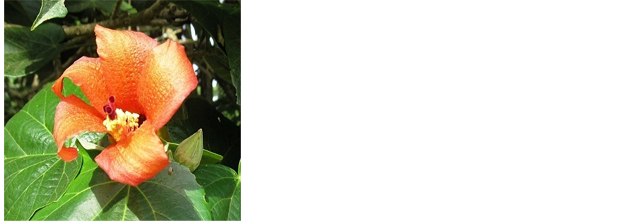
Figure 1. Flower of T. elatum.
For to the purification, 1 g of solid was dissolved in 25 mL of diethyl ether and the volume was completed to 100 mL with ethanol. The sample was refrigerated until an abundant solid appear and it was recuperated to filtration. This process was done twice, to obtain only a yellowish-green solid monitoring by TLC on silica gel with fluorescent indicator 254 nm on aluminum cards (layer thickness 0.2 mm) (10 × 20 cm) using n-butanol: acetic acid:water (4:1:5) as eluent (v/v/v).
2.4. NMR Procedures, Instrumentation and Parameters
NMR spectra were recorded on an Advance 500 spectrometer in DMSO d6 at 298˚K. Qualitative analyses were recorded by 1H (500 MHz) and 13C (125 MHz) and homonuclear and heteronuclear experiments like DEPT135, DEPT90, COSY, HSQC, HMBC, ROESY and TOCSY. Chemical shifts are reported in ppm relative to TMS and coupling constants in Hz.
3. Results and Discussion
1H-1H COSY, HSQC and HMBC correlations were used to assign all proton and carbon atoms within the corresponding substructures and established possible links to other parts of the molecule. Using 1H and 13C NMR, 1H-1H COSY, TOCSY, HSQC and HMBC experiments, identification of the sugar moiety was performed according to the strategy used for structure elucidation of flavonoid glucosides.
The 1H NMR spectrum of this flavonoid derivative showed four proton signals in the aromatic region; (δ = 8.08 ppm (d, 1H, J4 = 2.21 Hz, 2’-H), (δ = 7.92 ppm (dd, 1H, J3 = 8.72 Hz - J4 = 2.21 Hz, 6’-H), (δ = 6.99 ppm (d, 1H J3 = 8.71 Hz, 5’-H), consistent with a gossypetin derivative. The observed multiplicity (ABX system) is characteristic of a catechol. Observed chemical shift value of proton 6-H (δ = 6.25 ppm) and carbon 6-C (δ = 97.98 ppm) confirmed the presence of Hydroxyquinol (ring A). [7] [8] (Figure 2). The 13C NMR values for this flavonoid (Table 1) were assigned on the basis of 1JCH, 2JCH, 3JCH and 4JCH correlations observed in the HSQC and HMBC spectra.

Figure 2. 1H NMR spectrum of compound 1.

Table 1. NMR data of gossypetin, gossypitrin and gossypetin-3'-O-glucoside.
*Attribution realized accordance with literature data and the experiences by RMN (1H, 13C) and 2D (COSY, HSQC, HMBC, TOCSY and ROESY). **Indetermined.
The sugar region showed the presence of one unit. The 1H and 13C values of this sugar unit were assigned by a combination of 1D 1H NMR, 2D COSY, TOCSY and HSQC experiments. The 1H and 13C resonances were in accordance with β-glucopyranose confirmed by the presence of 6 carbons sp3 (5 CH and 1 CH2) where the protons at δ = 4.83 ppm (d, 1H, J = 7.32 Hz, 1’’-H), δ = 3.76 ppm (d, 1H, J = 11.96 Hz, 6’’-H), δ = 3.59 ppm (dd, 1H, J = 11.96 Hz, J = 4.10 Hz, 6’’-H) δ = 3.44 - 3.34 ppm (m, 4H, 2’’, 3’’, 4’’, 5’’-H) resonated in the characteristic zone of glycosylated flavonoid compounds indicates a glucose moiety (Figure 2). Measured coupling constants value for anomeric proton signal 1’’H (J = 7.32Hz) is according to one glycosylated structure type β. The substitution position was determined by ROESY and HMBC. HMBC correlations between 1’’H (δ = 4.83 ppm) and C3’ (δ = 144.93 ppm) and in ROESY between 2’H and 1’’H unequivocally confirmed the ring B substitution in 3' position. Therefore, the substance is consequently determined to be gossypetin-3'-O-β-glucopyranoside [9] (Figure 3 and Figure 4).
The principal difference between gossypitrin and gossypetin-3'-O-glucoside is the sugar moiety position in both flavonoids. Gossypetin-3'-O-glucoside, whose structure was unambiguously determined from the NMR

Figure 3. HMBC correlations in gossypetin-3'-O-glucoside.

Figure 4. HMBC spectrum of gossypetin-3'-O-glucoside, showing the correlation between 1’’H (δ = 4.83 ppm) and C3’ (δ = 144.93 ppm).
data, have the sugar moiety in C3' position according to the experiments, while gossypitrin have the sugar moiety in C7 position. Signal at 10.39 ppm (C-7) disappear in the spectrum 1H NMR of gossypitrin, while in 1H NMR spectrum of gossypetin-3'-O-glucoside the corresponding signal at 3'-C (9.34 ppm) disappear too. The UV, IR and MS of both flavonol glycosides do not allow differentiate the structures of the last mentioned compounds [10] - [12] .
4. Conclusion
In summary, the present study describes the identification of one of the major flavonoids from petals of the red flowering T. elatum as gossypetin-3'-O-β-glucoside. The identical sugar moiety of gossypetin-3'-O-β-D-gluco- side and gossypitrin (gossypetin-7-O-β-D-glucoside) may be due to broad specificities of glucosyl transferases involved in the biosynthesis of the glycosides from the respective aglycones. The aglycones of flavonols may be formed from a common C6-C3-C6 precursor, e.g. gossypetin flavonol. This is the first report with complete NMR data for this compound found in the flowers of Talipariti elatum that grows in Cuba.
Acknowledgements
The authors are indebted to Dr Juliette Smith-Ravin and Dr Odile Marcelin (BIOSPHERES group) at University of Antilles (UA), Martinica. Furthermore, we are grateful to Frédéric Verdeau and Loïk Sylvius for skillful experimental assistance. This work was supported by grants from the Regional Council of Martinica which has financed gossypitrin project.
Cite this paper
José González Yaque,Armando Cuéllar,Marc Gaysinski,Max Monan,Enmanuel Nossin,Frantz François-Haugrin, (2016) New Reported Flavonol Characterized by NMR from the Petals of Talipariti elatum S. w. in Cuba. American Journal of Plant Sciences,07,1564-1569. doi: 10.4236/ajps.2016.711148
References
- 1. Fossen, T. and Andersen, O.M. (2005) Spectroscopic Techniques Applied to Flavonoids. In: Andersen, O.M. and Markham, K.R., Eds., Flavonoids—Chemistry, Biochemistry and Applications, Taylor & Francis, USA, 37-142
http://dx.doi.org/10.1201/9781420039443.ch2 - 2. U.S. Department of Agriculture (2013) Hibiscus elatus Sw. “mahoe”. Natural Resources Conservation Service. Plants Database.
- 3. Cuéllar, A. and González, J. (2001) Phytochemistry Analysis of the Different Parts of the Flowers of Hibiscus elatus S. w. Revista Cubana de Farmacia, 35, 68-70.
- 4. Márquez, I., Cuéllar, A., Martínez, J., Alemán, A., Lora, J. and Castro, H. (1999) Estudio fitoquímico de la especie Hibiscus elatus S.w. Revista Cubana de Farmacia, 33, 127-131.
- 5. Cuéllar, A. and González Yaque, J. (2010) Obtención del glucósido flavonoide gossypitrina de los pétalos de flores de Talipariti elatum S.w y evaluación de su posible efecto antioxidante. Revista Colombiana de Ciencia Animal, 2, 338-348.
- 6. Parkin, A.G. (1916) The Coloring Matter of Cotton Flowers. Part III. Journal of the Chemical Society London, 109, 145-154.
http://dx.doi.org/10.1039/CT9160900145 Schliemann, W., Schneider, B., Wray, V., Schmidt, J., Nimitz, M., Porzel, A. and Bohm, H. (2006) Flavonols and an Indole Alkaloid Skeleton Bearing Identical Acylated Glycosidic Groups from Yellow Petals of Papaver nudicaule. Phytochemistry, 67, 191-201.
http://dx.doi.org/10.1016/j.phytochem.2005.11.002 - 7. Lee, S., Park, Y., Moon, B., Lee, E., Hong, S. and Lim, Y. (2008) Substitution Effect of Hydroxyl Groups on 1H and 13C Chemical Shifts in Hydroxyflavonols. Bulletin of the Korean Chemical Society, 29, 1597-1600.
http://dx.doi.org/10.5012/bkcs.2008.29.8.1597 - 8. Kaouadji, M., Ravanel, P., Tissut, M. and Creuzet, S. (1988) Novel Methylated Flavonols with Unsubstituted B Ring from Platanus acerifolia Buds. Journal of Natural Products, 51, 353-356.
http://dx.doi.org/10.1021/np50056a032 - 9. Francois-Haugrin, F. (2015) Extraction et purification de la gossypitrine des pétales de fleurs de Mahot bleu (Hibiscus elatus Sw.), et évaluation de ses propriétés antioxydantes par différentes méthodes. Perspectives en agroalimentaire, en cosmétique et en santé (confidentiel). Mémoire de diplome d'ingénieur CNAM, Paris.
- 10. Braunberger, C., Zehl, M., Conrad, J., Fischer, S., Adhami, H.-R., Beifuss, U. and Krenn, L. (2013) LC-NMR, NMR, and LC-MS Identification and LC-DAD Quantification of Flavonoids and Ellagic Acid Derivatives in Drosera pelttata. Journal of Chromatography B, 932, 111-116.
http://dx.doi.org/10.1016/j.jchromb.2013.06.015 - 11. Scoelly, T. and Kapetanidis, I. (1993) Flavonoids from Drosera rotundifolia. Scientia Pharmaceutica, 61, 277-282.
- 12. Wind, O., Christensen, S.B. and Molgaard, P. (1998) Colouring Agents in Yellow and White Flowered Papaver radicatum in Northern Greenland. Biochemical Systematics and Ecology, 26, 771-779.
http://dx.doi.org/10.1016/S0305-1978(98)00031-3
NOTES

*Corresponding author.