J. Biomedical Science and Engineering, 2009, 2, 280-286
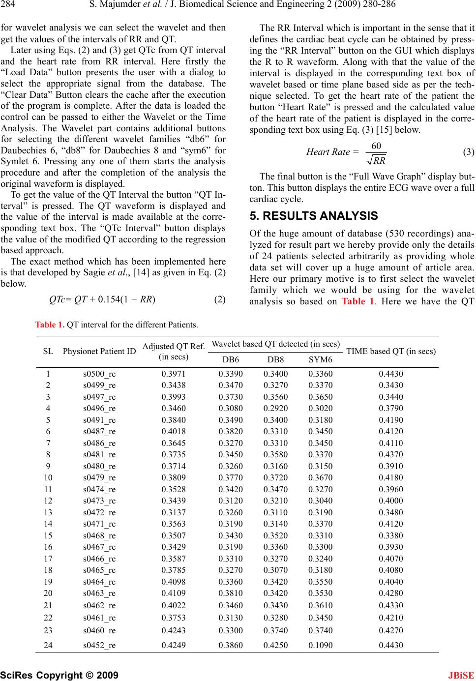
doi: 10.4236/jbise.2009.24042 Published Online August 2009 (http://www.SciRP.org/journal/jbise/
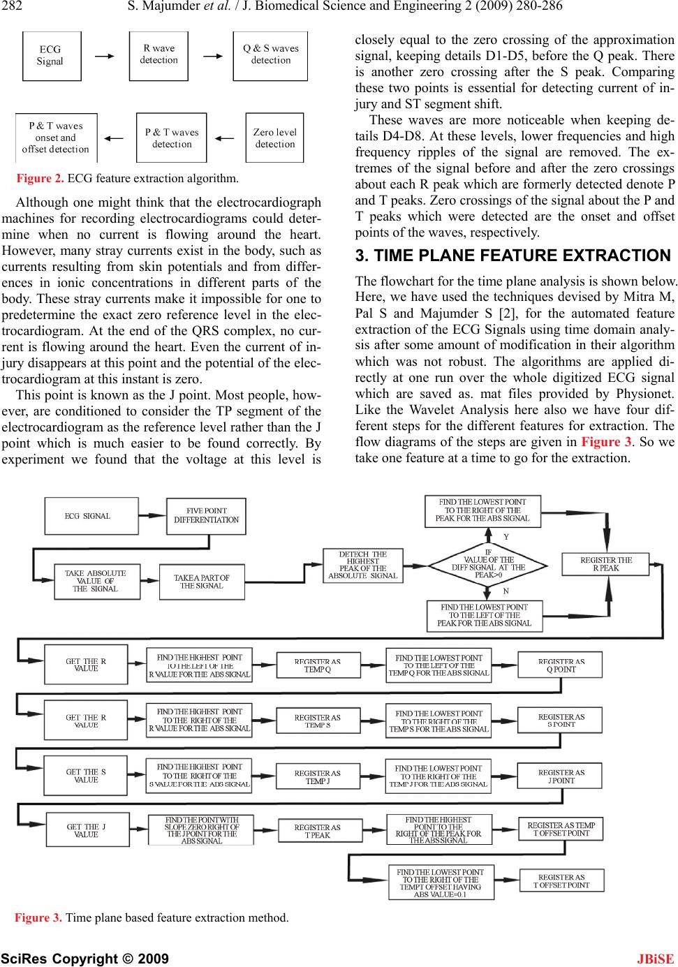
JBiSE
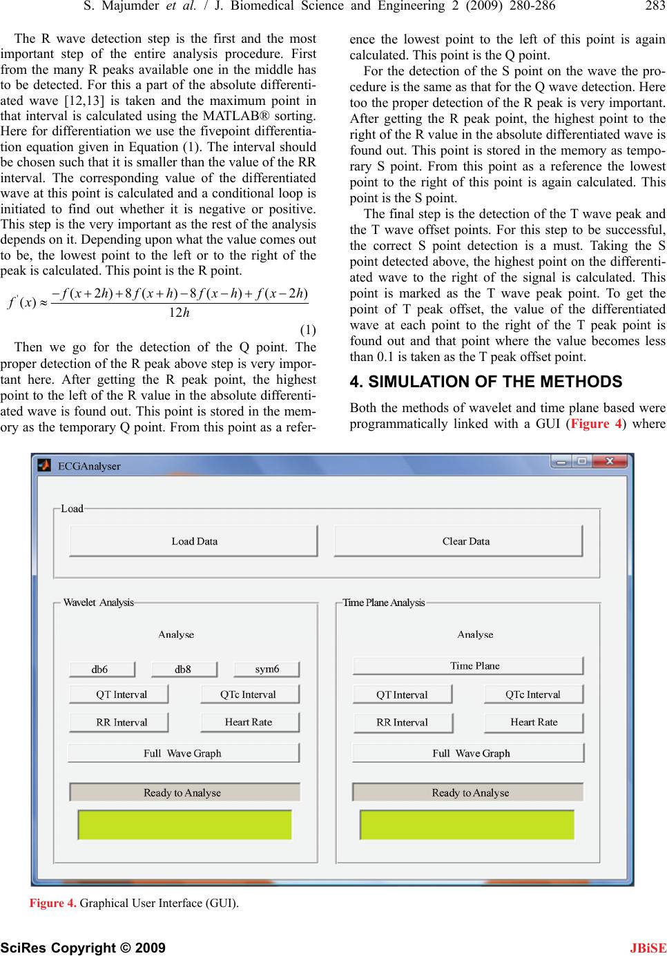
).
Published Online August 2009 in SciRes. http://www.scirp.org/journal/jbise
A hybrid wavelet and time plane based method for QT
interval measurement in ECG signals
Swanirbhar Majumder1, Saurabh Pal2, Sidhartha Dhar1, Abhijit Sinha1, Abhijit Roy1
1Dept of ECE, NERIST (Deemed University), Arunachal Pradesh, India, 2Haldia Institute of Technology, Haldia, West Bengal, India.
Email: swanirbhar@gmail.com, spal76@gmail.com, siddhar2911@gmail.com, rush2abhi_ec@yahoo.com, c4abhi@yahoo.co.in
Received 29 March 2009; revised 25 April 2009; accepted 6 May 2009.
ABSTRACT
Here we present a method of QT interval meas-
urement for Physionet's online QT Challenge
ECG database using the combination of wavelet
and time plane feature extraction mechanisms.
For this we mainly combined two previous
works one done using the Daubechies 6 wavelet
and one time plane based with modifications in
their algorithms and inclusion of two more
wavelets (Daubechies 8 and Symlet 6). But
found that out of these three wavelets Daube-
chies 6 and 8 gives the best output and when
averaged with the interval of time plane feature
extraction method it gives least percentage of
error with respect to the median reference QT
interval as specified by Physionet. Our modified
time plane feature extraction scheme along with
the wavelet method together produces best re-
sults for automated QT wave measurement as
its regular verification is important for analyzing
cardiac health. For the V2 chest lead particularly
whose QT wave is of tremendous significance
we have tested on 530 recordings of Physionet.
This is because delay in cardiac repolarization
causes ventricular tachyarrhythmias as well as
Torsade de pointes (TdP). A feature of TdP is
pronounced prolongation of the QT interval in
the supraventricular beat preceding the ar-
rhythmia. TdP can degenerate into ventricular
fibrillation, leading to sudden death.
Keywords: ECG; QT; Physionet; TdP
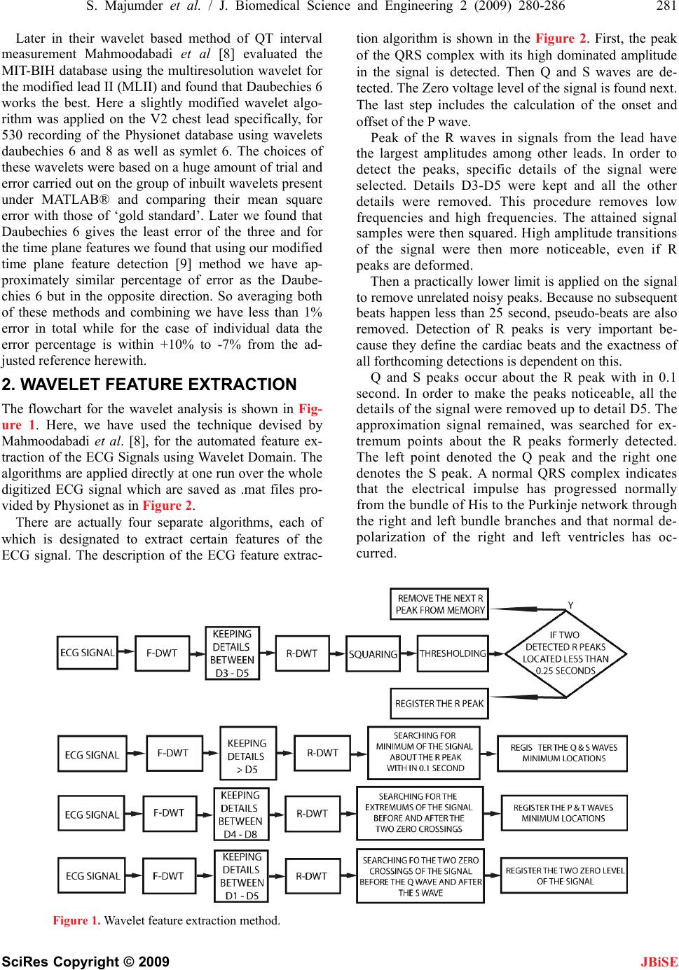
1. INTRODUCTION
The QT interval is measured as the time interval be-
tween the onset of the QRS complex and the end of the
T wave. At the end of the T wave repolarisation is com-
pleted and the T wave voltage amplitude returns to the
baseline [1]. The QT interval is thus a measure of the
duration of the ventricular depolarisation and repolariza-
tion. Some error may introduce in the QT interval meas-
urement due to the fact that it may not return back to the
base line or it may go below the base line along with the
onset of U wave occasionally.
Many drugs prolong cardiac repolarization which in
turn increases the QT interval. This might lead to ven-
tricular arrhythmia as severe as torsade de pointes
(TdP) in some critical cases [2,3]. Hence accurate
measurement of the QT interval is very important for
intensive cardiac care and also for pharmaceutical in-
dustry. A statistically significant increase in the mean
QT interval (corrected for heart rate) as small as 6 mil-
liseconds between baseline and maximal drug effect
may be important as a signal of repolarization abnor-
mality [4]. QT intervals can be detected manually, but
these are not so accurate as well as not repeatable in
general. Still we compare our results with the ‘gold
standard’ reference QT measurements taken from the
Physionet challenge 2006 because these were very
precisely taken to build the database for the challenge
so that the participants could compare their algorithms
with the manual methods. Rather automatic QT inter-
val measurement techniques are more accurate and
reproducible, except for the experience of the physi-
cian/doctor giving some extra suggestions which may
be beneficial in some particular special cases on ne-
glecting the time factor [5,6]. Moreover if a bit of
adaptiveness can be added to it via trained neural net-
work it might be a great effort. Many researchers have
performed several fundamental works on determina-
tion of QT interval along with other characteristic
waves. Yan Sun et al. have proposed a multiscale
morphological derivative (MMD) transform-based
singularity detector for the detection of fiducial points
in ECG signal, where these points are related to the
characteristic waves such as the QRS complex, P wave
and T wave [7]. Laguna P, Jane R, Caminal P have
developed a method where the intervals of clinical
importance can be detected by a multilead QRS detec-
tor that locates each beat, using a differentiated and
low-pass filtered ECG signal as input and the wave-
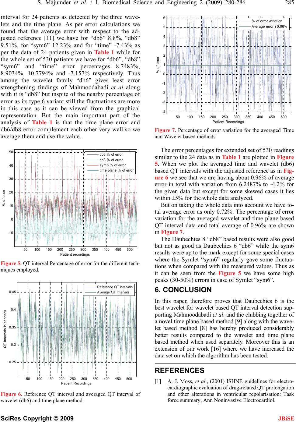
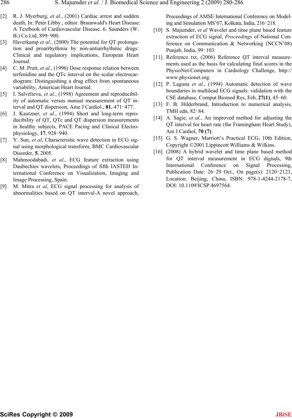
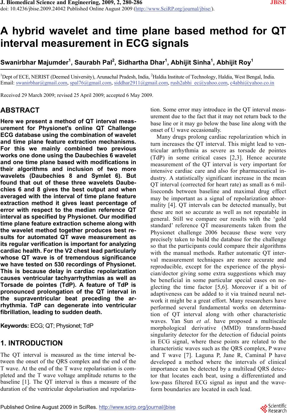
form boundaries are located in each lead.