Open Journal of Modern Neurosurgery, 2015, 5, 71-75
Published Online July 2015 in SciRes. http://www.scirp.org/journal/ojmn
http://dx.doi.org/10.4236/ojmn.2015.53012
How to cite this paper: Yip, C.-M., Tseng, H.-H., Hsu, S.-S. and Hsu, H.-I. (2015) Rathke Cleft Cyst with a Coexisting Gonado-
tropin Producing Pituitary Adenoma. Open Journal of Modern Neurosurgery, 5, 71-75.
http://dx.doi.org/10.4236/ojmn.2015.53012
Rathke Cleft Cyst with a Coexisting
Gonadotropin Producing Pituitary Adenoma
Chi-Man Yip1, Hui-Hwa Tseng2, Shu-Shong Hsu1, Huang-I Hsu1*
1Division of Neurosurgery, Department of Surgery, Kaohsiung Veterans General Hospital, Kaohsiung, Taiwan
2Department of Pathology, Kaohsiung Veterans General Hospital, Kaohsiung, Taiwan
Email: yip_chiman@yahoo.com, hhtseng@vghks.gov.tw, sshsu@vghks.gov.tw, *McCoyHsu@gmail.com
Received 29 April 2015; accepted 7 June 2015; published 10 June 2015
Copyright © 2015 by authors and Scientific Research Publishing Inc.
This work is licensed under the Creative Commons Attribution International License (CC BY).
http://creativecommons.org/licenses/by/4.0/
Abstract
Rathke cleft cyst is thought to arise from incomplete obliteration of the lumen of Rathke pouch.
The cells of the anterior pituitary lobe, from which pituitary adenomas develop, are also derived
from the cells of Rathke pouch. Although Rathke cleft cyst and pituitary adenoma have a shared
ancestry, they rarely occur coincidentally. Rathke cleft cysts have been found incidentally in 11% -
33% of post-mortem examinations, and were associated with 1.7% - 2.1% of the pituitary adeno-
ma cases. These coexisting lesions are difficult to diagnose pre-operatively due to the variable
signal intensity and position of the Rathke cleft cysts. Treatment of these lesions involves surgical
resection to decrease mass effect and medical management to normalize hormonal imbalances. To
our best knowledge, only 42 cases of Rathke cleft cyst with a coexisting pituitary adenoma have
been reported in the English or Japanese literatures, but none of the coexisting pituitary adenoma
is gonadotropin producing. We would like to report a case of Rathke cleft cyst with a coexis ti ng
gonadotropin producing adenoma that was successfully treated by endoscopic endonasal trans-
sphenoidal approach with the removal of the sellar lesion and temporary hormone replacement.
Keywords
Rathke Cleft Cyst, Gonadotropin Producing Pituitary Adenoma, Coexisting Lesions, Endoscopic
Endonasal Transsphenoidal Approach
1. Introduction
Rathke cleft cyst (RCC), first described by Luschka in 1860 [1] [2], is thought to arise from incomplete obliteration
of the lumen of Rathke pouch. This embryonic structure develops as a rostral outpouching of the stomodeum or
*Corresponding a uthor.
C.-M. Yip et al.
72
primitive oral cavity during the third or fourth week of gestation. It has an anterior and posterior wall, as well as
a central embryonic cleft. The anterior wall of the pouch proliferates to form the anterior lobe of the pituitary
gland and the pars tuberalis; the posterior wall becomes the pars intermedia [1]-[5]. The residual lumen of the
pouch is reduced to the narrow Rathke cleft, which usually regresses completely. The cells of the anterior pitui-
tary lobe, from which pituitary adenomas develop, are also derived from the cells of Rathke pouch [1]-[3]. Al-
though RCC and pituitary adenoma have a shared ancestry, they rarely occur coincidentally [2] [3] [5] [6]. To
our best knowledge, only 42 cases of Rathke cleft cyst with a coexisting pituitary adenoma has been reported in
the English or Japanese literatures, but none of the coexisting pituitary adenoma is gonadotropin producing [2]
[5] [6]. We would like to present a case of Rathke cleft cyst with a coexisting gonadotropin producing adenoma
which probably is the first reported combination.
2. Case Report
In December 2013, a 66-year-old man with past medical history of hypertension for five years, Legionella
pneumonia with adult respiratory distress syndrome (ARDS) post treatment in June 2013 presented to us with
the chief complaint of progressive blurred vision for more than two months. Traced back his past medical histo-
ry, he was diagnosed to have a sellar lesion in 2007. His magnetic resonance imaging (MRI) at that time showed
a cystic lesion measured about 1.7 cm × 1.3 cm × 1.2 cm in size at the sella and suprasellar region, manifesting
hyperintensity both on T1-weighted (T1WI) and T2-weighted (T2WI) images without obvious solid enhance-
ment (F igure 1). This lesion showed mild mass effect on the optic chiasm, however, he hesitated to have surgic-
al treatment. He was on cortisone replacement since the previous admission due to Legionella pneumonia. On
admission, his neurological examination was essentially normal except his visual field study showed bitemporal
hemianopia. Sellar MRI was repeated which disclosed a cystic mass lesion with some solid enhancement, about
3.2 cm × 2.5 cm × 2 cm in s ize located at sella with suprasellar extension, manifestin g iso- to hypo -intensity on
T1WI and hyperintensity on T2WI images, causing the compression of optic chiasm (Figure 2). Endo crinologi-
cal data were obtained; his cortisol level was normal (under regular cortisone replacement) but hypothyroidism
as well as hypogonadism were shown. The pre-operative tentative diagnosis was Rathke cleft cyst or pituitary
macroadenoma with cystic change.
On January 3, 2014, under general anesthesia, he underwent endoscopic endonasal transsphenoidal approach
with the removal of th e sellar lesion. A zero degree endoscope was introduced into the right side nostril to iden-
tify the sphenoid ostium. After the perpendicular plate of ethmoid bone was disarticulated and pushed to the left
side, the rostrum of the sphenoid sinus was exposed and drilled to complete the sphenoidotomy. Then the sellar
floor was exposed and drilled; the capsule of the lesion was opened, the solid tumor tissue rushed out followed
by the outflowing of the mucinous contents (Figure 3). After the removal of the solid and the cystic components
of the lesion, a piece of DuraGen was placed in the subdural space to cover the pituitary gland and a piece of
thin bone harvested from the resected sella turcica w as used to reconstruct the sellar floor follow ed by spraying
tissue glue on the reconstructive materials.
Figure 1. Coronal view of patient’s sellar MRI in 2007. (A) T1-weighted images (B) T2-weighted images (C) T1-
weighted with Gadolinium demonstrating a cystic lesion measured about 1.7 cm × 1.3 cm × 1.2 cm in size at t he
sella and suprasellar region, manifesting hyperintensity both on T1-weighted and T2-weighted images without
obvious solid enhancement.
C.-M. Yip et al.
73
Figure 2. Coronal view of patient’s sellar MRI in 2013. (A) T1-weighted images; (B) T2-weighted
images; (C) T1-weighted with Gadolinium disclosing a cystic mass lesion about 3.2 cm × 2.5 cm ×
2 cm in size located at sella with suprasellar extension, manifesting iso- to hypo-intensity on
T1-weighted and hyperintensity on T2-weighted images, with some solid enhancement, causing the
compression of optic chiasm.
Figure 3. Endonasal endoscopic view. Left side picture showed the rushing out of a solid tumor
tissue. Right side picture showed the outflowing of the mucinous contents.
Histology examination of the specimen showed a picture of pituitary tumor tissue with Rathke cleft cyst
which composed of cystic space containing myxoid/mucoid material, lined by cuboidal to low columnar epithe-
lium, with underlying collag enous stroma (Figure 4(A)). The remaining pituitary tissue consisted of monomor-
phic proliferations of cells with uniform round nuclei, delicate stippled chromatin, inconspicuous nucleoli, and
moderate quantities of cytoplasm, arranged in solid pa ttern (Fig ure 4(B)). Reticulin stain showed loss of normal
acinar structure (Figure 4(C)). By immunohistochemistry, the neoplastic cells were positive for FSH (Figure
4(D)), weakly positive for low molecular weight keratin (CAM5.2), and negative for prolactin, ACTH, TSH,
GH, and LH immunostains. There are also focal hemorrhage and neutrophil infiltration. Rathke cleft cyst with a
coexistent gonadotropin producing adenoma was diagnosed based on the morphology of the tumor cells and the
result of immunohistochemical stain s .
The patient’s postoperative course was uneventful and his vision was much improved after surgery. 1-year
post-operative follow-up sellar MRI done in January 2015 showed neither residual nor recurrent sellar lesion
(Figure 5) . The last endocrinological data checked in August 2014 were within normal limits and now he is not
on hormone replacement.
3. Discussion
Rathke cleft cysts are b enign, epithelial-lined cystic remnants of Rathke pouch and remain asymptomatic in the
majority of people [7]. In cases of symptomatic Rathke cleft cyst, the common symptoms are headache, visual
disorder, endocrine dysfunction probably caused by the mass effect on the adjacent structures [1] [2] [4] [7] [8].
Magnetic resonance imaging (MRI) is the preferred modality for preoperative evaluation and differentiation of
cystic sellar lesions including Rathke cleft cysts, cystic craniopharyngiomas, arachnoid cysts, cystic or hemorr-
hagic pituitary adenomas, pars intermedia cysts, epidermoid tumors, sellar abscesses and even intrasellar aneu-
C.-M. Yip et al.
74
Figure 4. Histology of the cystic mass lesion. (A) Hematoxylin and eosin (H&E) stain of the cystic
component demonstrating cystic space containing myxoid/mucoid material, lined by cuboidal to
low columnar epithelium, with underlying collagenous stroma. (B) H&E stain of the solid compo-
nent showing monomorphic proliferations of cells with uniform round nuclei, delicate stippled
chromatin, inconspicuous nucleoli, and moderate quantities of cytoplasm, arranged in solid pattern.
(C) Reticulin stain showing loss of normal acinar structure. (D) Immunostaining of the neoplastic
cells revealing positive for FSH.
Figure 5. Coronal view of patient’s 1-year post-operative follow-up sellar MRI. (A) T1-weighted
images; (B) T2-weighted images; (C) T1-weighted with Gadolinium showing neither residual nor
recurrent sellar lesion.
rys ms [1] [4] [7] [9]. On MRI, Rathke cleft cysts often appear as well-circumscribed, centrally located spherical
or ovoid lesions with little or no Gadolinium enhancement of the cystic wall in the sellar region [7]. However,
because of the diverse content in the Rathke cleft cysts, they may contain serous or mucoid fluid, mucopolysac-
charides, hemosiderin deposits, cellular sediments or particles of cholesterol and protein resulted in variable
signal intensity on MRI [1] [2] [7] [9]. A high T1-weighted image intensity has been interpreted to indicate a
high conten t of protein and mucopolysaccharides or hemorrhage. The cystic content of hyper- and iso-intensity
on T1 -weighted image is usually indicative of mucous with varying viscosity. Rathke cleft cysts with hypoin-
tensity on T1-weighted images usually conta in CSF-like transparent fluid with low vis cosity [2] [9].
C.-M. Yip et al.
75
Rathke cleft cysts have been found incidentally in 11% - 33% of post-mortem examinations, and were asso-
ciated with 1.7% - 2.1% of the pituitary adenoma cases [1]-[3] [8]. The relationship between Rathke cleft cyst
and pituitary adenoma is still co ntroversial [1] [3]. Associations between these two lesions have been reporte d to
be only occasional and coincidental [3]. Although Rathke cleft cyst and pituitary adenoma have a common em-
bryological ancestry, a pituitary adenoma and a concomitant Rathke cleft cyst rarely occurs. The diagnosis of
Rathke cleft cyst with a co existing pituitary adeno ma is difficult before surg ery because Rathke clef t cysts show
variable size, position and signal intensity on MRI. Nevertheless, the presence of a non-enhancing cyst with a
pituitary adenoma suggests the possibility of an accompanying Rathke cleft cyst [2] [9]. Treatment of this un-
usual coexisting lesion involves surgical resection to decrease mass effect and medical management to normal-
ize hormonal imbalances [2].
4. Conclusion
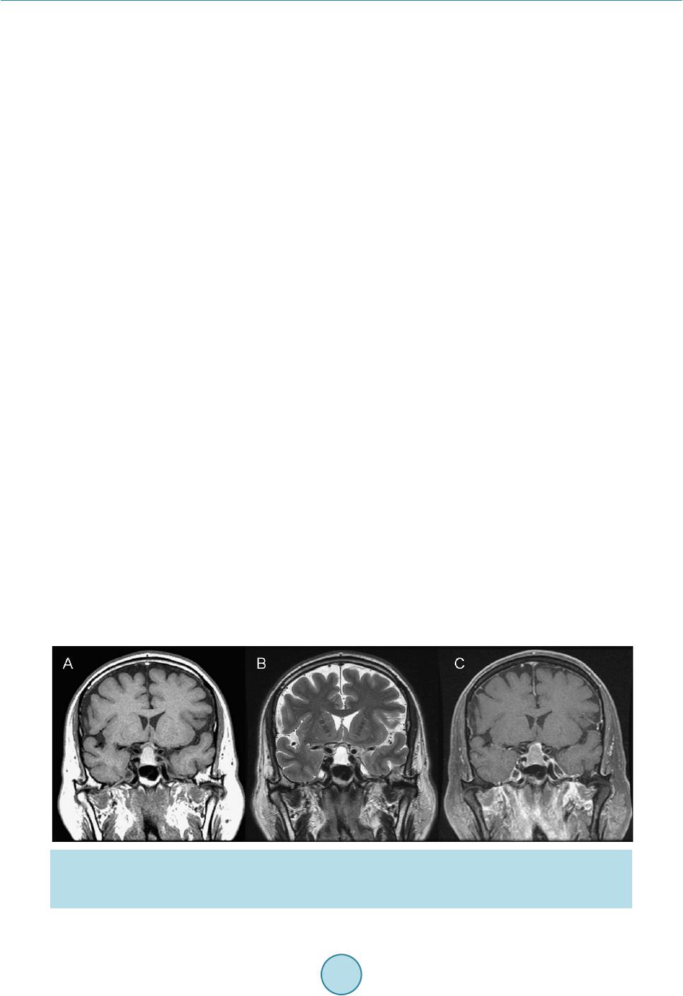
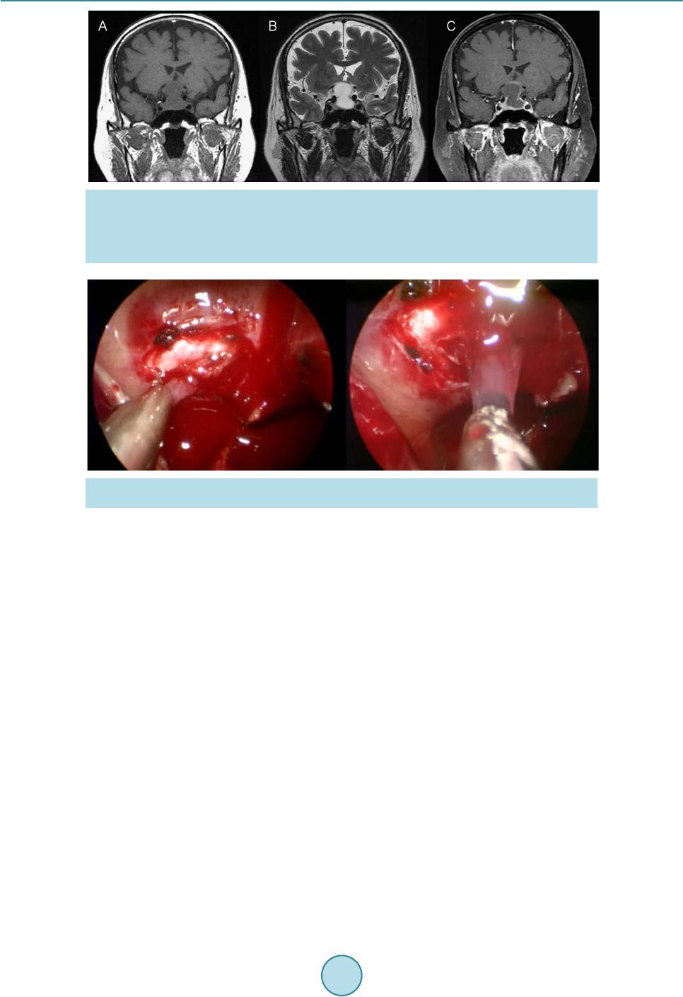
Our patient was diagnosed to have a sellar cystic lesion with suprasellar extension in 2007. This lesion pro-
gressed and let him to have significant visual and endocrine dysfunction in 2013. He was successfully treated by
endoscopic endonasal transsphenoidal approach with the removal of the sellar lesion and temporary hormone
replacement. His bitemporal hemianopia was resolved after surgery and his hormonal imbalance was recovered
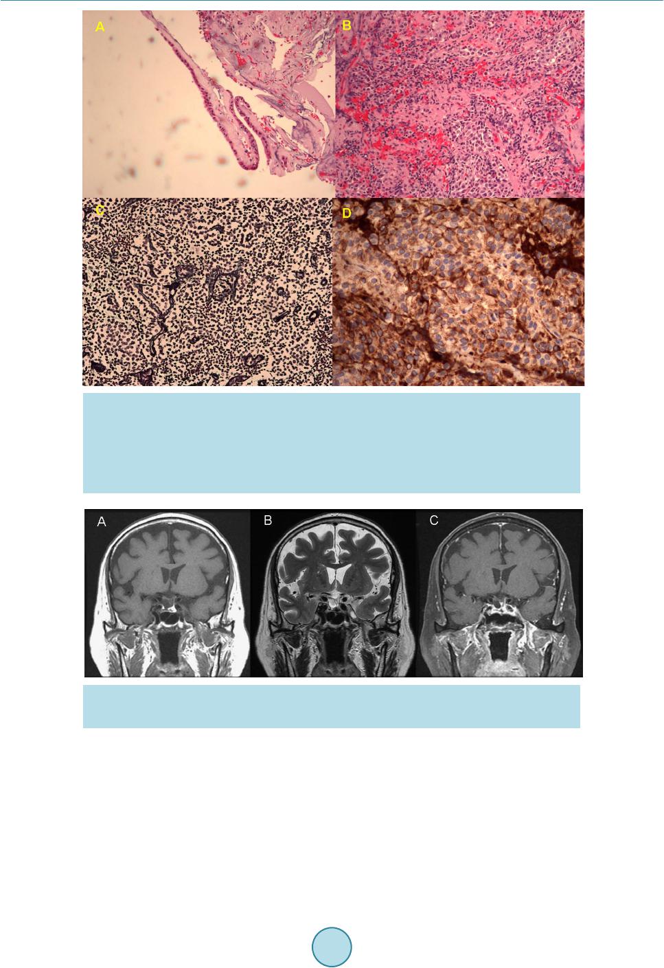
during the follow-up. The histological examination of the sellar cystic lesi on proved to be Rathke cleft cyst with
a coexistent gonadotropin producing adenoma which probably is the first reported combination.
References
[1] Riva, G., Meli, G.A., Chiaramonte, R., et al. (2007) Imaging of Rathke’s Cleft Cysts: Criteria for a Differential Diag-
nosis. The Neuroradiology Journal, 20, 617-621. http://dx.doi.org/10.1177/197140090702000602
[2] Babu, R., Back, A.G., Komisarow, J.M., et al. (2013) Symptomatic Rathke’s Cleft Cyst with a Co-Existing Pituitary
Tumor; Brief Review of the Literature. Asian Journal of Neurosurgery, 8, 183-187.
http://dx.doi.org/10.4103/1793-5482.125662
[3] Sumida, M., Arita, K., Migita, K., et al. (2001) Concomitant Pituitary Adenoma and Rathke’s Cleft Cyst. Neuroradi-
ology, 43, 755-759. http://dx.doi.org/10.1007/s002340100559
[4] Kasperbauer, J.L., Orvidas, L.J., Atkinson, J.L.D., et al. (2002) Rathke Cleft Cyst: Diagnostic and Therapeutic Con-
siderations. Laryngoscope, 112, 1836-1839. http://dx.doi.org/10.1097/00005537-200210000-00024
[5] You, C., Qiao, F., Jiang, S. and Xiao, A.Q. (2012) Growth Hormone Secreting Pituitary Adenoma Associated with
Rathke’s Cleft Cyst. Neurology India, 60, 310-311. http://dx.doi.org/10.4103/0028-3886.98521
[6] Wang, K., Ma, L. and You, C. (2012) Pituitary Adenoma and Concomitant Rathke’s Cleft Cyst: A Case Report and
Review of the Literature. Neurology India, 60, 309-310. http://dx.doi.org/10.4103/0028-3886.98520
[7] Zada, G. (2011) Rathke Cleft Cysts: A Review of Clinical and Surgical Management. Neurosurgical Focus, 31, 1-6.
http://dx.doi.org/10.3171/2011.5.FOCUS1183
[8] Gessler, F., Coon, V.C., Chin, S.S. and Couldwell, W.T. (2011) Coexisting Rathke Cleft Cyst and Pituitary Adenoma
Presenting with Pituitary Apoplexy: Report of Two Cases. Sku ll Base Reports, 1, 99-104.
http://dx.doi.org/10.1055/s-0031-1280737
[9] Noh, S.J., Ahn, J.Y., Lee, K.S. and Kim, S.H. (2007) Pituitary Adenoma and Concomitant Rathke’s Cleft Cyst. Acta
Neurochirurgica, 149, 1223-1228. http://dx.doi.org/10.1007/s00701-007-1295-x