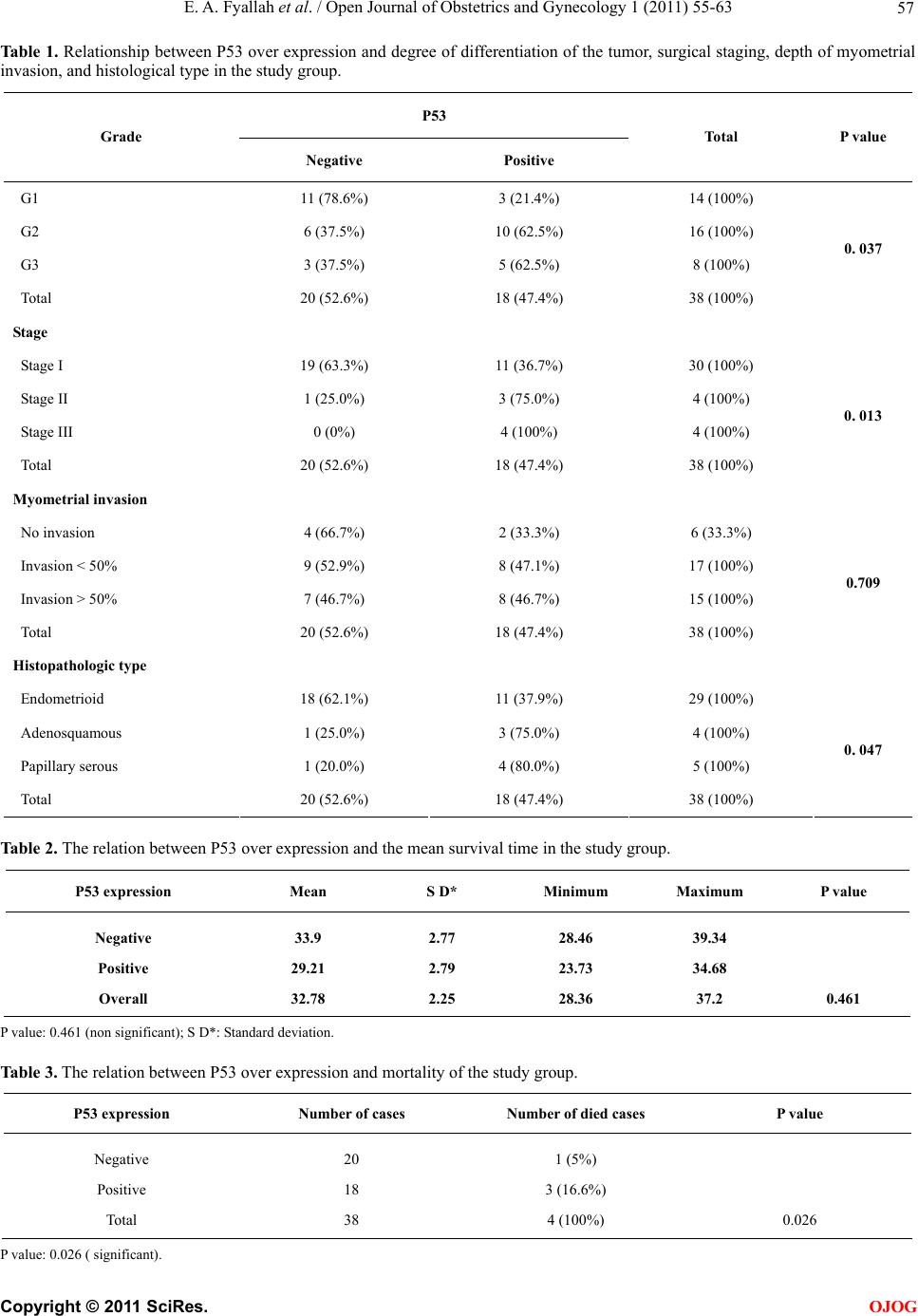
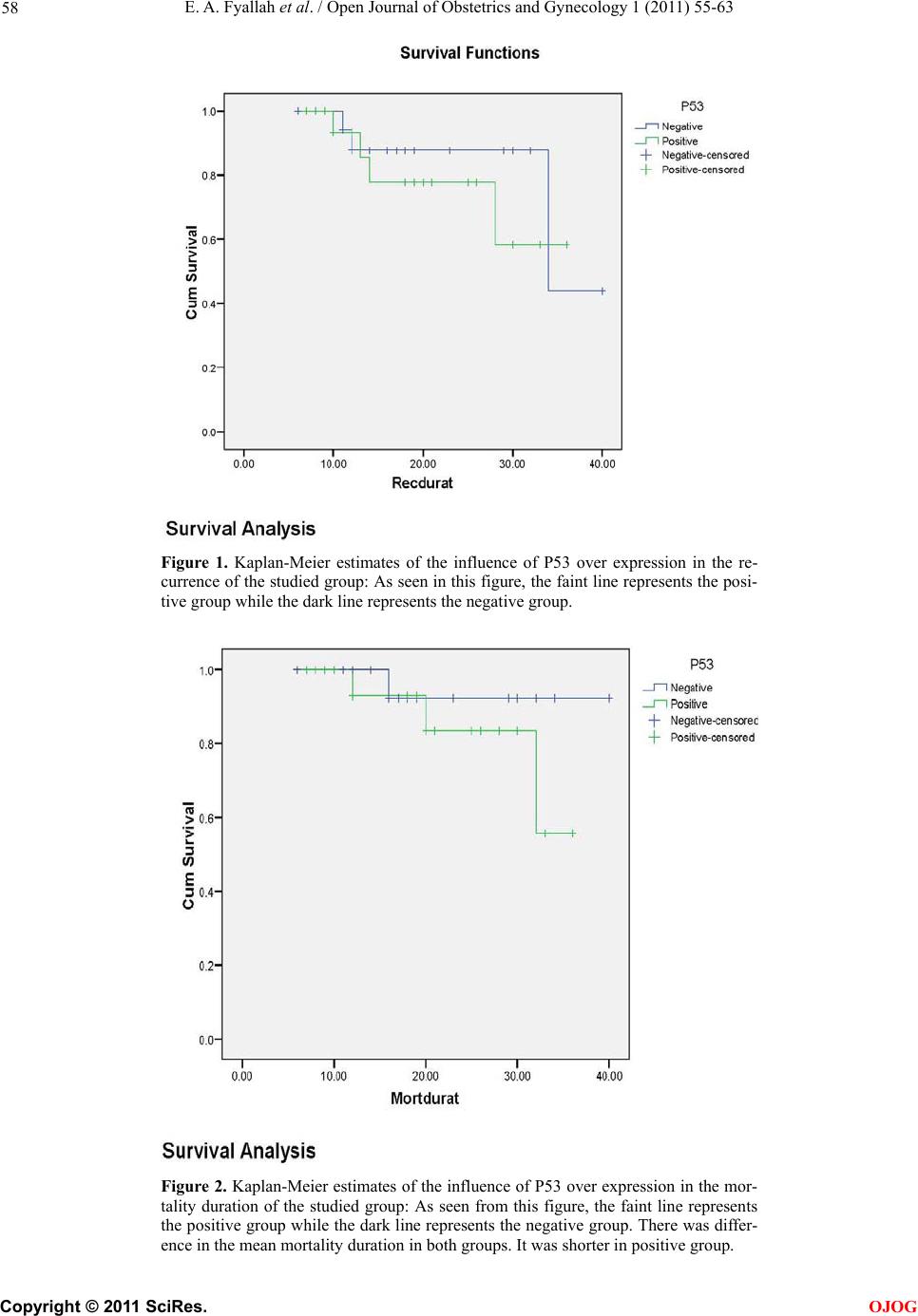
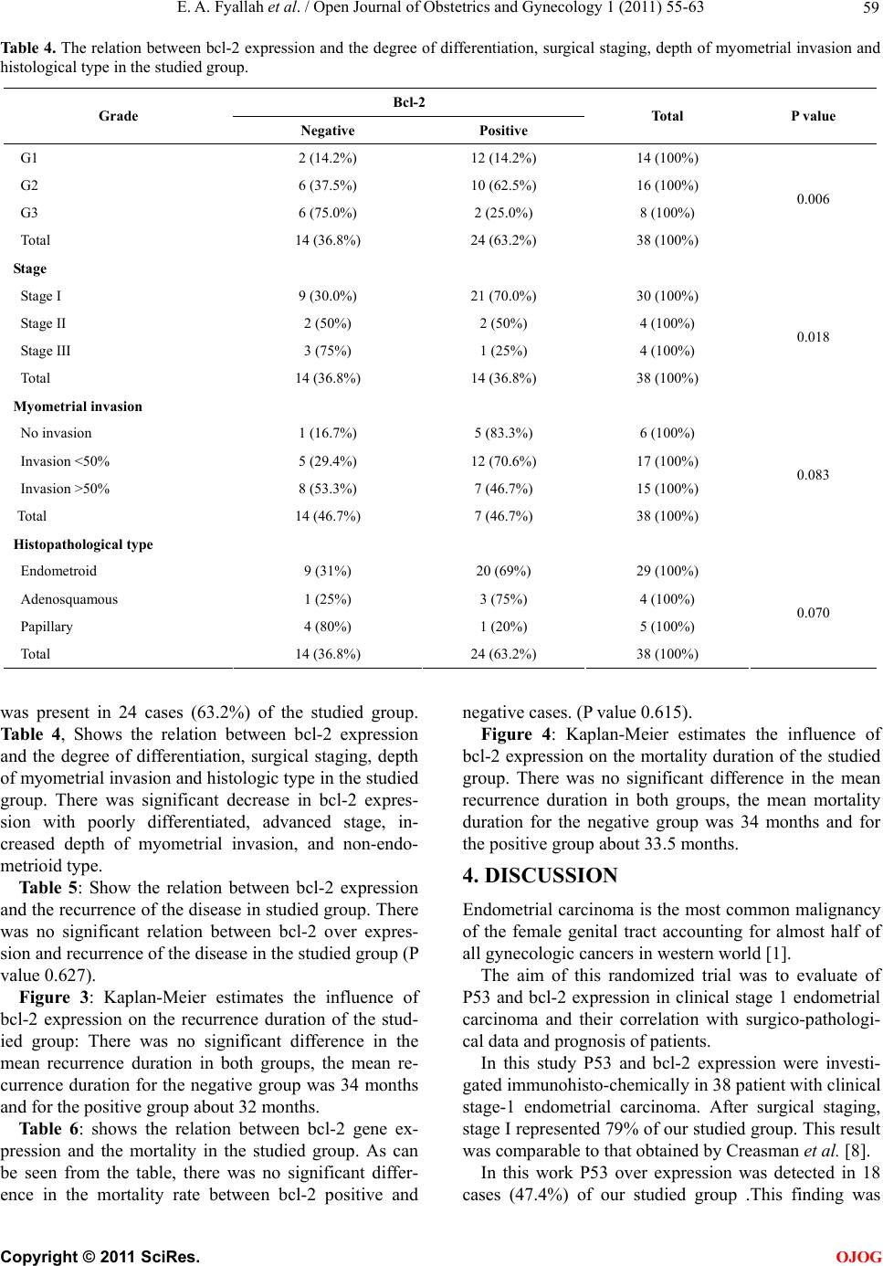
E. A. Fyallah et al. / Open Journal of Obstetrics and Gynecology 1 (2011) 55-63
62
plasm of tumor cells in 24 cases (63.2%). Nearly the
same results obtained by Erkanli et al. [15] and Appel et
al. [11].
There were negative correlation between bcl-2 ex-
pression and the grade of the tumor. These findings were
in agree with Halperin et al. [17]. There were also a sig-
nificant negative correlation between bcl-2 expression
and the stage of endometrial carcinoma (Table 2). These
findings were in agree with other authors [7,9].
Regarding the depth of myometrial invasion in our
study (Table 2), there were negative correlation between
bcl-2 over expression and the depth of myometrial inva-
sion, but the difference was not statistically significant
(P value 0.08). These findings were in agree with Marcia
et al. [14] and Appel et al. [11]. On the other hand, other
authors [7,9], reported a significant immuno-negativity
with increasing the depth of myometrial invasion.
Regarding the correlation between bcl-2 expression
and the histologic type of endometrial carcinoma in our
study (Table 2), there was high expression of bcl-2 in
endometrioid type than non-endometrioid typ es, this was
not statistically significant (P value 0.07). These findings
were in agree with that of Geisler et al. [18].
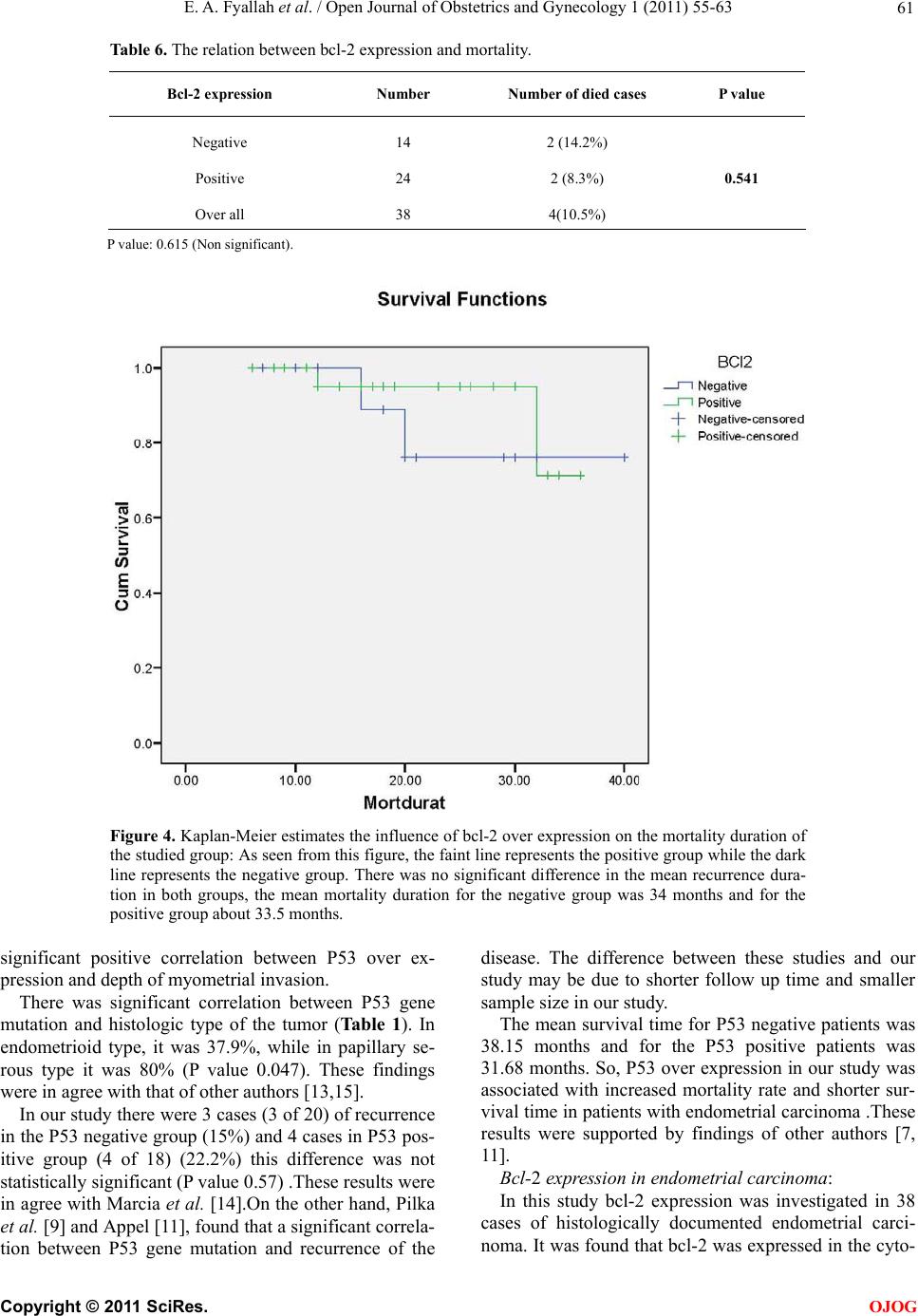
In the current study there was no significant correla-
tion between bcl-2 expression and recurrence or survival
of the patients with endometrial carcinoma (Ta b le s 6 &
7). These findings were supported by the findings ob-
tained by other authors [11,14,19].
5. CONCLUSIONS
P53 over expression in the D&C specimens was associ-
ated with adverse surgico-pathological criteria, increased
mortality rate, and shorter survival time in patients with
endometrial carcinoma.
A significant decrease in bcl-2 expression was associ-
ated with adverse surgico-pathological criteria, but it
was not significantly correlated with prognosis of the
patients.
REFERENCES
[1] Jamel, A., Thomas, A. and Murray, T. (2002) Cancer
statistics. Cancer Journal for Clinicians, 52, 23-57.
doi:10.3322/canjclin.52.1.23
[2] Mariani, A., Sebo, T.J., Katzmann, J.A., Rocke, P.C.,
Koeney, C.L., Lesnick, T.G. and Podratz, K.C. (2005)
Endometrial cancer: Can nodal status be predicted with
curettage? Gynecologic Oncology, 96, 594-600.
doi:10.1016/j.ygyno.2004.11.030
[3] Osmangaolu, M.A., Kadiglu, S. and Bozkaya, H. (2005)
The relationship between mutant P53 gene, DNA con-
tents and conventional clinico-pathological prognostic
variable in cases with endometrial carcinoma. European
Journal of Gynecological Oncology, 26, 64-70.
[4] Petros, A.M., John, J., Huang, Q., Nettesheim, D., Van
Dyk, L.F., Labrada, L., Speck, S.H., Levine, B., Olejnic-
zak, E.T. and Virgin, H.W. (2005) A surface groove es-
sential for viral bcl-2 function during chronic infection.
Plos Pathology, 1, 10.
[5] Mano, Y., Kikushi Yamamoto, K., Kita, T., Hirata, J. and
Tode, T. (1999) Bcl-2 as a predictor of chemosensitivity
and prognosis in primary epithelial ovarian cancer. Eu-
ropean Journal of Cancer, 35, 1214-1219.
doi:10.1016/S0959-8049(99)00124-0
[6] Kapucouglu, N., Aktepe, F., Kaya, H., Biracan, S., Kara-
han, N. and Cris, M. (2007) Immuno-histochemical ex-
pression of PTN in normal, hyperplastic and malignant
endometrium and its correlation with hormone receptors,
bcl-2, bax and apoptotic index. Pathology Research
Practice, 203, 153-162. doi:10.1016/j.prp.2007.01.003
[7] Ohkoushi, T., Sakuragi, N., Watari, H., Nomura, E. and
Tolo, Y. (2002) Prognostic significance of bcl-2 over ex-
pression and lymph node metastasis in surgically staged
endometrial carcinoma. American Journal of Obstetrics
& Gynecology, 187, 353-359.
doi:10.1067/mob.2002.123203
[8] Creasman, W.T., De Geest, K., Disaia, P.J. and Zaino, R.J.
(1999) Significance of true surgical pathologic staging. A
Gynecologic Oncology Group Study. American Journal
of Obstetrics & Gynecology, 181, 31-34.
doi:10.1016/S0002-9378(99)70431-X
[9] Pilka, R., Mickova, I., Lubusky, M., Duskova, M., Ri-
cankova, M. and Kudela, M. (2008) Expression of P53,
Ki 67, bcl-2, C-erb 2, estrogen and progesterone recep-
tors in endometrial cancer. Ceská Gynekologie, 73, 222-
227.
[10] Simionescu, C., Georgescu, C.V., Magaritescu, C., Bata,
S., Marinescu, M., Enachescu, V. and Patnu, E. (2006)
P53 and PCNA immunoexpression in endometrial carci-
nomas. Romanian Journal of Morphology and Embryol-
ogy, 97, 137-141.
[11] Appel, M.L., Edelweiss, M.I., Flek, J., Rivore, W.A.,
Monego, H.I. and Dos Reis, R. (2008) P53 and bcl-2 as
prognostic markers in endometrial carcinoma. Pathology
& Oncology Research, 14, 23-30.
doi:10.1007/s12253-008-9000-9
[12] Veralucia, L.B. and Liliana, A.L. (2003) P53, estrogen
and progesterone receptors in diagnostic curettage for
endometrial adenocarcinoma and their correlation with
morphological data and disease stage at hysterectomy.
Sao Paulo Medical Journal, 12, 163-166.
[13] Nicola, R., Simone, F., Federico, P., Barbara, M., Franco,
G., Marina, G. and Ezio, F. (2005) The association be-
tween P53 expression, stage and histological feature in
endometrial cancer. European Journal of Gynecological
Oncology, 123, 111-116.
doi:10.1016/j.ejogrb.2005.03.018
[14] Marcia, L.M., Appel Maria, I., Edelweiss, J. F., Luis, F.,
Heleusa, I. and Ricordo, R. (2008) P53 and bcl-2 as
prognostic markers in endometrial carcinoma. Pathology
& Oncology Research, 14, 23-30.
doi:10.1007/s12253-008-9000-9
[15] Erkanli, S., Eren, F., Pekin, S. and Bagis, T. (2004) Bcl-2
and P53 expression in endometrial carcinoma. Journal of
Experimental and Clinical Cancer Research, 23, 97-103.
[16] Cerchi, P.L., Marras, V., Capobianco, G., Amborosini, G.,
Piga, M.D., Fadda, G.M., Rosas, N. and Dessole, S.
C
opyright © 2011 SciRes. OJOG