Open Journal of Obstetrics and Gynecology, 2011, 1, 53-54 OJOG
doi:10.4236/ojog.2011.12011 Published Online June 2011 (http://www.SciRP.org/journal/ojog/).
Published Online June 2011 in SciRes. http://www.scirp.org/journal/OJOG
Tubal pregnancy with molar degeneration in concurrent
eutopic pregnancy: a case report
Jesus Joaquin Hijona Elosegui*, Antonio Carballo Garcia, Francisco Javier Frutos Arenas,
Juan Manuel Torres Marti
Department of Obstetrics and Gynecology, Hospital Materno Infantil, Complejo Hospitalario de Jaen, Jaen, Spain.
Email: *jesushijona@gmail.com
Received 8 March 2011; revised 11 March 2011; accepted 15 April 2011.
ABSTRACT
This research paper presents the infrequent case of a
heterotopic pregnancy based on a tubal ectopic preg-
nancy with molar degeneration in concurrent eutopic
pregnancy. Treatment with evacuation/suction curet-
tage and perlaparoscopic salpingectomy was required.
This case report confirms what is biologically valid in
the statistically unlikely.
Keywords: Heterotopic Pregnancy; Hydatiform Mole;
Molar Pregnancy; Pregnancy C omplications; Tubal
Pregnancy
1. INTRODUCTION
Vaginal bleeding in early pregnancy is the most frequent
reason for consultation by pregnant women. Differential
diagnosis in these cases is not always easy and ultra-
sound scan and determination of hCG are frequently
necessary to determine the pregnancy feasibility and to
discard other bleeding reasons.
The main reasons for metrorrhagia in the first quarter
of pregnancy are miscarriage, ectopic pregnancy and
gestational trophoblastic disease [1]. Coexistence of
these pathologies in the same patient, although infre-
quent, is possible; thus, it is always convenient to carry
out a thorough examination, including detailed case his-
tory, full physical examination and a thorough analytic
and ultrasound evaluation.
2. CASE REPORT
A 32 years old primiparous woman without a significant
gynecological case history came to the emergency ser-
vice in her sixth week of amenorrhea because of a pain-
less, dark and mild metrorrhagia, without any other re-
lated symptoms. When palpating, the abdomen was soft
and non-tender without masses, megalies, painful areas
or peritoneal irritation. The gynecological examination
did not reveal any significant findings and transvaginal
ultrasound showed a regular uterus with a 25 mm thick
endometrium. It was, hyperechogenic and showed small
anechoic images which did not confirm an image of
gestational sac. In the right adnexus, an anechoic forma-
tion with a diameter of 34 mm (compatible with a corpu s
luteum) was observed. In contralateral adnexal area, a
similar image with smaller diameter (23 mm), compati-
ble with another follicular formation, was also found.
The Douglas cul-de-sac was taken up by a small amount
of free liquid. Determination of hCG in serum was
7192 mUI/ml.
Due to these results and in view of the clinical suspi-
cion of a potential hydatiform mole or ectopic pregnancy,
it was decided to keep an expectant management with
new clinic, analytical and ultrasound control after 48
hours.
24 hours after the first examination, the patient re-
turned to the emergency service with abdominal pain.
The abdomen exploration did not show any changes,
except for the pain appearance after a deep palpation on
the left iliac fossa. The gynecological examination did
not reveal any significant modifications (except for the
painful mobilization of the cervix) and the transvaginal
ultrasound did not show any changes regarding the pre-
vious one. The analytical study resulted in a determina-
tion of beta-HCG of 8038 mUI/ml. In view of the clini-
cal and analytical progress and taking into consideration
the possibility of ectopic pregnancy with a potential
concurrent molar degeneration, we decided that the pa-
tient should be admitted, but she refused.
72 hours after the second examination, the patient re-
turned to the emergency services because she had ex-
perienced a pain increase. The abdominal examination
suggested a potential acute abdomen. The determination
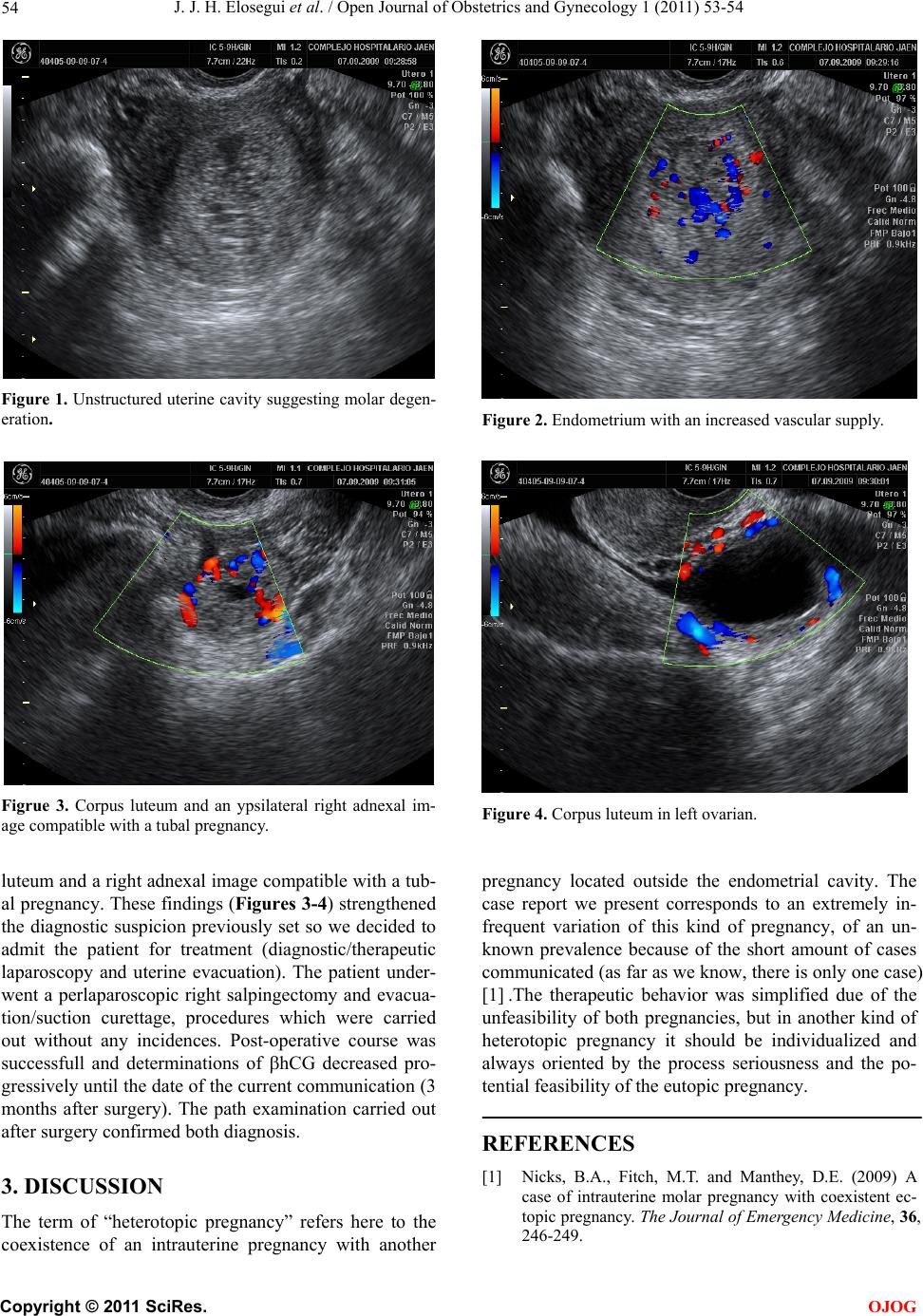
of hCG at that moment was 19262 mUI/ml. The ultra-
sound revealed an endometrium with increased vascular
supply and signs suggesting molar degeneration (Figures
1-2), as well as the presence in both ovaries of corpus