American Journal of Plant Sciences, 2011, 2, 175-179
doi:10.4236/ajps.2011.22019 Published Online June 2011 (http://www.SciRP.org/journal/ajps)
Copyright © 2011 SciRes. AJPS
Initial Stages in the Formation of Galls Induced by
Geoica utricularia in Pistacia Terebinthus Leaflets:
Origin of the Two Vascular Bundles Which
Characterize the Wall of the Galls
Álvarez Nogal
Biología Celular, Universidad de León, León, Spain.
Email: ralvn@unileon.es
Received April 9th, 2011; revised April 23rd, 2011; accepted May 5th, 2011.
ABSTRACT
Only a few species of aphids induce galls. Am ong these, Paracletus cimiciformis, Forda margina ta, Forda formicaria ,
Geoica utricularia and Baizongia pistaciae induce galls on Pistacia terebinthus leaflets. Prior to presen t study the au-
thor examined microscopically P. terebinthus leaflets. He also studied the microscopic morphology of galls induced by
the five species mentioned above. A clear microscopic differen ce between these galls is that in the wall of galls induced
by the genera Paracletus and Forda a single vascular bundle is seen. The interpretation is that these galls are laminae
of the modified leaflets. However, in the walls of the galls induced by the genera Geoica and Baizongia, two vascular
bundles are observed. In the present paper a study of the early stages of development of galls produced by G. utricu-
laria is described. The study was design ed to explai n the origin o f the two va scula r bundles presen t in the walls of these
galls. The findings indicate that the aphid induces a massive development of the two vascular bundles present in the
midvein of the leaflets of P. terebinthus: the main vascular bund le and the small supernum erary vascular bundle. Both
these extremely developed vascular bundles occupy the walls of the galls induced by G. utricularia.
Keywords: M icroscopi c S t u dy, Galls, Gallicolous Aphids, Geoica Utricularia, Pistacia Terebinthus, Vascular Bu n dles
1. Introduction
Aphids are insects that feed on the phloem sap that flows
through the plant [1,2]. Most aphids live in populations
in the open air. Only a few species (the Eriosomatinae)
form galls on host plants. The structure of the galls is
controlled by the aphids themselves [3]. Gall-inducing
aphids do not eat the tissue of the host, but they reorgan-
ize the vascular tissues to be able to access them easily
[4]. Among the species of gall-forming aphids, G. utricu-
laria induces galls on leaflets of plants of the genus Pis-
tacia.
P. terebinthus is found in the study area. On these
shrubs at least five different galls can be identified - ac-
cording to Inbar et al. [5]—induced by as many aphid
species (all belonging to the tribe Fordini): P. cimici-
formis, F. marginata, F. formicaria, G. utricularia, and B.
pistaciae. They are prosoplasmatic galls, i.e. they have a
defined size and shape and show a clear tissue differen-
tiation [6]. They are also monothalamic galls, i.e. they
have only one chamber [7,8]. The first three types of
galls are formed as a fold in the leaflet margins. The other
two types are voluminous galls and are balloon-shaped or
banana or goat horn-shaped, respectively [9,10]. Prior to
the present study, the author microscopically examined
the normal morphology of P. terebinthus leaflets [11].
Subsequently the same author microscopically studied
galls induced by P. cimiciformis, F. marginata, and F.
formicaria [9]. And recently, he studied the walls of the
galls induced by G. utricularia and B. pistaciae [10]. The
microscopic morphology of the galls induced in the leaf-
let margin is different from that induced by the genera
Geoica and Baizongia: in the wall of the galls of the first
three species there is only one vascular bundle, whereas in
the wall of the galls of the other two species two vascular
bundles are observed [9,10]. This structural difference
supports the existence of two clades within the tribe
Fordini, in agreement with several authors [5,12]. In ad-
dition, in the galls induced by the genera Paracletus and
Initial Stages in the Formation of Galls Induced by Geoica utricularia in Pistacia Terebinthus Leaflets:
176
Origin of the Two Vascular Bundles Which Characterize the Wall of the Galls
Forda the xylem is oriented toward the lumen of the gall.
Aphids inside the chamber of the gall must circumvent
the xylem or pass through it to reach the phloem. In galls
induced by the genera Geoica and Baizongia, the vascu-
lar bundle farthest removed from the lumen of the gall
positions itself in the same way as the one before it.
However, the second vascular bundle is positioned oppo-
site the first (xylem facing xylem), leaving the phloem
oriented toward the lumen of the galls [9,10].
In the present paper, newly formed and young galls of
G. utricularia are analyzed microscopically, in order to
learn the origin of the two vascular bundles present in the
walls of these galls.
2. Material and Methods
Essentially the same protocol was followed as used in 3
previous studies by the same author [9-11].
Galls on P. terebinthus induced by aphids of the spe-
cies G. utricularia were collected from the wild. Samples
were taken in April and May at sites in the west of the
province of León (Spain). Samples were collected from
the early stages of development of the galls, from incipi-
ent galls to young galls (Figures 1(a)-(e)). The corre-
sponding uninfested (control) leaflets were also collected.
Samples were placed in situ in FAA (formaldehyde, ace-
tic acid, and ethyl alcohol) and were fixed for 48 hours.
They were subsequently dehydrated through an increas-
ing ethyl alcohol series, passed through isoamyl acetate
as an intermediate liquid, impregnated with Paraplast for
90 minutes in an oven at 64˚C, and finally blocks were
formed. Of these blocks, 12 μm thick serial sections were
obtained using a paraffin microtome. The Safranin-Fast
Green staining method was used. The preparations were
mounted permanently on microscope slides with Entel-
lan.
Preparations were studied using a Nikon E600 micro-
scope under bright-field, epi-fluorescence, and polarized
light conditions.
3. Results
In the midvein of young P. terebinthus leaflets, 8 areas
can be distinguished (Figure 1(f)): 1/abaxial epidermis;
2/annular collenchyma (2 - 3 rows of cells); 3/storage
parenchyma; 4/main vascular bundle: collateral bundle
with conspicuous schizogenic ducts in the phloem and
abundant xylem elements; 5/storage parenchyma; 6/super-
numerary vascular bundle: collateral bundle with few xy-
lem elements and no schizogenic ducts in the phloem.
The 7/storage parenchyma; and 8/adaxial epidermis are
facing the main vascular bundle (Figure 1(g)).
In the first stages of development of the galls (Figure
1(h)) a higher cellularity is observed than in the control
leaflets. This increased number of cells is evident in the
parenchyma. In addition, the supernumerary vascular bun-
dle presents more xylem elements than the control leaf-
lets. Unicellular trichomes were observed in the epider-
mis lining the chambers of the galls (Figures 1(c)-(d)).
Subsequently (Figure 1(i)) an increase is observed in the
number of cells of the three parenchyma layers consid-
ered. The vascular bundles are very developed, and show
an evident procambium. At a later stage, the increased
cellularity is evident (Figure 1(j)). The development of
the two vascular bundles is also evident, and a prominent
procambium is observed in both. The generalized hyper-
trophy causes a displacement of the midvein with respect
to control leaflets. It can be seen how the two vascular
bundles enter the wall of the gall (Figure 2). Specifically,
it can be observed that the main vascular bundle occupies
the outermost part of the wall. The supernumerary vas-
cular bundle occupies the innermost part of the wall, i.e.
the part closest to the lumen of the gall. Unicellular
trichomes are located exclusively in the most proximal
part of the galls.
4. Discussion
Only a few species of aphids induce galls. Among them,
G. utricularia forms spherical galls on the abaxial side of
P. terebinthus leaflets.
Prior to the present study, the author did a histological
study of the leaflets on which these galls are found. Of
that study, it should be noted here that the leaflets of P.
terebinthus have pinnate venation, i.e. a main midvein or
midrib [11,13]. Specifically, in the midvein of the leaflets
of P. terebinthus two vascular bundles can be observed
microscopically: one primary vascular bundle and one
supernumerary vascular bundle with few elements. Both
are collateral vascular bundles. Xylem is seen facing xy-
lem, separated by few parenchyma cells. The phloem of
the supernumerary bundle is oriented toward the adaxial
side of the leaflet, and the phloem of the main vascular
bundle is oriented toward the abaxial side.
Subsequently, galls induced by P. cimiciformis, F.
marginata, and F. formicaria in the margins of the leaf-
lets of P. terebinthus were studied microscopically [9].
Such galls can be understood as laminae of the modified
leaflets. A single vascular bundle, developed to a greater
or lesser extent, is observed in them: the phloem is ori-
ented toward the outside of the gall, and the xylem is
oriented toward the lumen of the chamber. That is, the
veins, with varying degrees of hypertrophy, of the lamina
itself are observed in them. These are veins in which the
small supernumerary vascular bundle that characterizes
the midvein of the leaflets of P. terebinthus is not observed.
Next, the walls of the galls induced by G. utricularia
Copyright © 2011 SciRes. AJPS
Initial Stages in the Formation of Galls Induced by Geoica utricularia in Pistacia Terebinthus Leaflets:
Origin of the Two Vascular Bundles Which Characterize the Wall of the Galls
Copyright © 2011 SciRes. AJPS
177
Figure 1. Pistacia terebinthus leaflets bearing galls (arrows) induced by Geoica utricularia (a, b). An incipient gall is shown in
(a), and a young gall is shown in (b). Microscopic sections of galls induced by Geoica utricularia (c-e). The dot indicates the
evolution of the chamber of the gall. Transverse section of the midvein of the leaflet of Pistacia terebinthus (f). Detail of the
midvein shown in (f, g). Evolution of the midvein of leaflets bearing galls (h-j), from initial stages (h) to young galls (j). (c–j)
Safranin-Fast Green. Bright-field light microscopy. Numbers and abbreviations: 1, abaxial epidermis; 2, collenchyma; 3,
abaxial parenchyma; 4, main vascular bundle; 5, parenchyma between bundles; 6, supernumerary vascular bundle; 7,
abaxial parenchyma; 8, adaxial epidermis; pc, procambium; ph, phloem; sd, schizogenic duch; t, trichomes; x, xylem. Scale
bars (c, f, g) = 100 μm; (d, h, i) = 200 μm; (j) = 500 μm; (e) = 1000 μm.
Initial Stages in the Formation of Galls Induced by Geoica utricularia in Pistacia Terebinthus Leaflets:
178
Origin of the Two Vascular Bundles Which Characterize the Wall of the Galls
Figure 2. Young gall induced by Geoica utricularia. Note the
development of the primary vascular bundle (arrows) and
the supernumerary vascular bundle (arrowheads). Sa-
franin-Fast Green. Bright-field light microscopy. Numbers
and abbreviations: 1, abaxial epidermis; 3, abaxial paren-
chyma; 4, main vascular bundle; 5, parenchyma between
bundles; 6, supernumerary vascular bundle; 7, abaxial pa-
renchyma; 8, adaxial epidermis; pc, procambium; ph,
phloem; sd, schizogenic duch; t, trichomes; x, xylem. Scale
bar 500 μm.
and B. pistaciae were studied [10]. These walls have two
vascular bundles, and not just one as seen in the galls
studied before. The question that gave rise to the present
study related to the origin of the two vascular bundles
seen in the walls of the galls induced by G. utricularia.
To this end, newly formed galls and young galls were
analyzed microscopically and compared with the leaflets
of P. terebinthus.
The two -hypertrophied- vascular bundles present in
the midvein of the leaflets of P. terebinthus occupy the
walls of the galls induced by G. utricularia. This is
demonstrated not only by the detailed study of the serial
sections, but also by the study showing that the two vas-
cular bundles in the wall of the gall are facing each other.
Just like the supernumerary vascular bundle and the main
vascular bundle in the midvein of the leaflets of P. tere-
binthus are facing each other [11]. The fact that a promi-
nent procambium is observed in the two vascular bundles
provides further support.
Several authors have made comparisons between the
lamina of the leaflets of the genus Pistacia and the walls
of the galls induced by species of the genus Geoica
[4,14]. In view of the results of the present study, such a
comparison may not be correct. However, it does make
sense to make such a comparison regarding galls induced
by aphids in the genera Paracletus and Forda [9].
Therefore, the structure of galls induced by G. utricu-
laria should be compared with the midveins of the leaf-
lets of P. terebinthus. Or, to say it differently, the galls
induced by G. utricularia are structures depending on the
midvein of the leaflets. This observation is in agreement
with the suggestion of Inbar et al. [5] that the subtribe
Baizongiini (which includes the genus Geoica) evolu-
tionarily acquired the ability to develop large galls on the
midvein. In the absence of microscopic studies, these
findings do not contradict the suggestion of Remaudière
et al. [15]. These authors state that G. swirskii forms a
gall on P. atlantica in the leaf rachis but not on the mid-
vein of these leaflets, and not in the lamina of the leaflets
either.
Somehow the aphid causes hypertrophy of the vascular
bundles and of the accompanying parenchyma. Based on
1/the similarity between the microscopic composition of
the walls of galls induced by G. utricularia and B. pis-
taciae [10]; and 2/the field observation that the galls in-
duced by B. pistaciae concern the leaflet as a whole and
not a part of it, the following hypothesis can be formu-
lated: galls induced by B. pistaciae may follow a process
similar to that of galls induced by G. utricularia. But in
this process, B. pistaciae would act on the meristematic
masses of the bud primordia of P. terebinthus. This
means that B. pistaciae would settle on P. terebinthus
during earlier stages of development of the leaflets than
those in which G. utricularia acts. Microscopic studies
and field studies are required to reject or accept this hy-
pothesis.
Moreover, Al-Saghir et al. [16] state that species of the
genus Pistacia do not have trichomes. However, in the
first of the studies cited above [11] the existence of
glandular trichomes on young P. terebinthus leaves was
established. The same author also noted unicellular non-
glandular trichomes at the “entrance” of the galls induced
by F. formicaria [9]. In the present study trichomes similar
to those induced by F. formicaria are observed. In both
cases these should be interpreted as elements that are part
of the protection system of the galls against attack by
intruders on their interior [17-19].
To summarize the above, it should be underscored that
the origin of the two vascular bundles seen in the wall of
the galls induced by G. utricularia is found in the inter-
ference of the aphid with the normal development of the
Copyright © 2011 SciRes. AJPS
Initial Stages in the Formation of Galls Induced by Geoica utricularia in Pistacia Terebinthus Leaflets: 179
Origin of the Two Vascular Bundles Which Characterize the Wall of the Galls
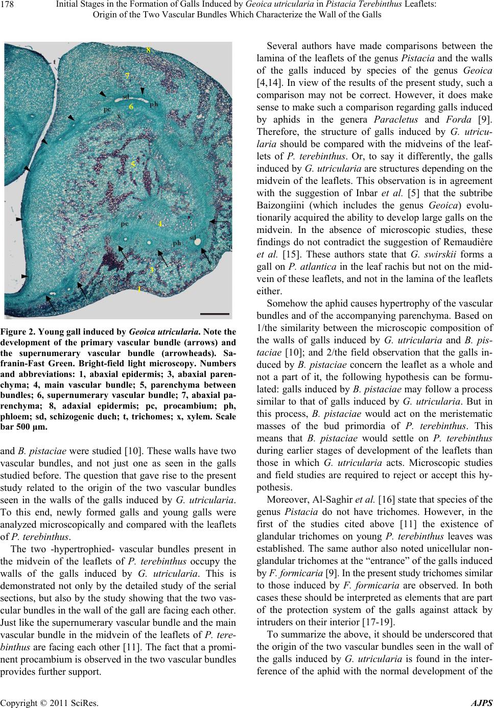
midvein of the leaflets, especially of the two vascular
bundles and the surrounding parenchyma cells.
5. Acknowledgements
The author wishes to thank to Nicolas Pérez Hidalgo,
Antonio Enzina García, Adoración Candelas González
and Juan Nieto Nafría for revising the manuscript and
Ronald Hartong of TECcientífica for his linguistic assis-
tance. I also thank the Junta de Castilla y León for fund-
ing project LE006A09.
REFERENCES
[1] N. J. Spiller, F. M. Kimmins and M. Llewellyn, “Fine
Structure of Aphid Stylet Pathways and Its Use in Host
Plant Resistance Studies,” Entomologia Experimentalis et
Applicata, Vol. 38, No. 3, 1985, pp. 293-295.
doi:10.1111/j.1570-7458.1985.tb03534.x
[2] W. F. Tjallingh and T. Hogen-Esch, “Fine Structure of
Aphid Stylet Routes in Plant Tissues in Correlation with
EPG Signals,” Physiological Entomology, Vol. 18, No. 3,
1993, pp. 317-328.
doi:10.1111/j.1365-3032.1993.tb00604.x
[3] M. Inbar, “The Evolution of Gall Traits in the Fordinae,”
In: K. Ozaki, J. Yukawa, T. Ohgushi and P. E. Price, Eds.,
Ecology and Evolution of Galling Arthropods and Their
Associates, Springer-Verlag, Tokyo, 2006, pp. 265-273.
doi:10.1007/4-431-32185-3_23
[4] D. Wool, R. Aloni, O. Ben-Zvi and M. Wollberg, “A
Galling Aphid Furnishes Its Home with a Built-in Pipe-
line to the Host Food Supply,” Entomologia Experimen-
talis et Applicata, Vol. 91, No. 1, 1999, pp. 183-186.
doi:10.1046/j.1570-7458.1999.00482.x
[5] M. Inbar, M. Wink and D. Wool, “The Evolution of Host
Plant Manipulation by Insects: Molecular and Ecological
Evidence from Gall-Forming Aphids on Pistacia,” Mo-
lecular Phylogenetics and Evolution, Vol. 32, No. 2, 2004,
pp. 504-511. doi:10.1016/j.ympev.2004.01.006
[6] J. Meyer, “Plant Galls and Gall Inducers,” Gebrüder
Borntraeger, Berlin, 1987.
[7] M. S. Mani, “Ecology of Plant Galls,” Dr. Junk Publisher,
The Hague, 1964.
[8] M. Arduin and J. E. Kraus, “Anatomia e Ontogenia de
Galhas Foliares de Piptadenia Gonoacantha (Fabales,
Mimosaceae) ,” Boletim de Botânica da Universidade de
São Paulo, Vol. 14, 1995, pp. 109-130.
[9] R. Álvarez, A. Encina and N. P. Hidalgo, “Histological
Aspects of Three Pistacia Terebinthus Galls Induced by
Three Different Aphids: Paracletus Cimiciformis, Forda
Marginata and Forda Formicaria,” Plant Science, Vol.
176, No. 2, 2009, pp. 303-314.
doi:10.1016/j.plantsci.2008.11.006
[10] R. Álvarez, “Microscopic Study of the Walls of Galls
Induced by Geoica utricularia and Baizongia Pistaciae in
Pistacia Terebinthus: A Contribution to the Phylogeny of
Fordini,” Arthropod-Plant Interactions, 2011, submitted
for publication.
[11] R. Álvarez, A. Encina and N. P. Hidalgo, “Pistacia Tere-
binthus L. Leaflets: An Anatomical Study,” Plant Sys-
tematic Evolution, Vol. 272, No. 1-4, 2008, pp. 107-118.
doi:10.1007/s00606-007-0640-0
[12] B. Ortiz-Rivas, D. Martínez-Torres and N. Pérez-Hidalgo,
“Molecular Phylogeny of Iberian Fordini (Aphididae:
Eriosomatinae): Implications for the Taxonomy of Genera
Forda and Paracletus,” Systematic Entomology, Vol. 34,
No. 2, 2009, pp. 293-306.
doi:10.1111/j.1365-3113.2008.00464.x
[13] M. Martínez-Millán and S. R. S. Cevallos-Ferriz, “Ar-
quitectura Foliar de Anacardiaceae. Leaf Architecture of
Anacardiaceae,” Revista Mexicana de Biodiversidad, Vol.
76, 2005, pp. 137-190.
[14] D. Wool and N. Bar-El, “Population Ecology of the Gall-
ing Aphid Forda Formicaria von Heyden in Israel:
Abundance, Demography, and Gall Structure,” Israel
Journal of Zoology, Vol. 41, 1995, pp. 175-192.
[15] G. Remaudière, M. Inbar, J. Menier and A. Sumida, “Un
Nouveau Geoica Gallicole sur Pistacia atlantica en Jor-
danie Hemiptera, Aphididae, Eriosomatinae, Fordini,”
Revue Française dEntomologie, Vol. 26, No. 1, 2004, pp.
37-42.
[16] M. G. Al-Saghir, D. M. Porter and E. T. Nilsen, “Leaf
Anatomy of Pistacia Species (Anacardiaceae),” Journal
of Biological Sciences, Vol. 6, No. 2, 2006, pp. 242-244.
doi:10.3923/jbs.2006.242.244
[17] J. Meyer and J. Maresquelle, “Anatomie des Galles,”
Ggebrüder Borntrager, Berlin, 1983.
[18] A. T. Simmons, G. M. Gurr, D. McGrath, H. I. Nicol and
P. M. Martin, “Trichomes of Lycopersicon spp. and Their
Effect on Myzus Persicae (Sulzer) (Hemiptera: Aphidi-
dae),” Australian Journal of Entomology, Vol. 42, 2003,
pp. 373-378. doi:10.1046/j.1440-6055.2003.00376.x
[19] M. Arduin, G. W. Fernandes and J. E. Kraus, “Morpho-
genesis of Galls Induced by Baccharopelma Dracunculi-
foliae (Hemiptera: Psyllidae) on Baccharis Dracunculifo-
lia (Asteraceae) Leaves,” Brazilian Journal of Biology,
Vol. 65, No. 4, 2005, pp. 559-571.
doi:10.1590/S1519-69842005000400002
Copyright © 2011 SciRes. AJPS