Surgical Science, 2011, 2, 177-182
doi:10.4236/ss.2011.24039 Published Online June 2011 (http://www.SciRP.org/journal/ss)
Copyright © 2011 SciRes. SS
Percutaneous Mesh Expansion and Fixation at the
Retro-Rectus Plane without Stabs by Using Redirecting
Suture Hook in Midline Hernias Repair
Ahmed E. Lasheen, Alaa N. El Sadek, Adel M. Tolba, Emad Salah, Ayman F. Mehanna
General Surgery Departme n t , Faculty of Medicine, Zagazig University, Zagazig, Egypt
E-mail: leniem@gmail.com, tgkim@chonnam.ac.kr
Received December 27, 2010; revised April 10, 201 1; accepted April 21, 201 1
Abstract
Background: Mesh expansion and fixation at retro-rectus plane through multiples stabs produces good results.
But these stabs cause cosmetic disorders for the patients and doctors. So, we find some modification to do
this procedure without these stabbing wounds in midline hernial repair. Patients and methods: This technique
was used to fix the mesh at retro-rectus plane in 50 patients suffering from midline hernias, from January
2008 through January 2010 at Zagazig university Hospital, Egypt. Laparotomy incision was done over the
hernial sac or at old incision; the contents were then released and reduced into peritoneal cavity without
much subcutaneous dissection. The suitable sheet of polypropylene mesh to cover the hernial defect and any
weak area was prepared and fixed at retro-rectus plane percutaneously without stabbing wounds by using
redirecting suture hook. The mean period of follow up was 26 months. Results: There was no recurrence
during the period of follow up. Five patients developed subcutaneous bluish discoloration at the site of some
stitches, which disappear within two weeks with conservative treatment. Conclusion: Percutaneous mesh
expansion and fixation at retro-rectus plane by using redirecting suture hook procedure has good results in
recurrence rate and cosmetic appearance.
Keywords: Percutaneous Fixation, Retro-Rectus Mesh, Redirecting Suture Hook
1. Introduction
The repair of incisional and ventral hernias continues to
be a surgical challenge. Reports published in the medical
literature indicate 3% to 13% of laparotomy patients de-
velop incisional hernias. [1] Moreover, clinical studies
indicate that the traditional, or open technique to repair
large abdominal wall defects is associated with recur-
rence rates ranging from 25% - 49%. [2,3] Multiple
studies have documented that traditional open ventral
hernioplasty has significant complication as infection,
hematoma, seroma, chronic sinus tract formation, mesh
extrusion, fistula formation and non healing wound. [4]
In addition, the wide dissection of soft tissue that is re-
quired for a Stoppa type retro-rectus repair or a Cherrel
type anterior repair leads to the many wound related
problems. [5] The stabbing technique for mesh expan-
sion and fixation at retro-rectus plane associated with
good results and avoided the all wound complications, as
this procedure not associated by much soft tissue dissec-
tion. [6] But, the multiple stab wounds in the anterior
abdominal wall through which every stitch was tied,
cause cosmetic disorder for patients and doctors. So,
some modification was done in stabbing technique to tie
the stitches at anterior plane to the anterior rectus sheath
without made these multiple stab wound s. What is called
percutaneous mesh expansion and fixation at retro-rectus
plane by using redirecting suture hook in midline hernias
repair.
2. Patients and Methods
Fifty patients with midline hernias were included in this
study at the Zagazig University Hospital, Egypt from
January 2008 through Janu ary 2010. Thirty patients have
incisional hernias after midline laparotomy incision and
twenty patients have big midline hernias, with diverca-
tion recti (37 female and 13 male). Full informed discus-
sion with all patients about the nature of this technique
and taken acceptance from every patient were done be-
178 A. E. LASHEEN ET AL.
fore beginning of the work including their data in this
study. The patients ages were ranged from 20 to 56 years
(mean age was 42.7 years). All patients were subjected to
percutaneous mesh expansion and fixation at retro-rectus
plane by using redirecting suture hook. Small incision is
placed on hernial sac or in old incision. The sac is dis-
sected and open without much subcutaneous dissection,
the sac contents are returned to abdominal cavity after
freeing of all adhesion, and peritoneum cavity is closed.
The retro-rectus plane is reached by longitudinal inci-
sions on the medial borders of rectus sheaths on both
side without subcutaneous dissection. The posterior rec-
tus sheaths on both sides are approximated together by
continuous suture using 2/0 polypropylene suture to ap-
proximate the two recti and correct divercation. Sheet of
polypropylene mesh to cover the hernial defect and any
weak area of the anterior abdominal sheath all around by
about 5 cm is prepared and suitable numbers of sutures
are placed on the edge of mesh using No. 0 non absorb-
able suture (polypropylene). The centre of mesh is fixed
in midline by using 2/0 polypropylene. The two tip hole
needles are passed through the skin of anterior abdomi-
nal wall without stab wound to retro-rectus plane and
then, came to the main wound to br ing the suture strands
outside the skin and caught them by artery forceps. After
completion of this procedure for all mesh sutures, the
expansion and fixation of mesh can be examined, new
sutures can be added if needed at this step. The redirect-
ing suture hook [7] is passed from the operation wound
at the anterior plane to the anterior rectus sheath to catch
the two strands of each suture, and then, it is withdrawn
to bring the two su ture strands from the operation wound.
Then, the two suture strands are tied through the opera-
tion wound, where the suture knot lies directly on the
anterior abdominal sheath (Figures 1(a)-(e)). The ante-
rior rectus sheaths on both sides are approximated by
some stitches and the wound is closed in layers with suc-
tion drainage (Figures 2(a)-(e)). Early ambulation was
allowed. Oral feeding was allowed 24 hours postopera-
tively unless the intestinal sounds were absent or there
was abdominal distension. The patients were discharged
from hospital 6 hours after starting the oral feeding, if
there was no vomiting or abdominal distension. The fol-
low up period was ranged from 6 to 30 months (Mean
period was 26 months).
3. Results
The operation time ranged from 60 to 120 minutes
(Mean 90 minutes). The length of the hospital stay aver-
aged 2.3 days (Range 1.5 to 2.5 days). The drainage was
removed after 5 to 9 days according to seroma amount
comes through it. Five pa tients show b luish discolor ation
at the skin corresponding to the site of passage of redi-
recting suture hook at anterior plane of anterior rectus
sheath (Figure 3). This bluish discoloration disappeared
with conservative treatment after about 2 weeks. No re-
currence or other complications have been recorded during
(a)
(b)
Copyright © 2011 SciRes. SS
A. E. LASHEEN ET AL.
179
(c)
(d)
(e)
Figure 1. (a)-(e) Diagram of mesh fixation with redirecting suture hook in midline hernias repair procedure. 1-Main opera-
tion wound. 2-Skin and subcutaneous tissue.3-Anterior rectus sheath. 4-Rectus muscle. 5-Posterior rectus sheath.
6-Abdominal cavity. 7-Sheet of polypropylene mesh. 8-Two tip hole needles. 9-Redirecting suture hook. (a) Limited dissection
to expose the medial borders of rectus sheath on both sides and two longitudinal incisions were done. Then, continuous suture
to approximate posterior layers of rectus sheaths on both sides in midline was done. The suitable mesh in dimensions was
prepared and putted suitable number of sutures on its edges. Then, the two tip hole needles passed from skin of anterior ab-
dominal wall to retro-rectal plane to appear from operation wound; (b) The strands of the corresponding suture were fixed to
the hole of each needle, then withdrawing the two needles back to bring the suture strands from the skin of anterior abdomi-
nal wall. The suture strands were detached from each needle and caught with artery forceps. Then, all the previous steps
were return with all mesh sutures to bring each suture strands from corresponding site of anterior abdominal wall; (c) Com-
pletion of putting all sutures in their corresponding skin site of anterior abdominal wall was done. Then, examined the mesh
fixation and expansion, and can be added new sutur es to produc e well fixed and e xpanded me sh. The redir ecting suture hook
was passed through the operation wound at anterior fascial plane to catc h the two strands of suture and withdraw n; (d) All
sutures strands were completed to bring from operation wound and caught by artery forceps; (e) All mesh sutures strands
were tied through the operation wound and their notes lied directly on anterior rectus sheath. Then, approximation of two
anterior rectus sheaths ove r mesh and wound was close d wi th dr ain.
Copyright © 2011 SciRes. SS
180 A. E. LASHEEN ET AL.
(a) (b)
(c) (d)
(e)
Figure 2. (a)-(e) Photo of mesh fixation with redirecting suture hook in midline hernias repair procedure. Male patient has
midline incisional hernia after exploratory laparotomy, where subjected to this procedure for repair. (a) Finishing of putting
all sutures which required producing good expanded and fixed mesh was complete. At first the each suture strands came
from the skin of anterior abdominal wall; (b) The redirecting suture hook w as passed through the operation wound at ante-
rior fascial plane to catch the tw o strands of suture and w ithdraw n to bring them fr om operation wound; (c) The tw o str ands
of each suture were came from the operation wound and caught by artery forceps. (d) Suture strands of each suture were tied
through the operation wound and the knot lied directly on the anterior rectus sheath; (e) Closure of the anterior rectus
heaths over the mesh is done. s
Copyright © 2011 SciRes. SS
A. E. LASHEEN ET AL.
Copyright © 2011 SciRes. SS
181
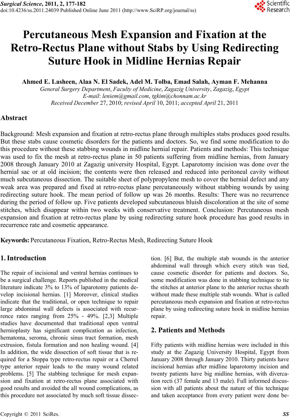
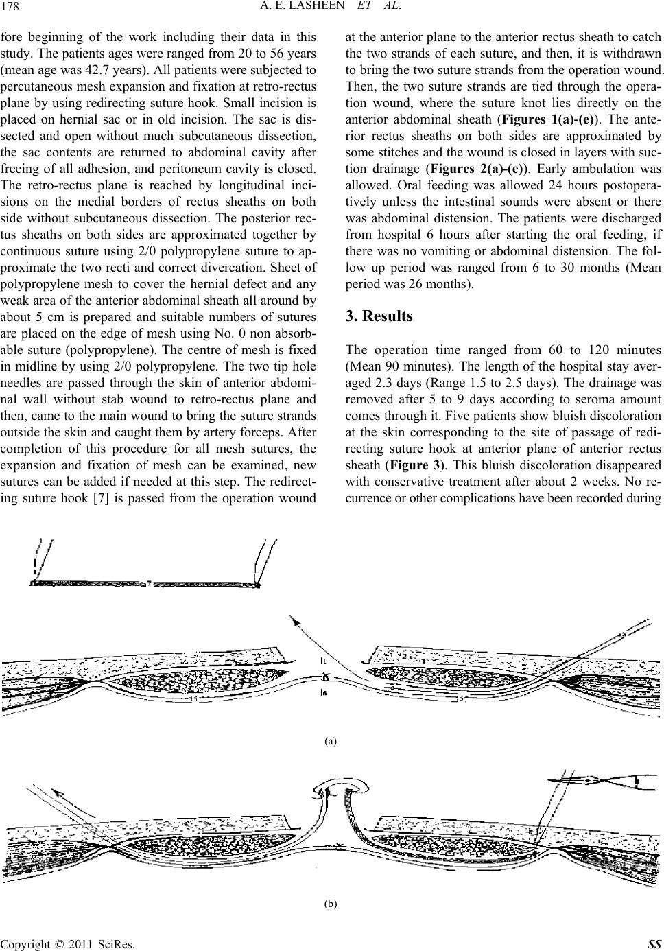
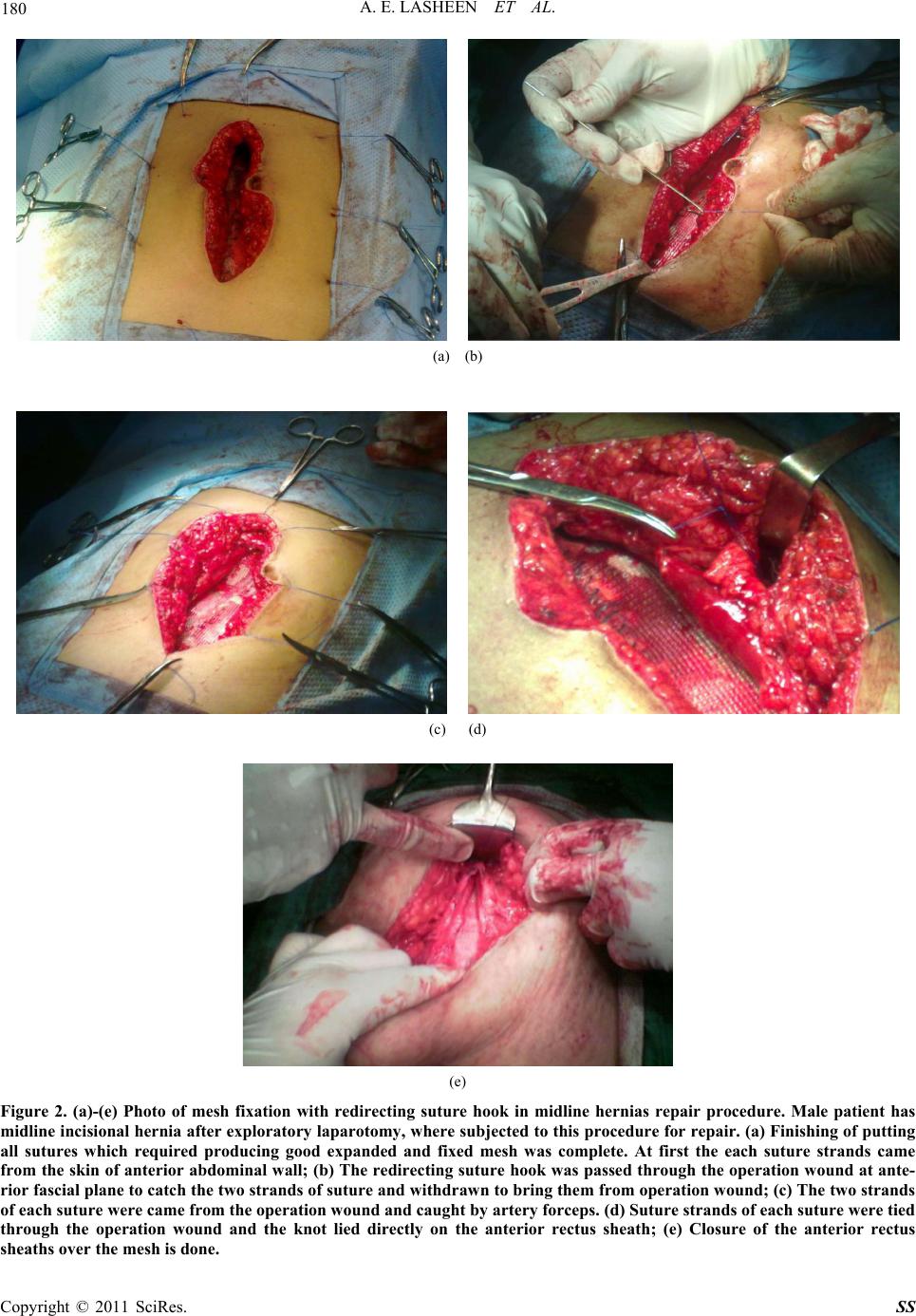
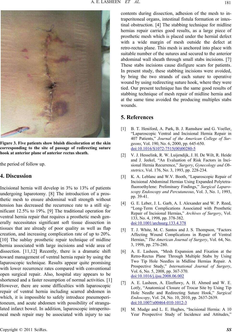
Figure 3. Five patients show bluish discoloration at the skin
corresponding to the site of passage of redirecting suture
hook at anterior plane of anterior rectus sheath.
the period of follow up.
4. Discussion
Incisional hernia will develop in 3% to 13% of patients
undergoing laparotomy. [8] The introduction of a pros-
thetic mesh to ensure abdominal wall strength without
tension has decreased the recurrence rate to a still sig-
nificant 12.5% to 19%. [9] The traditional operation for
ventral hernia repair that requires a prosthetic mesh gen-
erally necessitates significant soft tissue dissection in
tissues that are already of poor quality as well as flap
creation, and increasing complication rate of up to 20%.
[10] The sublay prosthetic repair technique of midline
hernia associated with large incisions and wide area of
dissection. [11,12] Recently, there is a dramatic shift
toward management of ventral hernia repair by using the
laparoscopic technique. Results appear quite promising
with lower recurrence rates compared with conventional
open surgical repair. Also, hospital stay appears to be
shortened and a faster resumption of normal activities. [1]
However, there are some difficulties with laparoscopic
repair of ventral hernia including scarred abdomen in
which, it is impossible to safely introduce pneumoperi-
toneum, and acute abdomen with possibility of strangu-
lated infarct bowel. In addition, laparoscopic intraperito-
neal mesh repair may be associated with injury to sac
contents during dissection, adhesion of the mesh to in-
traperitoneal organs, intestinal fistula formation or intes-
tinal obstruction. [4] The stabbing technique for midline
hernias repair carries good results, as a large piece of
prosthetic mesh which is placed under the hernial defect
with a wide margin of mesh outside the defect at
retro-rectus plane. This mesh is anchored into place with
suitable number of the sutures and secured to the anterior
abdominal wall sheath through small stabs incisions. [7]
These stabs incisions cause disfigure scars for patients.
In present study, these stabbing incisions were avoided,
by bring the two strands of each suture to operative
wound by using redirecting suture hook, where they were
tied. Our present technique has the same good results of
stabbing technique of mesh repair of midline hernia and
at the same time avoided the producing multiples stabs
wounds.
5. References
[1] B. T. Heniford, A. Park, B. J. Ramshaw and G. Voeller,
“Laparoscopic Ventral and Incisional Hernia Repair in
407 Patients,” Journal of the American College of Sur-
geons, Vol. 190, No. 6, 2000, pp. 645-650.
doi:10.1016/S1072-7515(00)00280-5
[2] V. J. Hesselink, R. W. Luijendijk, J. H. De Wilt, R. Heide
and J. Jeekel, “An Evaluation of Risk Factors in Inci-
sional Hernia Recurrence,” Surgery, Gynecology and Ob-
stetrics, Vol. 176, No. 3, 1993, pp. 228-234.
[3] K. A. Leblanc and W.V. Booth, “Laparoscopic Repair of
Incisional Abdominal Hernias Using Expanded Polytetra-
fluoroethylene: Preliminary Findings,” Surgical Laparo-
scopy Endoscopy and Percutaneous, Vol. 3, No. 1, 1993,
pp. 39-41.
[4] G. E. Leber, J. L. Garb, A. I. Alexander and W. P. Reed,
“Long-Term Complications Associated with Prosthetic
Repair of Incisional Hernias,” Archives of Surgery, Vol.
133, No. 4, 1998, pp. 378-382.
doi:10.1001/archsurg.133.4.378
[5] T. J. White, M. C. Santos and J. S. Thompson, “Factors
Affecting Wound Complications in Repair of Ventral
Hernias,” The American Journal of Surgery, Vol. 64, No.
3, 1998, pp. 276-280.
[6] A. E. Lasheen, “Mesh Expansion and Fixation at the
Retro-Rectus Plane Through Multiple Stabs by Using
Two Tip Hole Needles in Midline Hernias Repair. A
Prospective Study,” International Journal of Surgery,
Vol. 6, No. 5, 2008, pp. 367-370.
doi:10.1016/j.ijsu.2008.06.002
[7] A. E. Lasheen, A. Elzeftawy, A. H. Ahmed and W. E.
Lotfy, “Anatomical Closure of Trocar Site by Using Tip
Hole Needle and Redirecting Suture Hook,” Surgical
Endoscopy, Vol. 24, No. 10, 2010, pp. 2637-2639.
doi:10.1007/s00464-010-1012-3
[8] M. Mudge and L. E. Hughes, “Incisional Hernia: A 10
Year Prospective Study of Incidence and Attitudes,”
182 A. E. LASHEEN ET AL.
British Journal of Surgery, Vol. 72, No. 1, 1985, pp. 70-71.
doi:10.1002/bjs.1800720127
[9] J. J. Bauer, M. T. Harris, I. Kreel and I. M. Gelernt,
“Twelve-Year Experience with Expanded Polytetra-
fluoroethylene in the Repair of Abdominal Wall De-
fects,” Mount Sinai Journal of Medicine, Vol. 66, No. 1,
1999, pp. 20-25.
[10] D. Lomanto, S. G. Iyer, A. Shabbir and W. K. Cheah,
“Laparoscopic Versus Open Ventral Hernia Mesh Repair:
A Prospective Study,” Surgical Endoscopy, Vol. 20, No.
7, 2006, pp. 1030-1035. doi:10.1007/s00464-005-0554-2
[11] R. E. Stoppa, “The Treatment of Complicated Groin and
Incisional Hernias,” World Journal of Surgery, Vol. 13,
No. 5, 1989, pp. 545-554. doi:10.1007/BF01658869
[12] G. E. Wantz, “Incisional Hernioplasty with Mersilene,”
Surgery, Gynecology and Obstetrics, Vol. 172, No. 2, 1991 ,
pp. 129-137.
Copyright © 2011 SciRes. SS