Surgical Science, 2011, 2, 163-165
doi:10.4236/ss.2011.24035 Published Online June 2011 (http://www.SciRP.org/journal/ss)
Copyright © 2011 SciRes. SS
Tr icuspid Valve Reconstruction in a Patient with
Ventricular Septal Defect and Subacute Endocarditis
Hong-bin Wang1, Min Li1, Ming Zhang1, Yan-ling Zhu2, Hao Wen2
1 Department of Cardiac-Thoracic Surgery, Fengxian District Central
Hospital of Shanghai, Shanghai, China
2Department of Hepatobiliary Surgery, The First Teaching Hospital of
Xinjiang Medical University, Xinjiang, China
E-mail: whb627@126.com
Received April 24, 2011; revised May 13, 2011; accepted May 18, 2011
Abstract
Objective: Tricuspid valve reconstruction was advocated as the operative method for the treatment of tricus-
pid valve endocarditis recently. Many people accept that valve replacement therapy should be performed if
more than two valve leaflets are involved. The aim of the study to discuss if reconstructive surgery could be
done to treat two valve leaflets involved in tricuspid valve endocarditis. Methods: A 17-year-old boy with
ventricular septal defect (VSD) and tricuspid valve subacute endocarditis was surgical treated through extra-
corporeal circulation. two-thirds of the defective septal cusp, and half of the defective anterior cusp were ex-
cised during operation. The tricuspid valve was reconstructed with autologous pericardial strip, cusp com-
missuroplasty and 4-0 prolene sutures made as chordae tendineaes. VSD was repaired using a pericardial
patch. Results: The patient was discharged post-operation with excellent restoration of the tricuspid valve
activity. Cardiac ultrasound revealed normal tricuspid valve activity and low degree of regurgitation two
years follow-up. Conclusions: It seems that tricuspid valve reconstruction could be performed for two defec-
tive leaflets or half of the tricuspid valve.
Keywords: Tricuspid Valve Subacute Endocarditis, VSD, Reconstruction Technique
1. Introduction
Infective endocarditis often induce acute valvular insuf-
ficiency, and cause significant hemodynamic variations.
Surgical therapy is indicated if this disorder is associated
with other hear t ma l formations.
2. Material and Methods
A 17-year-old patient (Kirkiz nation ality) was transferred
to our hospital after experiencing recurrent fever for 4
months. The patient did not undergo regular anti-infec-
tive therapy. He had no history of drug addiction. Physi-
cal examination revealed a ystolic murmur (Grade 3/4)
existed in the left third and fourth intercostals. Blood
culture was negative. Cardiac ultrasound examination
revealed a tumor-like bulge in the membranous septum;
Color Doppler flow imaging revealed left to right shunt.
In the verge of ventricular septal defect, septal tricuspid
valve, and the chordae, abscission of the vegetation with
distinct sized of high echogenic mass induced septal
valve defect and considerable tricuspid regurgitation
(Figures 1 and 2). The electrocardiogram showed sinus
tachycardia. From above the diagnosis of congenital
VSD with subacute endocarditis was made, and antibio t-
ics were started.
A median sternotomy was performed 5 days after ad-
mission, after aortobicaval cannulation, cardiopulmonary
bypass was instituted at mild h ypoth ermia. The ao rta was
cross-clamped and cold blood cardioplegia was delivered
antegradely. After cardioplegic arrest, a right transverse
atriotomy allowed for an examination of the VSD and
the tricuspid valve. A perimembranous VSD was found
located near the aortic valve ring; the size of the defect
was about 1.2 × 0.8 cm, and a large vegetation had ac-
creted in the septal leaflet nearby the anterior valve. A
large vegetation had also accreted in the anterior leaflet
next to the septum leaflet, and another vegetation (di-
ameter, 0.6 cm) was seen on the anterior leaflet. In addi-
tion, a part of the chordae in the anterior and septal leaflets
164 H. B. WANG ET AL.
Figure 1. Aortic short axis view: anechoic region in the in-
terventricular septal periphery, vegetations adhesion to the
inter-stump gap.
Figure 2. Two-dime nsional color flow mapping in the apical
four-chamber view: abscission of the septal leaflet of the
tricuspid valve in the right atrium, significant vegetations
formation was seen in the septal and anterior leaflets. The
color graph showed a high degree of tricuspid regurgita-
tion.
was ruptured. The excess tissue was excised from the
annular edge of the anterior and the septal leaflet,
Therefore, we excised two-thirds of the defective septal
cusp, half of the defective anterior cusp, and the invo lved
chordae; the 0.6 cm vegetation in th e anterior leaflet was
also excised (Figure 3). VSD was repaired using a peri-
cardial patch and sutured continuously using 4-0 prolene
sutures. The small defective anterior cusp was patched
using a pericardial patch and sutured continuously using
6-0 prolene sutures. The brick-shaped pericardial strip
was ideal for the patch, and it was sheared into sizes
corresponding to anterior and septal leaflets. Then the
patch was stitched to anulus using running 4-0 sutures.
Because of the patient’s poor economic condition, we
constructed chords with prolone rather than gortex. After
Figure 3. Perioperative findings revealed significant vegeta-
tion formation in the tricuspid valve and anterior leaflets.
repeating ligaturing the same 4-0 prolene suture which
stitched in the anterior and posterior papillary muscles,
we made three artificial chordae to fix the free edge of
the pericardial prosthetic cusp. (Figure 4). One was
placed at the point of “the anterior and the spetal leaflet
commissure”, the other two were inserted at the suture
intersection of the natural and prosthetic cusps. The op-
timal length of the artificial chorda was determined by
filling the right ventricle with saline. The water filling
test showed slight regurgitation at the intersection of the
septum and the posterior valve. Using 3-0 prolene su-
tures, the tricuspid ring in the intersection of the septum
and the posterior tricuspid valve was shortened with a
figure-of-eight suture (cusp commissuroplasty), and the
water filling test revealed absence of regurgitation. The
patient was in normal sinus rhythm after the aortic
cross-clamp removed.
Extracorporeal circulation was successfully discontin-
ued, the bypass was maintained for 147 minutes and the
aortic clamp time was 94 minutes.
3. Results
The patient was treated with Cefazolin (2g/d) for 4
weeks after discharging from hospital and got recovered
successfully. The central venous pressure (CVP) postop-
eration was less than 6.5 cm H2O. Cardiac ultrasound
examination performed the ninth day postoperation re-
vealed restored apposition of the artificial septal and an-
terior leaflets of the tricuspid valve, low degree of tricus-
pid regurgitation in the systolic phase, and significant
restoration of the enlarged right atrium and ventricle
(Figure 5). Cardiac examinations during the 2-year-fol-
low-up period revealed normal tricuspid valve activity
and low degree of regurgitation. No recurrent symptoms
of subacute endo carditis were observed.
Copyright © 2011 SciRes. SS
H. B. WANG ET AL.
Copyright © 2011 SciRes. SS
165
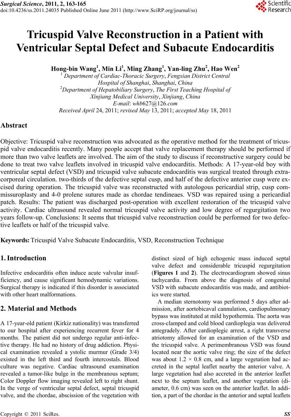
Figure 4. Operative procedures: (A) two large and one
small vegetations were presented at the anterior and septal
leaflets of the tricuspid valve, (B) two-thirds of the septal
cusp and half of the anterior cusp were excised, (C) peri-
cardial patch was used to replace the defected leaflets, arti-
fical chordae tendineae and Cusp commissuroplasty were
used to reconstruct the tricuspid valve.
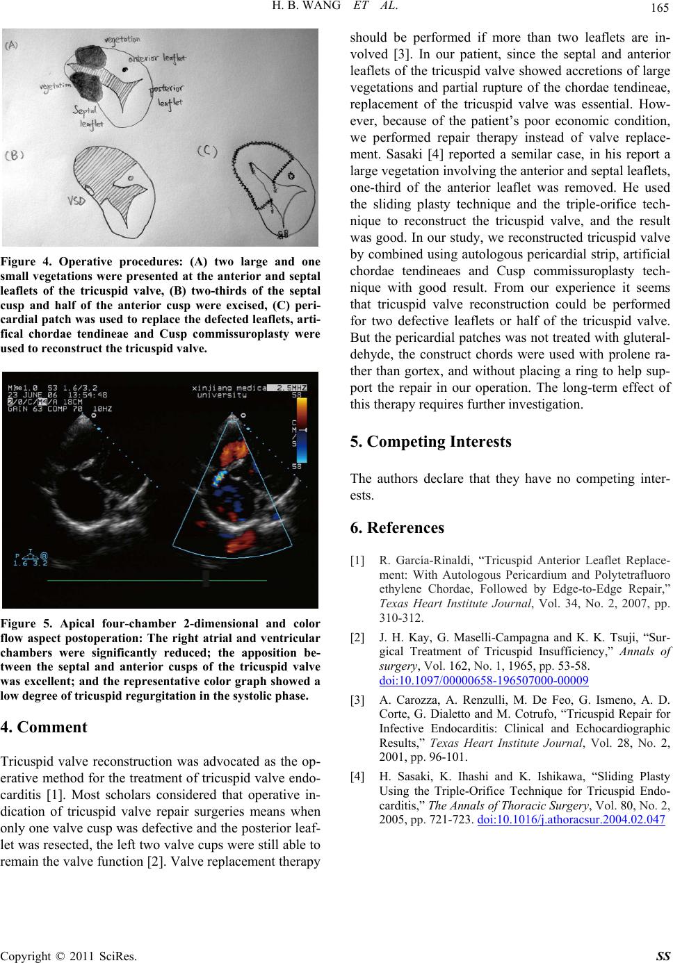
Figure 5. Apical four-chamber 2-dimensional and color
flow aspect postoperation: The right atrial and ventricular
chambers were significantly reduced; the apposition be-
tween the septal and anterior cusps of the tricuspid valve
was excellent; and the representative color graph showed a
low degree of tricuspid re g ur gi tation in the systolic phase.
4. Comment
Tricuspid valve reconstruction was advocated as the op-
erative method for the treatment of tricuspid valve endo-
carditis [1]. Most scholars considered that operative in-
dication of tricuspid valve repair surgeries means when
only one valve cusp was defective and the posterior leaf-
let was resected, the left two valve cups were still able to
remain the valve function [2]. Valve replacement therapy
should be performed if more than two leaflets are in-
volved [3]. In our patient, since the septal and anterior
leaflets of the tricuspid valve showed accretions of large
vegetations and partial rupture of the chordae tendineae,
replacement of the tricuspid valve was essential. How-
ever, because of the patient’s poor economic condition,
we performed repair therapy instead of valve replace-
ment. Sasaki [4] reported a semilar case, in his report a
large vegetation involv ing the an terior and septal leaflets,
one-third of the anterior leaflet was removed. He used
the sliding plasty technique and the triple-orifice tech-
nique to reconstruct the tricuspid valve, and the result
was good. In our study, we reconstruc ted tricuspid valve
by combined using autologous pericardial strip, artificial
chordae tendineaes and Cusp commissuroplasty tech-
nique with good result. From our experience it seems
that tricuspid valve reconstruction could be performed
for two defective leaflets or half of the tricuspid valve.
But the pericardial patch es was not treated with gluteral-
dehyde, the construct chords were used with prolene ra-
ther than gortex, and without placing a ring to help sup-
port the repair in our operation. The long-term effect of
this therapy requires further investigation.
5. Competing Interests
The authors declare that they have no competing inter-
ests.
6. References
[1] R. García-Rinaldi, “Tricuspid Anterior Leaflet Replace-
ment: With Autologous Pericardium and Polytetrafluoro
ethylene Chordae, Followed by Edge-to-Edge Repair,”
Texas Heart Institute Journal, Vol. 34, No. 2, 2007, pp.
310-312.
[2] J. H. Kay, G. Maselli-Campagna and K. K. Tsuji, “Sur-
gical Treatment of Tricuspid Insufficiency,” Annals of
surgery, Vol. 162, No. 1, 1965, pp. 53-58.
doi:10.1097/00000658-196507000-00009
[3] A. Carozza, A. Renzulli, M. De Feo, G. Ismeno, A. D.
Corte, G. Dialetto and M. Cotrufo, “Tricuspid Repair for
Infective Endocarditis: Clinical and Echocardiographic
Results,” Texas Heart Institute Journal, Vol. 28, No. 2,
2001, pp. 96-101.
[4] H. Sasaki, K. Ihashi and K. Ishikawa, “Sliding Plasty
Using the Triple-Orifice Technique for Tricuspid Endo-
carditis,” The Annals of Thoracic Surgery, Vol. 80, No. 2,
2005, pp. 721-723. doi:10.1016/j.athoracsur.2004.02.047