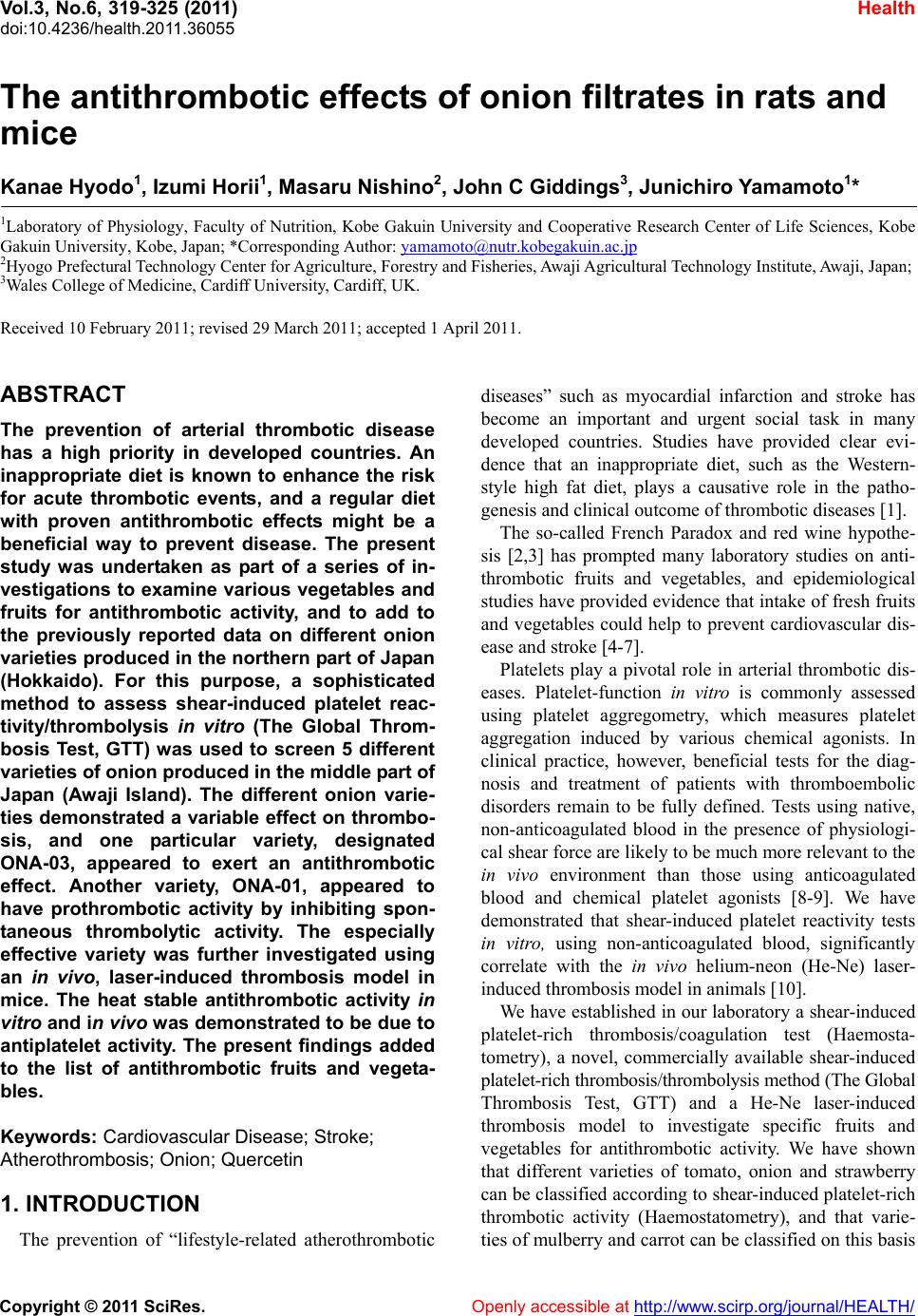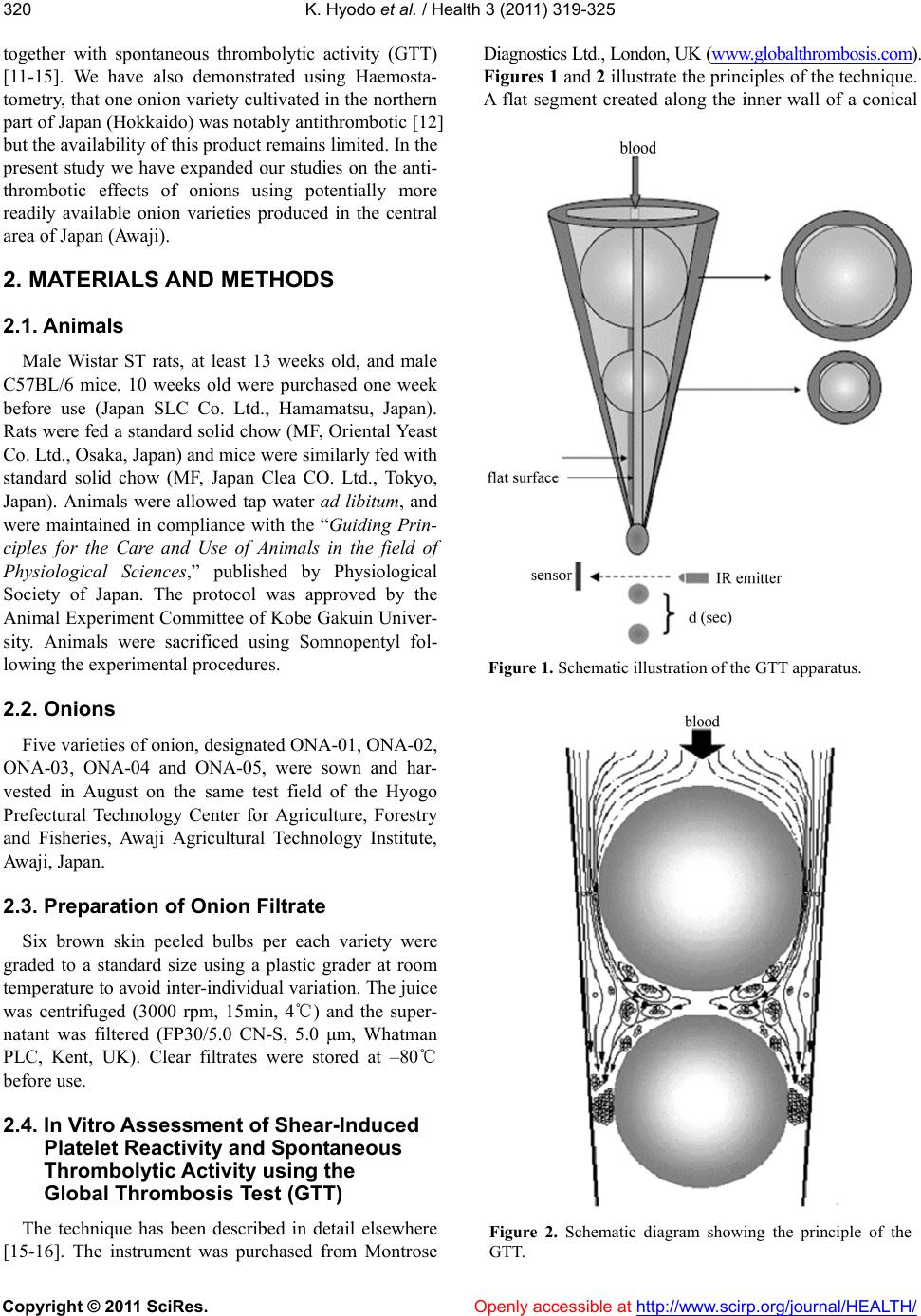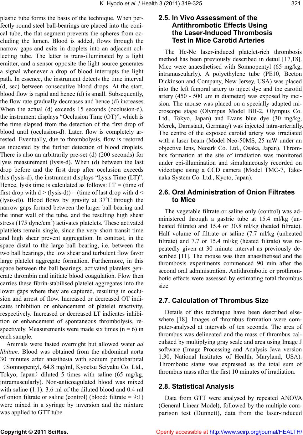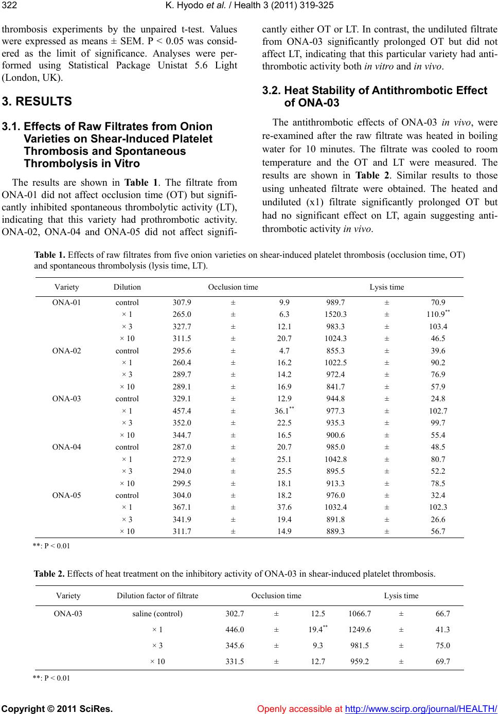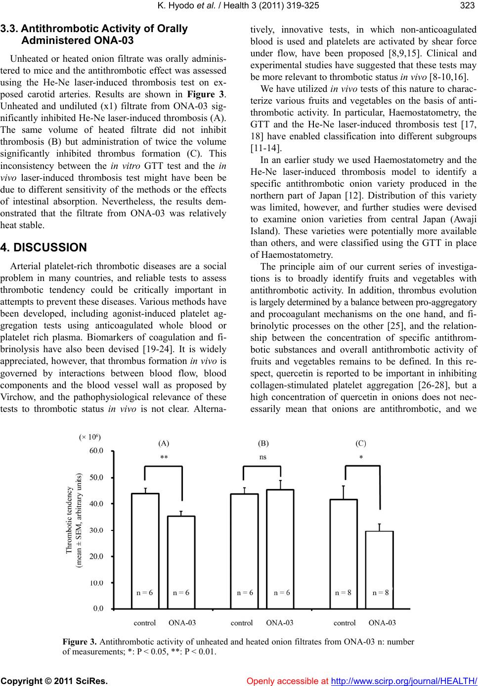
K. Hyodo et al. / Health 3 (2011) 319-325
Copyright © 2011 SciRes. Openly accessible at http://www.scirp.org/journal/HEALTH/
324
have previously demonstrated that there was no signifi-
cant correlation between quercetin concentration of on-
ions and overall antiplatelet activity using shear-induced
platelet function tests in vitro (Haemostatometry; 12).
Furthermore, the fibrinolytic properties of onions have
been the focus of some antithrombotic studies [29] but
significant fibrinolytic activity was not evident in any of
the five varieties of onions used in the present investiga-
tions. Moreover, for clinical purposes it is important to
note that the amounts of onion extracts given to animals
in the present studies would be approximately equivalent
to 40 kg onion given to a person with a body weight 70 kg.
This is clearly impracticable, and further studies are re-
quired to identify and isolate antithrombotic substances
that could be scientifically important and lead to signifi-
cant clinical benefit. Detailed analysis of this nature is
outside the scope of the present studies, but nevertheless,
our characterization of particular prothrombotic and an-
tithrombotic fruits and vegetables could lead to the de-
velopment of novel therapeutic products to help to pre-
vent arterial thrombotic diseases.
REFERENCES
[1] Lichtenstein, A.H., Appel, L.J., Brands, M., Carnethon,
M., Daniels, S., Franch, H.A., Franklin, B., Kris-Etherton,
P., Harris, W.S., Howard, B., Karanja, N., Lefevre, M.,
Rudel, L., Sacks, F., Van Horn, L., Winston, M. and
Wylie-Rosett, J. (2006) Summary of american heart as-
sociation diet and lifestyle recommendations revision
2006. Arteriosclerosis Th rombos is and Vascular Biology,
26, 2186-2191.
doi:10.1161/01.ATV.0000238352.25222.5e
[2] Ulbricht, T.L. and Southgate, D.A. (1991) Coronary heart
disease: seven dietary factors. Lancet, 338, 985- 992.
doi:10.1016/0140-6736(91)91846-M
[3] Renaud, S. and de Lorgeril, M. (1992) Wine, alcohol,
platelets, and the French paradox for coronary heart dis-
ease. Lancet, 339, 1523-1526.
doi:10.1016/0140-6736(92)91277-F
[4] Joshipura, K.J., Ascherio, A., Manson, J.E., Stampfer,
M.J., Rimm, E.B., Speizer, F.E., Hennekens, C.H., Spie-
gelman, D. and Willett, W.C. (1999) Fruit and vegetable
intake in relation to risk of ischemic stroke. Journal of
the American Medical Associat ion , 282, 1233-1239.
doi:10.1001/jama.282.13.1233
[5] Liu, S., Manson, J.E., Lee, I.M., Cole, S.R., Hennekens,
C.H., Willett, W.C. and Buring, J.E. (2000) Fruit and
vegetable intake and risk of cardiovascular disease: the
women’s health study. American Journal of Clinical Nu-
trition, 72, 922-928.
[6] Joshipura, K.J., Hu, F.B., Manson, J.E., Stampfer, M.J.,
Rimm, E.B., Speizer, F.E., Colditz, G., Ascherio, A.,
Rosner, B., Spiegelman, D. and Willett W.C. (2001) The
effect of fruit and vegetable intake on risk for coronary
heart disease. Annals of Internal Medicine, 134, 1106
-1114.
[7] Bazzano L.A, He J, Ogden L.G, Loria C.M, Vupputuri S,
Myers L. and Whelton P.K (2002) Fruit and vegetable
intake and risk of cardiovascular disease in US adults:
the first national health and nutrition examination survey
epidemiologic follow-up study. American Journal of
Clinical Nutrition, 76, 93-99.
[8] Ratnatunga, C.P., Edomondson, S.F., Rees, G.M. and
Kovacs, I.B. (1992) High-dose aspirin inhibits shear-in-
duced platelet reaction involving thrombin generation.
Circulation, 85, 1077-1082.
[9] Gorog, D.A. and Kovacs, I.B. (1995) Thrombotic status
analyser. Journal of Thrombosis and Haemostasis, 73,
514-520.
[10] Yamamoto, J. (2007) Prediction of thrombotic status by a
new test and prevention of thrombotic disorders by foods
with antithrombotic activity. The 7th TTM Forum Report
2007, 203-220, (Japanese).
[11] Yamamoto, J., Taka, T., Yamada, K., Ijiri, Y., Murakami,
M., Hirata, Y., Naemura, A., Hashimoto, M., Yamashita,
T., Oiwa, K., Seki, J., Suganuma, H., Inakuma, T. and
Yoshida, T. (2003) Tomatoes have natural antithrombotic
effects. British Journal of Nutrition, 90, 1031-1038.
doi:10.1079/BJN2003994
[12] Yamada, K., Naemura, A., Sawashita, N., Noguchi, Y. and
Yamamoto, J. (2004) An onion variety has natural anti-
thrombotic effect as assessed by thrombosis/thrombolysis.
Thrombosis Research, 114, 213-220.
doi:10.1016/j.thromres.2004.06.007
[13] Naemura, A., Mitani, T., Ijiri, Y., Tamura, Y., Yamashita,
T., Okimura, M. and Yamamoto, J. (2005) Anti-throm-
botic effect of strawberries. Blood Coagulation & Fibri-
nolysis, 16, 501-509.
doi:10.1097/01.mbc.0000184737.50594.a8
[14] Yamamoto, J., Naemura, A., Ura, M., Ijiri, Y., Yamashita,
T., Kurioka, A. and Koyama, A. (2006) Testing various
fruits for anti-thrombotic effect: I. Mulberries. Platelets,
17, 555-564.
doi:10.1080/09537100600759295
[15] Yamamoto, J., Yamashita, T., Ikarugi, H., Taka, T.,
Hashimoto, M., Ishii, H., Watanabe, S. and Kovacs, I.B.
(2003) Gorog Thrombosis Test: a global in-vitro test of
platelet function and thrombolysis. Blood Coagulation &
Fibrinolysis, 14, 31-39.
doi:10.1097/00001721-200301000-00007
[16] Saraf, S., Wellsted, D., Sharma, S. and Gorog, D.A.
(2009) Shear-induced global thrombosis test of native
blood: pivotal role of ADP allows monitoring of P2Y12
antagonist therapy. Thrombosis Research, 124, 447- 451.
doi:10.1016/j.thromres.2009.04.013
[17] Kovacs, I.B., Tigyi-Sebes, A., Trombitas, K. and Gorog, P.
(1975) Evans blue: an ideal energy-absorbing material to
produce intravascular microinjury by He-Ne gas laser.
Microvascular Research, 10, 107-124.
doi:10.1016/0026-2862(75)90025-4
[18] Ijiri, Y., Miura, M., Hashimoto, M., Fukunaga, C., Wata-
nabe, S., Kubota, A., Oiwa, K., Okuda, T. and Yamamoto,
J. (2002) A new model to evaluate the diet-induced
prothrombotic status, using He-Ne laser-induced throm-
bogenesis in the carotid artery of apolipoprotein
E-deficient and low-density lipoprotein receptor-deficient
mice. Blood Coagulation & Fibrinolysis, 13, 497-504.
doi:10.1097/00001721-200209000-00004