Vol.2, No.2, 146-157 (2011)
doi:10.4236/as.2011.22021
C
opyright © 2011 SciRes. Openly accessible at http://www.scirp.org/journal/AS/
Agricultural Science s
Optimization and production of antifungal hydrolysis
enzymes by Streptomyces aureofaciens against
Colletotrichum gloeosporioides of mango
Wafaa Mohamed Haggag1*, Enas Mostafa Mohamed2, Ahamed Mohamed El Azzazy2
1Department of Plant Pathology National Research Center, Cairo, Egypt; *Corresponding Author: wafaa_haggag@yahoo.com
2Biotechnology and Genetic Engineering Unit, National Research Center, Cairo, Egypt.
Received 9 March 2011; revised 15 March 2011; accepted 28 March 2011.
ABSTRACT
We isolated naturally occurring actinomycetes
with an ability to produce metabolites having
antifungal property against, Colletotrichum glo-
eosporioides, the causal agent of mango anth-
racnose. One promising strain was strong an-
tifungal activity, was selected for further studies.
Based on the physiological and biochemical ch-
aracteristics, the bacteria l strain was identical to
Streptomyces aureofaciens. Culture filtrate col-
lected from the exponential and stationary ph-
ases inhibited the growth of fungus tested, in-
dicating that growth suppression was due to
extracellular antifungal metabolites present in
culture filtrate. Isolate highly produced extrace-
llular chitinase and β-1,3-glucanase during the
exponential and late exponential phases, re-
spectively. In order to standardize the metabo-
lite production some cultural conditions like di-
fferent incubation time in hours, pH, carbon
sources and concentratio ns and nitro gen s o ur c e
were determined. During fermentation, growth,
pH and hydrolysis enzymes production were
monitored.Treatment with bioactive components
exhibited a significantly high protective activity
against development of anthracnose disease on
mango trees and increased fruit yi eld.
Keywords: Antifungal; Colletotrichum
Gloeospori oi des; Mango Anth ra cno se A nd
Streptomyces Aureofaciens
1. INTRODUCTION
Mango suffers from several diseases at all stages of its
life. Anthracnose disease caused by Colletotrichum
gloeosporioides (Glomerella cingulata Spauld & Sch-
renk) is one of the most common and serious diseases of
mango (Mangifera indica L.) in the tropics [1]. The dis-
ease occurs at any stage of fruit growth and the pathogen
causes the disease on a wide range of hosts such as apple,
pear, guava and mango [2]. Flower blight, fruit rot, and
leaf spots are among the symptoms of this disease [3].
Severe infection destroys the entire inflorescence result-
ing in no setting of fruits. Young infected fruits develop
black spots, shrivel and drop off. Fruits infected at ma-
ture stage carry the fungus into storage and cause con-
siderable loss during storage, transit and marketing . The
most visible evidence of disease occurs on postharvest
mango fruit by latent infection which usually results in
commercial losses [4]. Disease control methods include
the prophylactic use of fungicides such as benomyl, pro-
chloraz, mancozeb, carbendazim, iprodione and thia-
bendazol. Chemical fungicides not only may pollute the
atmosphere but also can be environmentally harmful, as
the chemicals spread out in the air and accumulate in the
soil, level and development of pathogen resistance [5].
Therefore, microbe-based biocontrol methods are one
alternative way to control diseases in place of agroche-
micals. Various microbial antagonists have been investi-
gated as potential antifungal biocontrol agents of plant
diseases. Many species of actinomycetes, especially those
belonging to the genus Streptomyces (Gram-positive,
mycelia-forming soil bacteria), are well known as bio-
control agents that inhibit or lyse several soilborne and
airborne plant pathogenic fungi [6,7]. The antifungal
potential of extracellular metabolites of Streptomyces
strains against some fungi was previously reported from
different locations of the world. The antagonistic activity
of Streptomyces to fungal pathogens is usually related to
the production of extracellular hydrolytic enzymes [8].
Chitinase and β-1,3-glucanase are considered to be im-
portant hydrolytic enzymes in the lysis of fungal cell
walls [9-12]. However, data related to the antagonistic
ability of the extracellular metabolites of Streptomyces
strains to suppress the growth of the fungal pathogens C.
W. M. Haggag et al. / Agricultural Sciences 2 (2011) 146-157
Copyright © 2011 SciRes. Openly accessible at http://www.scirp.org/journal/AS/
147
gloeosporioides having a broad host range are limited.
Thus ,this study investigated to study i) antifungal activ-
ity of the cell-free culture filtrate of this antagonist to
determine whether the production of the extracellular
hydrolytic enzymes is involved in its observed effect ii)
the optimization of antifungal metabolites production
and using selected antagonistic bacteria for the control of
pre- and post-harvest anthracnose on mango fruit.
2. MATERIAL AND METHODS
2.1. Organisms and Media
Isolation of the pathogen from mango fruit with
anthracnose symptom was performed by tissue trans-
planting technique using potato dextrose agar (PDA).
Stock cultures of C. gloeosporioides was maintained
on PDA slants and stored at 4˚C. Actinomycetes were
isolated from the root tissues of mango trees by the
surface-sterilization technique [13]. All cultures were
purified by streak plate technique and confirmed by
colony morphology and screened for their antifungal
activity [14].
2.2. In Vitro A ntifungal Activity of
Extracellular Metabolites in
Cell-Free Culture Filtrates
To prepare the cell-free culture filtrate, the antagonist
was cultured into broth medium and incubated on an
incubator shaker (150 rpm) at 28˚C. The fermentation
broth was collected during the exponential and station-
ary phases. Cells were removed by centrifugation at
8,000 rpm for 20 min at 4˚C. Cell-free supernatant was
filtered aseptically through a sterile membrane with 0.45-
μm pore size and stored at 4˚C. The growth inhibitory
effects of the assay as described previously by Prapag-
dee et al. [15] with some modifications. Minimum In-
hibitory Concentration (MIC) of these compounds were
determined.
2.3. Identification of Isolated Antagonist
One promising isolate which showed a unique, stable
and interesting property of inhibiting only dermatoph-
ytes was selected and characterized. Identification of the
isolate to species level was based on morphological,
cultural, physiological and biochemical characteristics as
described by Taechowisan & Lumyong [16].
2.4. Effect of Cultivation Conditions on
Enzyme Production
Chitinase assay. The reaction mixture contained 1 ml
of 0.1% colloidal chitin in sodium acetate buffer (0.05 M,
pH 5.2) and 1 ml culture filtrate was incubated at 37˚C
for 2 h in a water bath with constant shaking. Suitable
substrate and enzyme blanks were included. Chitinase
activity was assayed by the colorimetric method of [16].
The reaction was terminated by adding 0.1 ml of 0.08 M
potassium tetraborate, pH 9.2 to 0.5 ml of reaction mix-
ture and then boiled in a water bath for 3 min. Then 3 ml
of diluted pdimethyl amino benzaldehyde (p-DMAB,
Sigma Chemicals Company, USA) reagent was added
and again incubated at 37˚C for 15 min. The released
product in the reaction mixture was read at 585 nm in a
spectrophotometer (Hitachi, Japan). One unit of chitinase
activity was defined as the amount of enzyme, which
produces 1 μ mole of N-acetylglucosamine in 1 ml of
reaction mixture under standard assay condition.
β-1,3-Glucanase activity was assayed by colourimet-
ric method of Nelson [17]. Reaction mixtures were in-
cubated at 37˚C for 30 min and were stopped by boiling
for 5 min. One unit of B-1,3-glucanase activity was de-
fined as the amount of enzyme that releases 1 μmol of
reducing sugar equivalents (expressed as glucose) per
min.
2.5. Optimization of Incubation Period, PH
and Carbon and Nitrogen Sources
Concentration for A ntifungal Met abolite
Production
The optimization of composition of incubation period,
and cultural conditions was carried out based on step-
wise modification of the governing parameters for anti-
fungal production. The cultures were transferred to seed
broth (200 mL of Medium) contained in a 500 mL Er-
lenmeyer flask and incubated at 30˚C on a rotary shaker
(175 rpm) for 6 - 8 hours. A 500 mL Erlenmeyer flask
containing 200 mL of the same seed medium was incu-
bated as specified above. The seed culture was trans-
ferred to a 5 liter fermenter containing each one 3.5 liter
of the three liquid media
2.6. Assay in Liquid Cultures
Bioagents growth was estimated directly by spectro-
photometric measurement of the OD600 (Amax) using a
PM 2A spectrophotometer and dry biomass concentra-
tion (bmax). Changing the pH 3 to 10 in the production
medium the effect of pH was observed. The effect of
cultivation temperature on the antifungal production was
examined at different temperatures starting from 25˚C to
60˚C with 5˚C intervals.
2.7. Effect of Incubation Period on
Antifungal (Enzymes) Production
The effects of incubation period were evaluated by 24
h interval by checking the antifungal activity were also
W. M. Haggag et al. / Agricultural Sciences 2 (2011) 146-157
Copyright © 2011 SciRes. http://www.scirp.org/journal/AS/Openly accessible at
148
done. Hydrolysis enzymes was determined as previous
above. Culture optimization was carried out based on
stepwise modification of the governing parameters for
metabolites production and bioassay test at incubation
time in hours (24, 48, 72, 96 and 120); pH (6.0, 7.0, 7.5,
8.0 and 9.0); carbon sources and concentrations (glucose,
Cellulose, fructose, starch and sucrose) and nitrogen
source (KNO3, NaNO3, (NH4)2SO4, NH4NO3, NH4Cl,
urea, casein, yeast extract and peptone) in three repli-
cates. Fermentation studies were carried out in three
stages. To prepare inocula (b), a loopful spore from a
well-sporulated plate added each 40.0 ml respective seed
medium in 250 ml Erlenmeyer flasks and incubated at
28.0˚C for 48 h on a rotary shaker (150 rpm). After op-
timization of the fermentation parameters, 2.0 ml of the
seed culture (5.0%, v/v) was transferred to 250 ml Er-
lenmeyer flask containing 40.0 ml of the production me-
dium. The yield of the antifungal metabolite was moni-
tored in terms of arbitrary units (AU). Antifungal me-
tabolite production was carried out in 100 ml starch ca-
sein medium (starch 1%, casein 0.1%, KH2PO4 0.05%,
0.05%, MgSO4.7H2O, pH-7) in 500 ml Erlenmeyer
flasks.
2.8. In Vivo Evaluation of Antifungal
Activity
To determine the efficacy of the antifungal metabolite
against anthracnose pathogen, a field experiment was
conducted under natural infested conditions, using the
susceptible cultivars i.e. Sadekia (8˚ yr-old) at Noubaria
region, El Behera and Ismailia Sedekia (15˚ yr-old)
Governorates. Two sprayers were applied at 30 d inter-
vals starting from 1 st March (about one month before
normal flowering) on mango trees in one year. The bio-
active components at active concentration mixed with
0.1% carboxymethyl cellulase (CMC) as sticker was
sprayed using a knapsack sprayer. Trees were sprayed
till run-off with approximately 2˚ L of spray solution per
tree. Treatments were assigned in a randomized com-
plete block design. Trees sprayed with water and fungi-
cide served as a check treatment. Plots consisting of
three mango trees were replicated three times. Irrigation,
fertilization and other cultural practices were carried out
as recommended. The disease incidence was determined
as percentage of infected flowers at 30 days interval
during the growing season (March to July). Fruit yield
(kg/tree) were determined at harvesting date for treat-
ments.
2.8.1. Statistica l Analysis
The collected data were evaluated statistically using
the software SPSS for Windows (release 7.5.1, 20 De-
cember 1996; SPSS Inc., Chicago, IL). Data were sub-
jected to analyses of variance and treatment mean values
were compared by a modified Duncan’s multiple test (P
> 0.05).
3. RESULTS
3.1. Antifungal Activity
Preliminary screening for antifungal production was
done by streak on agar medium .Subsequent screening of
10 isolates that showed inhibitory activity against test
organism (Table 1). In particular, S. aureofaciens fol-
lowed by S. griseofuscus S2 showed significant activities
against pathogen. The antifungal activity of the purified
active substance against pathogen was determined. The
minimum inhibitory concentration (MIC) was deter-
mined by the diffusion method. The nutrient agar plates,
seeded with test organisms were used for the MIC de-
termination. The response was observed as a clear zone
(mm) around the paper discs (diameter 0.5 mm) loaded
with different concentration of active compound (20 μl)
of each concentration were spotted on paper discs. In
most cases, purified active substance of S. aureofaciens
showed antifungal activity against C. gloeosporioides
growth expressed as zone inhibition. The highest reduc-
tion was recorded at 1:3 concentration. The results in
Tabl e 1 showed the isolate S. aureofaciens was the best
isolate produces antifungal substances than the other
Streptomyces isolates. So, S. aureofaciens was chosen in
this study.
3.2. Enzymes Assays
The general ability of tested S. aureofaciens to pro-
duce secondary metabolites include hydrolysis enzymes
was determined (Figure 1). Exochitinase and β-1,3-glu-
canase appeared to be conman metabolites produced by
the tested BCAs. Maximum production of chitinase
and β-1,3-glucanase by the tested S. aureofaciens in
shaken broth culture occurred after 150 hrs (1.98 Unit/
ml) and (2.9 Unit/ml), respectively.
3.3. Optimization Condition for Antifungal
Metabolite Production
The time course for pH shows slight increase until 72
hrs then a negligible decrease through the remaining
time. The dry weight increases with time through all the
incubation period. The substrate decreases radically from
the beginning of incubation time till 120 hr then almost
stabilize. In the inhibition zone, enzymes curves, Colle-
totrichum follow the same pattern whereas the inhibition
zone, chitinase and β-1,3-glucanase increases with incu-
bation period till it reaches its maximum at 69 hr. The
time course for optical density shows that the maximum
W. M. Haggag et al. / Agricultural Sciences 2 (2011) 146-157
Copyright © 2011 SciRes. Openly accessible at http://www.scirp.org/journal/AS/
149
Table 1. Antifungal activity of Streptomyces spp. against Colletotrichum gloeosporioides.
Spore suspension Purified active substance
Isolate Inhibition zone (cm)Inhibition zone (cm) MIC
Streptomyces aureofaciens 2.6a 2.4a 1:32
S. albidoflavu 1.8c 1.6c 1:2
S. albidoflavus 1.9bc 1.7b 1:2
S. cyanocolor 1.8c 1.7b 1:2
S. griseofuscus S1 1.8c 1.3d 1:1
S. griseofuscus S2 2.1b 1.8b 1:16
S. nodosus, 1.8c 1.5bc 1:2
S. alanosinicus 2.0b 1.7b 1:2
S. rochei 1.8c 1.3d 1:1
S. mutabilis, 1.8c 1.3d 1:1
Values represent the mean percentage of six replicates. Values in each column followed by the same letter are not significantly
different (P < 0.05)
Figure 1. Hydrolysis enzymes activities in different days.
value is obtained at 36 hr incubation time, then a decline
occurs. This is explained in the residual substrate curve
as we notice that a gradual consumption of glucose
occurs till 36 hrs then the glucose concentration re-
mains constant for the rest of incubation time (Figure
2). From the experimental results it was noticed that by
using different types of carbon source, the production
of chitinase and β, 1-3 glucanse (unit/ml) were maxi-
mum with starch as carbon sources. Also, the zone in-
hibition of Colletotrichum was maximum after 48 h
incubation with starch as carbon sources. So, the starch
was chosen as an optimum carbon sources (Figure 3).
By using different concentration of starch for the
growth of Streptomyces, the dry weight concentration
increased after 24 h with increasing the glucose and
fructose concentrations till 5 g/l then decreased (Figure
3). Also, the production of β, 1-3 glucanse (unit/ml)
and Chitinase increased after 48 h by increasing the
stach concentration till 5 g/l then decreased again
(Figure 3). The zone inhibition of Colletotrichum in-
creased gradually by increasing the glucose concentra-
tion till 5 g/l then remained almost constant. From the
previous observations, 5 g/l was chosen as the optimum
glucose concentration (Figure 4). By using different
types of nitrogen source for the growth of Streptomyces
it was noticed that the maximum biomass growth was
obtained using malt extract and soyabean as nitogen
source while the maximum zone inhibition for Colleto-
tricum were obtained with malt extract and CSL. The
maximum production of Chitinase and β, 1-3 glucanse
Enzymes were obtained with peptone and peptone in
equal ratio in the media (Figure 5).
3.4. Field Experiments
More attention in the last decade has focused on re-
placing synthetic fungicides with new strategies, such as
the use of biological control agents. The efficacies of the
spraying mango trees cv. Sadekia with culture filtrate of
Streptomyces aureofaciens on controlling of anthracnose
disease were determined in season of 2010 in Boheria and
Ismailia Governorates under nature infection conditions.
In control treatment, the diseases incidence was higher on
W. M. Haggag et al. / Agricultural Sciences 2 (2011) 146-157
Copyright © 2011 SciRes. Openly accessible at http://www.scirp.org/journal/AS/
150
leaves and flowers blossom clusters in both regions. Sig-
nificant suppression of diseases incidence were achieved
by applying of culture filtrate of S. aureofaciens compared
to the fungicide and untreated control (Table 2). Applica-
tion of bioactive components of S. aureofaciens gave
completely reduced in all diseases, increased in flowering
and yield of both regions (Table 2).
5. DISCUSSION
In recent years, anthracnose has become a major chal-
lenge to both the pathologists working with the pathogen
and to mango researchers in general. The routine control
measure involving chemical pesticide application leads
to toxicity, residual effect and resistance development by
pathogens. The current situation is mainly focused on
biological control. Since all the commercial mango va-
rieties are susceptible to the disease, biological control
provides an effective, persistent and durable protection
Streptomyces aureofaciens had the ability to exhibit high
[18].
Figure 2. Effect of different nitrogen sources on the growth, inhibition activity and production
of hydrolysis enzymes by S. aureofaciens.
W. M. Haggag et al. / Agricultural Sciences 2 (2011) 146-157
Copyright © 2011 SciRes. Openly accessible at http://www.scirp.org/journal/AS/
151
Figure 2. Cont.
B,1-3 glucanase
W. M. Haggag et al. / Agricultural Sciences 2 (2011) 146-157
Copyright © 2011 SciRes. Openly accessible at http://www.scirp.org/journal/AS/
152
Figure 3. Effect of different carbon sources on the growth, inhibition activity and production
of hydrolysis enzymes by S. aureofaciens.
W. M. Haggag et al. / Agricultural Sciences 2 (2011) 146-157
Copyright © 2011 SciRes. Openly accessible at http://www.scirp.org/journal/AS/
153
0
0.2
0.4
0.6
0.8
1
1.2
1.4
1.6
1.8
2
Glucose. FructoseSucrose CelluloseStarch
B 1-3 glucanse Enzymes (uni
t
/ ml)
0
0.2
0.4
0.6
0.8
1.2
Ch itinase Enzym es (unit/ m
1
1.4
1.6
Glucose. FructoseSucroseCelluloseStarch
l)
Figure 3. Cont.
R2 = 0.9975
0.0000
5.0000
10.0000
15.0000
20.0000
25.0000
01234567
Starch conc. g/l
D ry weig h t g/l
R
2
= 1
5.8
5.9
6
6.1
6.2
6.3
6.4
6.5
6.6
6.7
6.8
01234567
Starch conc. g/l
pH
Figure 4. Effect of different carbon concentration on the growth, inhibition activity and production of hydrolysis enzymes by S.
aureofaciens.
R
2
= 0.9949
2.85
2.9
2.95
3
3.05
3.1
3.15
3.2
3.25
3.3
3.35
01234567
Starch conc. g/l
Inhibition zone (mm) 72h
R
2
= 0.9616
0
0.2
0.4
0.6
0.8
1
1.2
1.4
1.6
1.8
2
01234567
Starch co nc . g/ l
Chitinase Enzymes (unit/ ml)
R
2
= 1
0
0.5
1
1.5
2
02468
Starch conc. g/l
B 1-3 glucanse Enzymes
(un it / ml )
Figure 4. Cont.
B,1-3 glucanse Enzymes (unit/ml)
Chitinase Enzymes (unit/ml)
Inhibition zone (mm) 72 h
Chitinase Enzymes (unit/ml)
B,1-3 glucan se Enzymes (unit/ml)
W. M. Haggag et al. / Agricultural Sciences 2 (2011) 146-157
Copyright © 2011 SciRes. Openly accessible at http://www.scirp.org/journal/AS/
154
antifungal activity in vitro against Colletotrichum gloeo-
sporioides. The culture filtrate of this strain had also the
ability to in vivo suppress infection of Colletotrichum
gloeosporioides on mango trees. Many species of actin-
omycetes, especially those belonging to the genus Strep-
tomyces , are well known as biocontrol agents that inhibit
or lyse several soilborne and airborne plant pathogenic
fungi [7]. It is well known that Streptomyces sp. can
produce industrially useful compounds, notably wide
spectrum of antibiotics, as secondary metabolites, and
continues to be screened for new bioactive compounds
[12]. Optimization of fermentation media ingredients and
environmental factors for enzyme production is a more
convenient and effective strategy, compared to other re-
cent approaches like molecular techniques, to manifest
the physiological characteristics to synthesis enzymes.
Biological control of plant pathogens could reduce of
this concern. More recently an increased number of re-
searches focused on the potential of some types of bacte-
ria, yeast, and actinomycetes as a biocontrol agent
[9,11,6]. Regardless of the fermentation process that is
used to grow cells, it is necessary to monitor and control
parameters starting from the selection of optimum carbon
and nitrogen sources and including inoculum volume,
moisture content, pH, temperature, incubation period etc.
Changes in one of these parameters can have a dramatic
effect on the yield of cells and the stability of protein
product. The meaning of optimization in this context
needs careful consideration of the environmental and
nutritional parameters for growth and production. Me-
dium formulation is the foremost step for designing suc-
cessful laboratory experiments for yield enhancement.
0
1
2
3
4
5
6
7
8
9
M
alt extract(10 g
)+Yeast extract (4 g
)
M
alt extract(10 g
)+ Peptone (2.61 g
)
M
alt extract(10 g
)+ Soya beans (2.8 g
)
M
alt extract(10 g
)+ C
SL
(11.88 m
l)
Yeast extract (4 g
) + Peptone (0.2 g
)
Yeast extract (4 g
) + C
SL
(0.91 m
l)
Yeast extract (4 g
) + Soya B
eans
( 0.214 g
)
Peptone ( 1.41 g
) + Soya beans (1.51 g
)
Peptone ( 1.41 g
) + C
SL
(6.394 m
l)
Soya beans (1.51 g
) + C
SL
(6.394 m
l)
Yeast extract (4.31 g
)
M
alt extract (140.67 g
)
Peptone (2.81 g
)
C
SL
(12.79 g
)
Soya beans
(3.014 g
)
p
H
0
5
10
15
20
25
30
35
40
Malt extract(10 g)+Yeast extract (4 g)
Malt extract(10 g)+ Peptone (2.61 g)
Malt extract(10 g)+ Soya beans (2.8 g)
Malt extract(10 g)+ CSL (11.88 ml)
Yeast extract (4 g) + Peptone (0.2 g)
Yeast extract (4 g) + CSL (0.91 ml)
Yeast extract (4 g) + Soya Beans ( 0.214 g)
Peptone ( 1.41 g) + Soya beans (1.51 g)
Peptone ( 1.41 g) + CSL (6.394 ml)
Soya beans (1.51 g) + CSL (6.394 ml)
Yeast extract (4.31 g)
Malt extract (140.67 g)
Peptone (2.81 g)
CSL (12.79 g)
Soya beans (3.014 g)
Dry wei g ht g / l
Figure 5. Effect of different nitrogen sources on the growth, inhibition activity and produc-
tion of hydrolysis enzymes by S. aureofaciens.
W. M. Haggag et al. / Agricultural Sciences 2 (2011) 146-157
Copyright © 2011 SciRes. Openly accessible at http://www.scirp.org/journal/AS/
155
0
0. 5
1
1. 5
2
2. 5
Malt extract (10 g)+Yeast extract (4 g)
Malt extract(10 g)+ Peptone (2.61 g)
Malt extract(10 g)+ Soya beans (2.8 g)
Malt extract (10 g)+ CSL (11.88 ml)
Yeast extract (4 g) + Peptone (0.2 g)
Yeast extract (4 g) + CSL (0.91 ml)
Yeast extract (4 g) + Soya Beans ( 0.214 g)
Peptone ( 1.41 g) + Soya beans (1.51 g)
Peptone ( 1.41 g) + CSL (6.394 ml)
Soya beans (1.51 g) + CSL (6.394 ml)
Yeast extract (4.31 g)
Malt extract (140.67 g)
Peptone (2. 81 g)
CSL (12.7 9 g)
Soya beans (3. 014 g)
C olletotricum Zone inhibition (c m
)
0
0. 1
0. 2
0. 3
0. 4
0. 5
0. 6
0. 7
0. 8
0. 9
1
Malt ext ract(10
g)+Yeast extract (4 g)
Malt extract(10
g)+ Peptone (2.61 g)
Malt extract(10
g)+ Soya beans (2.8 g)
Malt ext ract(10
g)+ CSL
(11.88
ml)
Yeast extract (4 g) +
Peptone (0.2 g)
Yeast extract (4 g) + CSL
(0.91
ml)
Yeast extract (4 g) + Soya Beans ( 0.214
g)
Peptone ( 1.41
g) +
Soya beans (1.51 g)
Peptone ( 1.41 g) + CSL
(6.394
ml)
Soya beans (1.51
g) +
CSL
(6.394
ml)
Yeast extract (4.31
g)
Malt extract (140.67
g)
Peptone (2.81
g)
CSL
(12.79
g)
Soya beans (3.014
g)
Chitinase Enzymes (unit/ ml
)
0
0. 2
0. 4
0. 6
0. 8
1
1. 2
1. 4
1. 6
Malt extract(10
g)+ Yeast extract (4 g)
Malt extract(10
g)+ Peptone (2.61 g)
Malt extract(10
g)+ Soya beans (2.8 g)
Malt extract(10
g)+ CSL
(11.88
ml)
Ye ast extract (4 g) +
Peptone (0.2 g)
Yeast extract (4 g) +
CSL
(0.91
ml)
Yeast extract (4 g) + So ya Beans ( 0.214
g)
Peptone ( 1.41
g) +
Soya beans (1.51
g)
Peptone ( 1.41 g) + CSL
(6.394
ml)
Soya beans (1.51
g) +
CSL
(6.394
ml)
Ye ast extract (4.31
g)
Malt extract (140.67
g)
Peptone (2.81
g)
CSL
(12.79
g)
Soya beans (3.014
g)
B 1-3 glucanse Enzymes (u nit/ ml
)
Figure 5. Cont.
B,1-3 glucanse Enzymes (unit/ml)
W. M. Haggag et al. / Agricultural Sciences 2 (2011) 146-157
Copyright © 2011 SciRes. http://www.scirp.org/journal/AS/
156
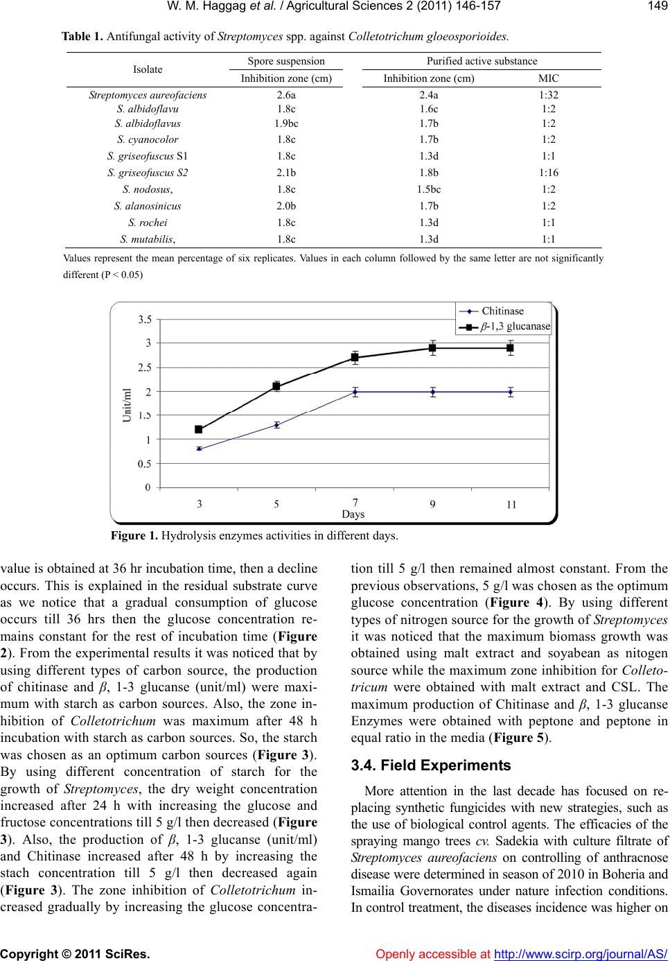
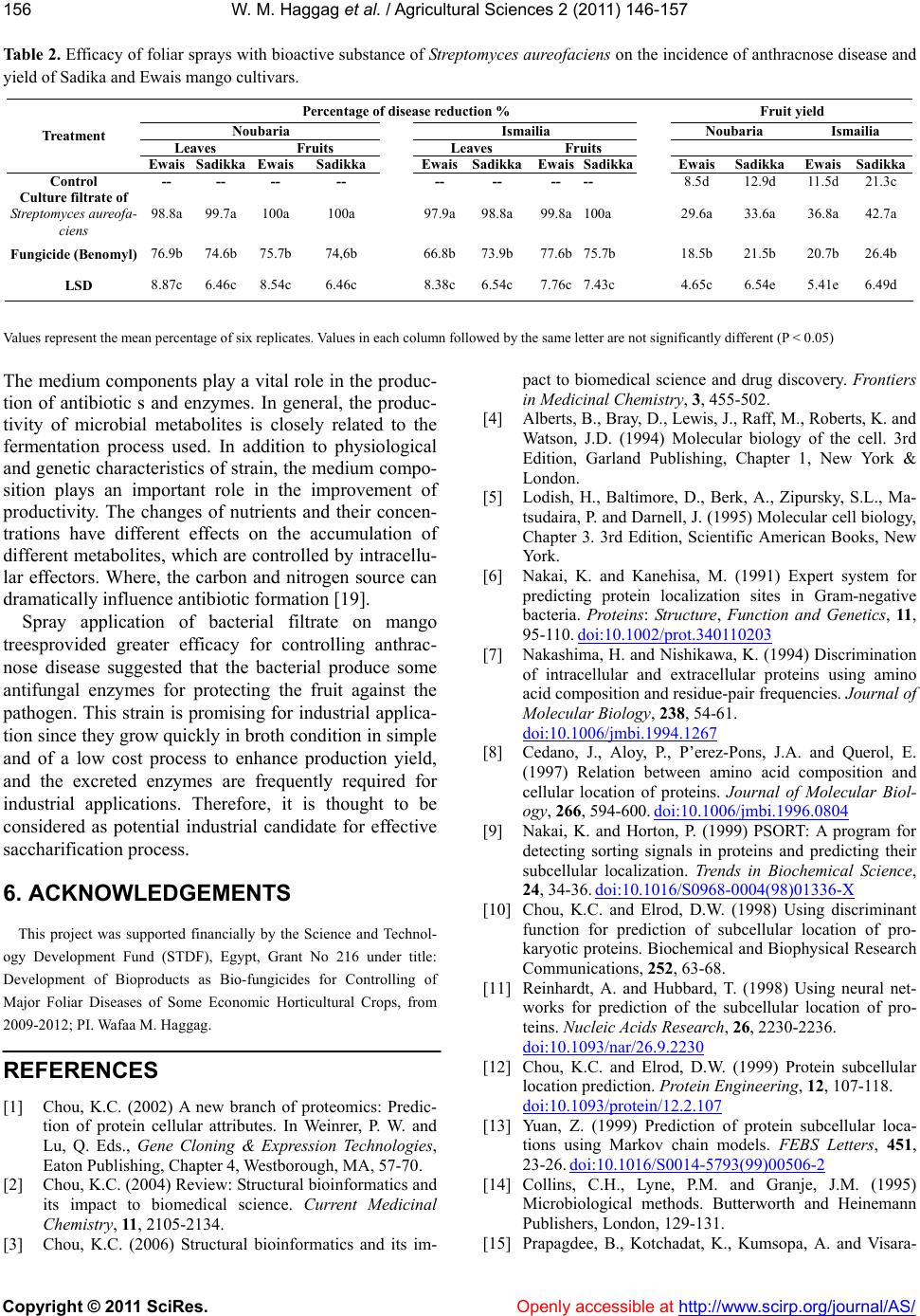
Table 2. Efficacy of foliar sprays with bioactive substance of Streptomyces aureofaciens on the incidence of anthracnose disease and
yield of Sadika and Ewais mango cultivars.
Percentage of disease reduction % Fruit yield
Noubaria Ismailia Noubaria Ismailia
Leaves Fruits Leaves Fruits
Treatment
Ewais Sadikka Ewais Sadikka EwaisSadikkaEwaisSadikka Ewais Sadikka EwaisSadikka
Control -- -- -- -- -- -- -- --
8.5d 12.9d 11.5d21.3c
Culture filtrate of
Streptomyces aureofa-
ciens 98.8a 99.7a100a 100a 97.9a98.8a 99.8a100a 29.6a 33.6a 36.8a42.7a
Fungicide (Benomyl) 76.9b 74.6b 75.7b 74,6b 66.8b73.9b 77.6b75.7b 18.5b 21.5b 20.7b26.4b
LSD 8.87c 6.46c 8.54c 6.46c 8.38c6.54c 7.76c7.43c 4.65c 6.54e 5.41e6.49d
Values represent the mean percentage of six replicates. Values in each column followed by the same letter are not significantly different (P < 0.05)
The medium components play a vital role in the produc-
tion of antibiotic s and enzymes. In general, the produc-
tivity of microbial metabolites is closely related to the
fermentation process used. In addition to physiological
and genetic characteristics of strain, the medium compo-
sition plays an important role in the improvement of
productivity. The changes of nutrients and their concen-
trations have different effects on the accumulation of
different metabolites, which are controlled by intracellu-
lar effectors. Where, the carbon and nitrogen source can
dramatically influence antibiotic formation [19].
Openly accessible at
Spray application of bacterial filtrate on mango
treesprovided greater efficacy for controlling anthrac-
nose disease suggested that the bacterial produce some
antifungal enzymes for protecting the fruit against the
pathogen. This strain is promising for industrial applica-
tion since they grow quickly in broth condition in simple
and of a low cost process to enhance production yield,
and the excreted enzymes are frequently required for
industrial applications. Therefore, it is thought to be
considered as potential industrial candidate for effective
saccharification process.
6. ACKNOWLEDGEMENTS
This project was supported financially by the Science and Technol-
ogy Development Fund (STDF), Egypt, Grant No 216 under title:
Development of Bioproducts as Bio-fungicides for Controlling of
Major Foliar Diseases of Some Economic Horticultural Crops, from
2009-2012; PI. Wafaa M. Haggag.
REFERENCES
[1] Chou, K.C. (2002) A new branch of proteomics: Predic-
tion of protein cellular attributes. In Weinrer, P. W. and
Lu, Q. Eds., Gene Cloning & Expression Technologies,
Eaton Publishing, Chapter 4, Westborough, MA, 57-70.
[2] Chou, K.C. (2004) Review: Structural bioinformatics and
its impact to biomedical science. Current Medicinal
Chemistry, 11 , 2105-2134.
[3] Chou, K.C. (2006) Structural bioinformatics and its im-
pact to biomedical science and drug discovery. Frontiers
in Medicinal Chemistry, 3, 455-502.
[4] Alberts, B., Bray, D., Lewis, J., Raff, M., Roberts, K. and
Watson, J.D. (1994) Molecular biology of the cell. 3rd
Edition, Garland Publishing, Chapter 1, New York &
London.
[5] Lodish, H., Baltimore, D., Berk, A., Zipursky, S.L., Ma-
tsudaira, P. and Darnell, J. (1995) Molecular cell biology,
Chapter 3. 3rd Edition, Scientific American Books, New
York.
[6] Nakai, K. and Kanehisa, M. (1991) Expert system for
predicting protein localization sites in Gram-negative
bacteria. Proteins: Structure, Function and Genetics, 11,
95-110. doi:10.1002/prot.340110203
[7] Nakashima, H. and Nishikawa, K. (1994) Discrimination
of intracellular and extracellular proteins using amino
acid composition and residue-pair frequencies. Journal of
Molecular Biology, 238, 54-61.
doi:10.1006/jmbi.1994.1267
[8] Cedano, J., Aloy, P., P’erez-Pons, J.A. and Querol, E.
(1997) Relation between amino acid composition and
cellular location of proteins. Journal of Molecular Biol-
ogy, 266, 594-600. doi:10.1006/jmbi.1996.0804
[9] Nakai, K. and Horton, P. (1999) PSORT: A program for
detecting sorting signals in proteins and predicting their
subcellular localization. Trends in Biochemical Science,
24, 34-36. doi:10.1016/S0968-0004(98)01336-X
[10] Chou, K.C. and Elrod, D.W. (1998) Using discriminant
function for prediction of subcellular location of pro-
karyotic proteins. Biochemical and Biophysical Research
Communications, 252, 63-68.
[11] Reinhardt, A. and Hubbard, T. (1998) Using neural net-
works for prediction of the subcellular location of pro-
teins. Nucleic Acids Research, 26, 2230-2236.
doi:10.1093/nar/26.9.2230
[12] Chou, K.C. and Elrod, D.W. (1999) Protein subcellular
location prediction. Protein Engineering, 12, 107-118.
doi:10.1093/protein/12.2.107
[13] Yuan, Z. (1999) Prediction of protein subcellular loca-
tions using Markov chain models. FEBS Letters, 451,
23-26. doi:10.1016/S0014-5793(99)00506-2
[14] Collins, C.H., Lyne, P.M. and Granje, J.M. (1995)
Microbiological methods. Butterworth and Heinemann
Publishers, London, 129-131.
[15] Prapagdee, B., Kotchadat, K., Kumsopa, A. and Visara-
W. M. Haggag et al. / Agricultural Sciences 2 (2011) 146-157
Copyright © 2011 SciRes. Openly accessible at http://www.scirp.org/journal/AS/
157
thanonth, N. (2007) The role of chitosan in protection of
soybean from sudden death syndrome caused by Fusa-
rium solani f. sp. glycines. Bioresource Technology, 98,
1353-1358. doi:10.1016/j.biortech.2006.05.029
[16] Taechowisan, T. and Lumyong, S. (2003) Activity of
endophytic actinomycetes from roots of Zingiber offici-
nale and Alpinia galanga against phytopathogenic fungi.
Annals of Microbiology, 53, 291-298.
[17] Nelson, N.J. (1955) Colorimetric analysis of sugars.
Methods Enzymol, 3, 85-86.
[18] Koomen, I. and Jeffries, P. (1993) Effects of antagonistic
microorganism on the post harvest development of Col-
letotrichum gloeosporioides on mango. Plant Pathology,
42, 23-237.
doi:10.1111/j.1365-3059.1993.tb01495.x
[19] Vaidya, R.J., Vyas, P. and Chhatpar, H.S. (2003) Statisti-
cal optimization of medium components for the produc-
tion of chitinase by Alcaligenes xylosoxydans. Enzyme
and Microbial Technology, 33, 92-96.
doi:10.1016/S0141-0229(03)00100-5