Journal of Biomedical Science and Engineering
Vol.5 No.1(2012), Article ID:16935,4 pages DOI:10.4236/jbise.2012.51005
Development of laser-based heart sound detection system
![]()
1Department of Electrical and Computer Engineering, University of Minnesota Duluth, Duluth, USA
2Department of Internal Medicine, Chicago College of Osteopathic Medicine, Midwestern University, Downers Grove, USA
3Department of Biomedical Sciences, University of Minnesota Duluth, Duluth, USA
Email: jingbai@d.umn.edu
Received 1 November 2011; revised 20 November 2011; accepted 7 December 2011
Keywords: Heart Sound Detection; Stethoscope; Laser
ABSTRACT
In this paper, we demonstrate the prototype of a new stethoscope using laser technology to make the heartbeat signal “visible”. This heartbeat detection technique could overcome the limitation of the acoustic stethoscope brought by the poor ability of human ear to hear low frequency heart sounds. This is important, as valuable information from sub-audio sounds is present at frequencies below the range of human hearing. Moreover, the diagnostic accuracy of the acoustic stethoscope is also very sensitive to noise from immediate environment. In the prototype of laser-based stethoscope, the heartbeat signal is correlated to the optical spot of a laser beam reflected from a thin mirror attached to the patient’s chest skin. The motion of the mirror with the chest skin is generated by the heart sounds. A linear optical sensor is applied to detect and record the motion of the optical spot, from which the heartbeat signal in time-domain is extracted. The heartbeat signal is then transformed to frequency domain through digital signal processing. Both time-domain and frequency-domain signals are analyzed in order to classify different type of heart murmurs. In the prototype of the laser-based stethoscope, we use a heart-sound box to simulate the chest of a human being. The heart-sounds collected from real patients are applied to activate the vibration of the heart-sound box. We also analyze different heart murmur patterns based on the time-domain and frequency-domain heartbeat signals acquired from the optical system.
1. INTRODUCTION
The acoustic stethoscope is one of the most commonly used instruments in health clinics nowadays [1]. They operate on the transmission of heart sound from the chest piece, via air-filled hollow tubes, to the ears of the healthcare worker. However, even for very experienced healthcare workers, the accuracy of diagnosis of heart murmurs with the acoustic stethoscope is limited in part by the hearing range of human ears [2]. With the aid of acoustic stethoscopes, the healthcare workers can identify systolic and diastolic heart murmurs, extra heart sounds, rubs, and other anomalies. However, in-depth tests are needed in order to further distinguish different types of murmurs, i.e., mitral stenosis or mitral regurgitation, and whether or not these murmurs indicate real pathology. One important limitation of the acoustic stethoscope is owing to the poor ability of the human ears to low frequency heart sounds. This is important, as valuable information from sub-audio sounds is present at frequencies below the range of human hearing. To surmount this weakness of acoustic stethoscopes, recent research on the heart-beat detection has been advanced to direct visual observation and video recording, to make the heart sound “visible”. Major activities focus on the Radar heart-beat detection systems based on Doppler effect [3-6], which is the change in frequency of a wave for an observer moving relative to the source of the wave. This heartbeat detection technique is non-contact and wireless. However, most of these systems operate around 2.4 GHz, the band busy with industrial applications and wireless communications, which make the heartbeat detection very vulnerable to the interferences from environmental noises. To make the heartbeat detection system visually display the heart sound signals and robust to environmental noises, we develop a new type of stethoscope using laser technology. In this laser-based stethoscope, the heartbeat signal is correlated to the optical spot of a laser beam reflected from a small mirror attached to the patient’s chest. Motion of the mirror with the chest skin is generated by the acoustic pressure from the heartbeat and thus induces the movement of the optical spot. The movement of the optical spot is detected and recorded by an optical position sensor as continuous waveforms in real time, which is correlated with the heartbeat signal. The time-domain signal is then transformed to frequency-domain through signal processing techniques to reveal more details on heart murmurs. This proposed laser-based stethoscope could be helpful in reducing errors in heart sound classification by practitioners and reduce unnecessary referrals. Additionally, the visual aspect of this stethoscope will provide subaudio (below the human hearing range) and images of sounds to the practitioner. For the prototype setup, we built a heart-sound box to simulate the chest of a human being. The vibration diaphragm on top of the sound box acts as the chest skin. The vibration of the heart-sound box is activated by the heart-sound waves collected from real patients. With the prototype of the system, we successfully acquired the display of heartbeat signals in both time-domain and frequency-domain, which aid in indepth heart murmur investigation.
The paper is organized as follows: in Section 2, the design methodology and the setup of prototype is presented. Section 3 presents the detection results using the prototype of the system. Heart sound signals of different pathological cases acquired are compared in time-domain and frequency domain. Lastly, conclusions are drawn in Section 4.
2. SYSTEM DEVELOPMENT OF LASER-BASED STETHOSCOPE
2.1. Design Methodology
The system design is shown in Figure 1. The reflective mirror with double-side fashion tape on the back will be attached to the chest skin of the patient. Movement of the mirror with the chest skin is produced by the acoustic pressure from the heartbeat transmitted through chest tissues and skin. The light beam from a fixed laser source arrives at the chest mirror and then bounces off toward the linear optical position sensor. The movement of the optical spot reflected from the chest skin can be detected by the two-dimensional (2D) linear position sensor module and recorded as the visible vibration waveform in time-domain by the data acquisition (DA) system of the sensor. Such movement is directly correlated to the vi-
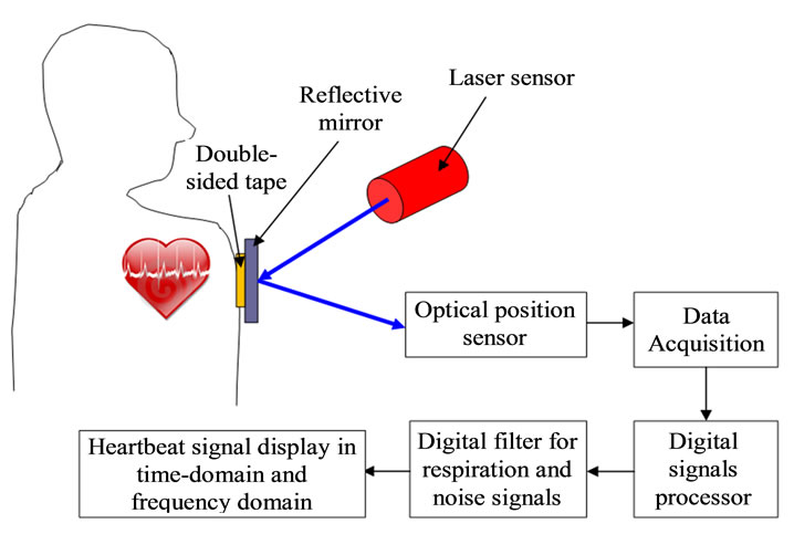
Figure 1. The system design of the laser-based stethoscope.
bration amplitude of the chest skin. According BoricLubecke et al. [5], the average peak-to-peak chest motion due to respiration is 4 - 12 mm, whereas the chest displacement due to heartbeat alone is about 0.3 mm. The resolution of the position sensor is 5.7 µm, which is sufficient for the detection accuracy of chest movement. The signal detected can be displayed in real time on the computer connected with the DA system. In addition to the heartbeat, various other activities of the human body could also cause the movement of the chest skin. These activities include the pulmonary respiration, regular body movement, etc. In order to extract the signal solely for heartbeat, digital frequency filter is designed to filter out the signals from other movements based on the fact that both the body movement and pulmonary respiration occur at much lower frequency than the heartbeat [6]. Both time-domain and frequency-domain heartbeat signals can be acquired and displayed by the system and aid in the in-depth analysis of heart murmur problems.
2.2. System Setup
The optical system setup is shown in Figure 2. We use the Aerotech 110SF 5.0 mW red laser as the laser source. The laser source is fixed on the optical table. We built a heart-sound box to simulate the chest of a human being. The heart-sound box is constructed with a speaker at the center and a rubber sheet covering the top. In order to simulate the tiny movement of chest skin with the heart sound [7,8], we chose rubber material with thickness at 1.6 mm and tensile strength of 1000 Psi, which is close to the mechanical properties of human chest skin [9]. The vibration of the sound-box is generated by the heartsound waves collected from the real patients through the iStethoscope Pro (the iPhone app). The displacement of the optical spot is detected by the linear position sensor module manufactured by Edmund Optics Inc. [10]. The detected signal is recorded in time-domain by the USB DA
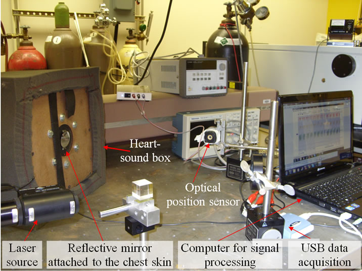
Figure 2. The prototype setup of the laser-based stethosocpe.
system associated with the linear position sensor [10]. Heart sound signal in time-domain is displayed on a personal computer connected with the DA system of the sensor. Signals in frequency-domain are obtained by the Fast Fourier transform (FFT) technique.
3. RESULTS AND DISCUSSION
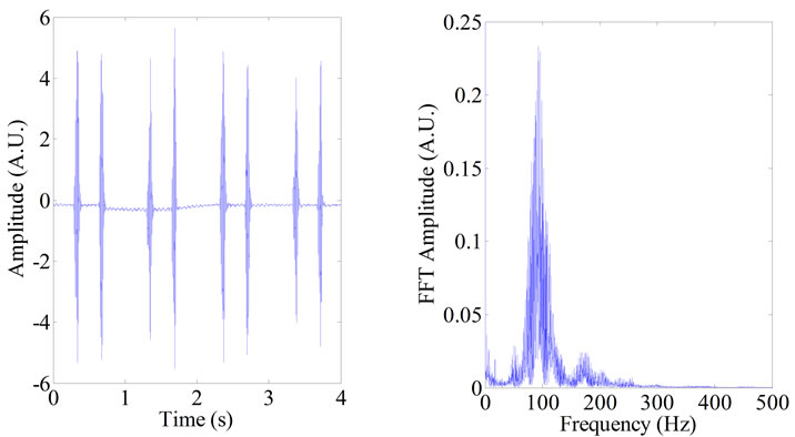
The sampling frequency of the optical sensor is chosen to be 8000 Hz. The sampling duration of each heart sound is 4 seconds. The system is applied to analyze two different pathological cases of the heart sound, i.e., mitral stenosis and mitral regurgitation, as well as the normal case. The time-domain heart signals and frequency-domain signals acquired from the system are displayed in Figure 3. The frequency scan is 1 to 500 Hz, which reasonably cover the heart sound range of a human being [11]. The vibration amplitude of the diaphragm on the sound box can be adjusted through the volume of the speaker. We set it to very low volume in order to approximate the tiny movement of the chest skin of human being with heartbeat.
Mitral stenosis and mitral regurgitation are two cases of heart valve disorder that involves the mitral valve. Comparing to the normal heart sound, in time-domain, heartbeat signals of both these two diseases shows big difference with the normal heartbeat in the first heart sound (lub or S1); in the frequency domain, the major differences lie in spectrum around 100 Hz. Mitral stenosis is characterized by the narrowing of the orifice of the mitral valve of the heart. The blood flow through the narrowed valve opening results in a flow pattern of higher velocity and greater turbulence. As seen from Figure 3, comparing to the normal heartbeat, the timedomain signal of the mitral stenosis shows a bursting of first heart sound S1, which corresponds to the presystolic reinforcement; in the frequency domain, the spectrum is broadened around 100 Hz compared to the normal heartbeat, extra heart sound presents at 0 - 50 Hz, which is below the human hearing range. The mitral regurgitation is a disorder of the heart in which the mitral valve does not close properly when the heart pumps out blood. This causes the blood to flow backwards into the atrium during ventricular systole. Figure 3(c) shows the time-domain and frequency-domain heartbeat patterns for mitral regurgitation. The time-domain signal shows less intensive and more extended S1 sound which corresponds to the incompetence in closing of mitral valve. In the frequency-domain, the peak frequency around 100 Hz in the normal heartbeat is missing in the mitral regurgitation case; instead, two side peak frequencies exist. The heart sound signals displayed are also very distinguishable between the mitral stenosis and mitral regurgitation. In time domain, the S1 sound in mitral regurgitation shows lower pitch and better smoothness than that in the mitral
 (a)
(a)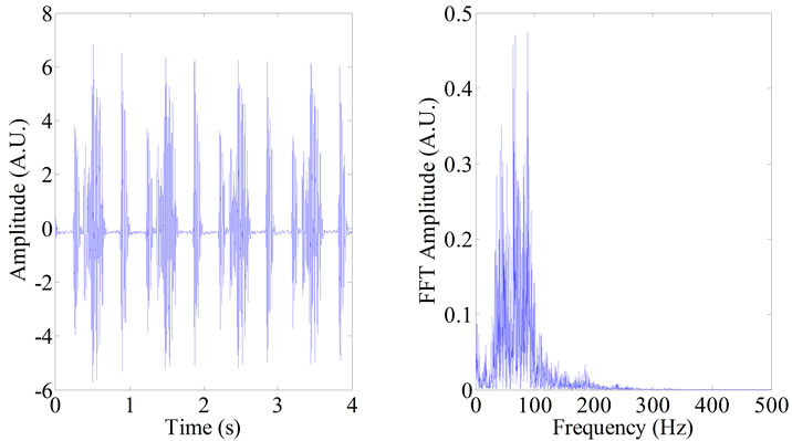 (b)
(b)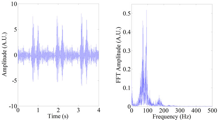 (c)
(c)
Figure 3. Time-domain and frequency-domain heart-sound signals acquired from the system: (a) Normal heartbeat; (b) Mitral stenosis; (c) Mitral regurgitation.
stenosis; while in the frequency domain, the mitral stenosis shows a broader spectrum around 100 Hz and lower peak around 200 Hz than that in the mitral regurgitation.
Here we reported a conceptual design and prototype setup of a new heart sound detection system using laser technology. We have planned to perform clinical trial to as our next research step. According to our institutional policy, approval from the Institutional Review Board (IRB) is required prior to the clinical trial with real patients. The application for IRB approval usually takes several months. Now we are in the middle of IRB application toward the clinical trial. Eventually, the discrete system will be integrated to be portable using the micro-optical-electromechanical (MOEM) technology.
4. CONCLUSION
In summary, we develop the prototype of a newly proposed heartbeat detection system based on laser technology. We take two pathological cases as well as the normal heartbeat as examples to illustrate the analysis on heartbeat patterns acquired by the system. Both timedomain and frequency-domain heart-beat signals can be displayed by the system for in-depth analysis of heart murmurs. This proposed optical stethoscope system will surmount the weaknesses of the conventional stethoscope caused by the limitation of human ear’s ability to detect low frequency heart sound (out of the human hearting range) and the limitation of not providing the practitioner visual representations of the sounds. In the next step, the system will be tested in clinical trials on real patients. The long-term goal of this research is to integrate the discrete components into a compact, portable system using the MOEM technology.
5. ACKNOWLEDGEMENTS
This research is supported by the research award from the Whiteside Institute for Clinical Research. Especially, we sincerely thank Prof. Michael Sydor for informative discussions and providing us the optical table, laser and some other optical elements in his Advanced Physics Lab at the Physics Department of University of Minnesota Duluth.
![]()
![]()
REFERENCES
- Christe, B.L. (2009) Introduction to biomedical instrumentation: The technology of patient care. Cambridge University Press, Cambridge.
- Cutnell, John, D. and Johnson, K.W. (1998) Physics. 4th Edition, John Wiley & Sons Ltd., New York.
- Xiao, Y., Li, C. and Lin, J. (2007) A portable noncontact heartbeat and respiration monitoring system using 5-GHz radar. IEEE Sensors Journal, 7, 1042-1043. doi:10.1109/JSEN.2007.895979
- Ivashov, S.I., Razevig, V.V., Sheyko, A.P. and Vasilyev, I.A. (2004) Detection of human breathing and heartbeat by remote radar. Progress in Electromagnetic Research Symposium, Pisa, 28-31 March 2004, 663-666.
- Boric-Lubecke, O., Droitcour, A.D., Lubecke, V.M., Lin, J. and Kovacs, G.T.A. (2003) Wireless IC Doppler for sensing of heart and respiration activity. IEEE Proceedings of International Conference on Telecommunication in Modern Satellite, Cable and Broadcasting Services, Serbia and Montenegro, Nis, 1-3 October 2003, 337-344.
- Lu, G., Yang, F., Jing, X. and Wang, J. (2010) Contactfree measurement of heartbeat signal via a Doppler radar using adaptive filtering. IEEE Proceeding of International Conference on Image Analysis and Signal Processing, Hangzhou, 12-14 April 2010, 89-92.
- Dalmay, F., Antonini, M.T., Marquet, P. and Menier, R. (1995) Acoustic properties of the normal chest. European Respiratory Journal, 8, 1761-1769. doi:10.1183/09031936.95.08101761
- Leduc, D., Brunko, E. and De Troyer, A. (2000) Response of the Canine inspiratory intercostal muscles to chest wall vibration. American Journal of Respiratory and Critical Care Medicine, 161, 510-516.
- Gennisson, J.-L., Baldeweck, T., Tanter, M., Catheline, S., Fink, M., Sandrin, L., Cornillon, C. and Querleux, B. (2004) Assessment of elastic parameters of human skin using dynamic elastography. IEEE Transactions on Ultrasonics, Ferroelectrics, and Frequency Control, 51, 980-989. doi:10.1109/TUFFC.2004.1324402
- Edmund optics, linear position sensor module (NT57- 058). http://www.edmundoptics.com/products/displayproduct.cfm? Productid=2476
- Debbal, S.M. and Bereksi-Reguig, F. (2008) Frequency analysis of the heartbeat sounds. Biomedical Soft Computing and Human Sciences, 13, 85-90.