Chinese Medicine
Vol.06 No.01(2015), Article ID:54545,19 pages
10.4236/cm.2015.61005
A 180-Day Subchronic Oral Toxicity Study of Total Flavones of E. leptorrhizum Stearn in Rats
Ting Liu, Lianqiang Hui, Chunyu Cao*, Ran Hao, Shuangrong Gao, Rong He, Baoqiang Dai, Lifang Wang
Institute of Chinese Materia Medica, China Academy of Chinese Medical Sciences, Beijing, China
Email: *chunyu.cao@gmail.com
Copyright © 2015 by authors and Scientific Research Publishing Inc.
This work is licensed under the Creative Commons Attribution International License (CC BY).
http://creativecommons.org/licenses/by/4.0/


Received 3 November 2014; accepted 8 March 2015; published 11 March 2015
ABSTRACT
A subchronic oral toxicity was conducted to evaluate the safety of total flavones of E. leptorrhizum Stearn in Sprague-Dawley rats. The test article was administered once daily by gavage in male and female rats at dose levels of 24, 48, and 96 mg/kg body weight/day for 180 days. 90 and 180 days after administration, ten and tweedy animals (each half of male and female) of each group were tested. 28 days after withdrawal, five male and female rats were tested. There were no significant toxicological changes shown in daily clinical signs, body weight, food consumption, hematology parameters, blood biochemistry, organ weights and histopathological examination except leukocyte differential count. It was concluded that the no-observed-effect level (NOEL) for total flavones of E. leptorrhizum Stearn was >96 mg/kg in SD rats.
Keywords:
Total Flavones of E. leptorrhizum Stearn, Safety, Subchronic Toxicity

1. Introduction
Herba epimedium is dried aerial parts of E. brevicornum Maxim. or E. sagittatum Maxim. or E. pubescens Maxim. or E. wushanense T. S. Ying or E koreanum Nakaide [1] . It was earliest recorded about 2000 years ago in the “Shen Nong’s herbal classic”, which is China’s earliest existing medical monograph. Modern pharmacology studies prove that Epimedium can regulate immune function, increase male reproductive function, delay renal failure [2] -[6] . Therefore, herba epimedium is widely used as food additives in health products. The extraction and active ingredients are an important source of healthy foods and medicine. Many research was done on epimedium extract and active ingredient, more than twenty related patents have been approved by the US Patent and Trademark Office and the World Intellectual Property Organization [7] -[11] .
The pharmacological activity of icariin is most explicit, it has multiple pharmacological effects, such as anti- tumor, enhance immune function, improve cardiovascular function, endocrine regulation, prevention and treatment of osteoporosis, anti-aging, etc. [12] . Epimendin C is also from epimedium, having a chemical structure and pharmacological activity similar to Icariin, such as anti-tumor, enhance immune function, against osteoporosis, etc. [13] -[16] , therefore, expected to become the new food additives.
Icariin is used as a control standard in Chinese Pharmacopoeia [1] , but very little research has been done on epimendin C, a flavonoids in epimedium. Some researchers believe that epimendin C has definite pharmacological activity, and it is another important active ingredient in epimedium. Epimendin C should be used as an important indicator to control epimedium quality [2] [3] . E. leptorrhizum Stearn grow mostly in Guizhou region, which is widely distributed locally. Only little amount of E. leptorrhizum Stearn is used as medicine because icariin content is lower than the criteria in Chinese Pharmacopoeia, while the content of epimendin C is higher. Wei Deng [17] reported that significant improvement was observed when E. leptorrhizum Stearn applied on osteoporosis model mice induced by dexamethasone. Our research also showed that the total flavones of E. leptorrhizum Stearn could promote hFOB1.19 osteoblast proliferation and ALP secretion [18] . It indicated that the promotion of osteoblast activity by total flavones of E. leptorrhizum Stearn maybe a pathway of prevention and treatment of osteoporosis.
Epimedium is a traditional Chinese medicine, and no adverse reactions were reported from clinical practice. In animal studies, the median lethal dose (LD50) of epimedium water extract is more than 80 g/kg, and all the results including mouse bone marrow micronucleus test, Ames test, TK Gene mutation test were negative [19] . The total flavones of Epimedium 1.0, 2.0 and 4.0 g/kg showed no distinct toxicity, taken orally once a day, continuous for 12 weeks [20] . MTD of acute toxicity test in mice was 1440 times of Human clinical dose, it showed a very mild toxicity [21] . The herb used for above research is not from E. leptorrhizum Stearn. There are no animals and no clinically relevant reports about the safety of E. leptorrhizum Stearn whether raw material or extract. It is likely to be useful as an alternative medicine as epimedium if it can be confirmed to be safe.
A 180-day repeated oral dose toxicity study in rats was conducted to evaluate the safety of excessive intake of total flavones of E. leptorrhizum Stearn. The present study was conducted in compliance with technical specifications for examination and assessment of health food as set forth in Ministry of Health of the People’s Republic of China, issued 2003, which meet OECD guidelines.
2. Materials and Methods
2.1. Test Article
E. leptorrhizum Stearn which was provided by Guizhou Tongjitang pharmaceutical CO. LTD. Add 20 times of 70% ethanol into E. leptorrhizum Stearn, reflux 3 times, each time for 1 hour, combine extracts, recycling to concentrated liquid. Add distilled water (1:1) into concentrated liquid, allow the solid to settle, discard the precipitation, oncentrate the supernatant. Dry the concentrated extraction under vacuum condition at 80˚C, crushing, that is flavonoids part of E. leptorrhizum Stearn (YYH-C), which content 4.96 mg/g of flavonoids, 1.17% of Icariin and 8.87% of Epimendin C [22] . YYH-C was dissolved with distilled water to 2.4, 4.8, and 9.6 mg/ml to provide administered doses of 24, 48, and 96 mg/kg/day.
2.2. Aminals
Male and female Sprague-Dawley rats at 5 weeks of age purchased from Beijing Weitonglihua experimental animal technique Co. Ltd (Licence number: SCXK (Jing) 2012-0001.). The animals were acclimatized to housing conditions for a period of one week, and the treatment started at 6 weeks of age to health animals. The animals were kept in a room maintained at a temperature 23˚C ± 3˚C and a relative humidity of 40% - 70% with 12 hours light and 12 hours dark cycle and with approximately 15 times air change per hour. Animals were bred in bottom-meshed stainless steel cages, with filtrated tap water and the fixed-formula rat granula feed. This experiment was approved by the Animal Care & Welfare Committee of Institute of Chinese Materia Medica, China Academy of Chinese Medical Sciences.
2.3. Grouping and Administration
The rats were randomly divided into four groups: a control group and three treatment groups of YYH-C 24, 48, 96 mg/kg. Each group consisted of 20 males and 20 females. The test articles were administered daily by gavage to rats for 180 days at dose of 24, 48, and 96 mg/kg, and the control group rats were given the same volume of distilled water. Individual dose volume of 10 ml/kg was calculated according to the latest measured body weight.
2.4. Observation and Clinical Examination
2.4.1. General Conditions, Body Weight, and Food Consumption
General conditions of rats such as behavior, gait and general activity, appearance, hair luster, stool and urine secretion and so on were observed before and after dosing each day. The body weights of all rats and the remaining granules in each crib were weighed once a week during study under the condition of no fasting.
2.4.2. Hematology and Serum Biochemistry
After the treatment, all animals were fasted overnight and then anesthetized by phenobarbital sodium, and blood samples were collected from the abdominal aorta. The following parameters for hematology analysis using anticoagulated whole blood with EDTA 2 K were measured: red blood cell count (RBC), white blood cell count (WBC), hemoglobin (Hb), haematocrit (Ht), blood platelet number (PLT), mean corpuscular volume (MCV), mean corpuscular hemoglobin (MCH), mean corpuscular hemoglobin concentration (MCHC), leukocyte differential count, and reticulocytes(Ret) were analyzed by using an automatic blood cell analyser (Siemens ADVIA- 2120,made in Germany). The following parameters for blood biochemical analysis using serum were measured: total protein (TP), albumin (ALB), aspartate aminotransferase (AST), alanine amiotransferase (ALT), alkaline phosphatase (ALP), γ-glutamyl transpeptidase (γ-GT), creatine kinase (CK), urea nitrogen (UREA), creatinine (CRE), glucose (GLU), total bilirubin (T-bil), total cholesterol (T-cho), triglyceride (TG) were determined by automatic biochemistry analyzer (TOSHIBA 40FR, made in Japan), and Na+, K+, Cl− were determined by electrolytes analyzer (Easy Lyte PLUS Na/K/Cl NALYZER, made in America).
2.4.3. Histomorphological Assessment
The animals were sacrificed by exsanguinations from the abdominal aorta. At necropsy, the brain, thymus, lung with bronchi, heart, liver, spleen, kidney, adrenal gland, testicle, epididymis, ovary and uterus were removed and weighted. The relative organ weights were calculated using the animals, fasted body weights. In addition, pituitary, the spinal cord, thyroid glands, salivary glands, optic nerve, eyes, stomach, pancreas, small intestine (duodenum, jejunum and ileum), large intestine (cecum, colon and rectum), urinary bladder, epididymis, prostate and bone marrow were also removed. All organs and tissues were examined grossly and then fixed in 10% buffered formalin solution. Histomorphological assessment was first performed on all tissues of control group and the highest dose group. If any treatment-related changes appeared at the highest dose group, the relavant tissues from the lower dose groups were then examined. The organs and tissues were routinely processed for paraffin- embedding, sliced, HE (Haematoxylin & Eosin) stained, and then histomorphological observation was performed under microscopes.
2.5. Statistical Analysis
Data of males and females in each group were separately calculated, and expressed as mean ± SD (standard deviation). Variance in data for body weights, food consumption, hematology, serum biochemistry, and relative organ weights was checked for homogeneity and analyzed with one-way analysis of variance (ANOVA). If the variance was homogeneous, the data were analylyzed by LSD test, if not, the Tamhane’s T2 test was applied. Probability values of P < 0.05 were considered significant.
3. Result
3.1. In-Life Parameters
No deaths and no clinical signs to the treatment were observed during the administration period.
Body weights (Figure 1 & Figure 2) and food consumption (Figure 3 & Figure 4) in both sexes in each

Figure 1. Mean body weight of male rats.

Figure 2. Mean body weight of female rats.
treatment group were comparable to those in the control group. Male rats of 24 mg/kg group of YYH-C decreased significantly on food consumptions in 20 - 23 weeks, as well as 96 mg/kg dose group in 21 week (P < 0.05, P < 0.01). In 25 - 26 week, 24 mg/kg group of YYH-C reduced significantly compared with the control group both on body weights and food consumptions (P < 0.05). The rest of the time points of the remaining treatment group no statistically difference was observed.

Figure 3. Mean food consumption of male rats.

Figure 4. Mean food consumption of female rats.
3.2. Hematology Parameters
Hematology data are shown in Tables 1-3.
A dose-depended obvious decrease of LYM% and increase of MID% were seen in male rats of all three dose groups after 90 days administration (P < 0.05, P < 0.01), moreover, decrease of LYM% and increase of MID% were also seen after 180 days administration with no obviously dose-depended manner (P > 0.05). However, an obvious increase of LYM% and decrease of MID% were seen in female rats of 48, 96 mg/kg dose groups after 180 days administration (P < 0.05, P < 0.01, P < 0.001).
Though the results showed an obvious decrease of RBC, Ht , Hb of female rats in 48 mg/kg group, and an obvious increase of Hb in 96 mg/kg groups after 90 days administration; an obvious increase of PLT of male rats both in 24 and 96 mg/kg groups, and an obvious decrease of Ret of female rats in all treatment groups after 180 days administration; an obvious increase of RBC, Ht, Hb and obvious decrease of PLT of female rats in all treatment groups after 28 days withdrawal (P < 0.05, P < 0.01), no toxicological significance were indicated

Table 1. Hematological examination of rats after 90 days administration (n = 5).
Values are presented as mean ± standard variation. *Significantly different from the controls at levels of P < 0.05. **Significantly different from the controls at levels of P < 0.01.
Table 2. Hematological examination of rats after 180 days administration (n = 10).
Values are presented as mean ± standard variation. *Significantly different from the controls at levels of P < 0.05. **Significantly different from the controls at levels of P < 0.01. ***Significantly different from the controls at levels of P < 0.001.
as the values of all above were fluctuated within the scopes of historical data of our laboratory and no obvious dose effect relationship. No abnormalities were seen in other hematology parameters of male and female rats in three YYH-C groups.
3.3. Blood Biochemistry
Serum biochemistry data are shown in Tables 4-6.
An obvious increase of UREA level after 90 days administration and GGT content after 180 days administration were seen in female rats of all three dose groups (P < 0.05, P < 0.01), the content of ALB of female rats in 48, 96 mg/kg groups decreased significantly after 180 days administration (P < 0.05), moreover, the content of ALP of female rats in 24, 48 mg/kg groups increase significantly after 28 days withdrawal (P < 0.05, P < 0.001).
The serum Cl− level showed an obviously increase of male rats in all treatment groups and decrease of female rats in 24, 48 mg/kg groups after 90 days administration (P < 0.05, P < 0.01), The serum K+ level decreased significantly of male rats in 96 mg/kg group and increased of female rats in 48mg/kg groups after 180 days admin-
Table 3. Hematological examination of rats after 28 days withdrawal (n = 5).
Values are presented as mean ± standard variation. *Significantly different from the controls at levels of P < 0.05. **Significantly different from the controls at levels of P < 0.01.
istration (P < 0.05), but the change had no physiological significance because the value of these group was fluctuated in a tight range and within the scopes of historical data of our laboratory.
Some other indicators, including the content of AST, UREA, CK of male rats and CRE of female rats after 90 days administration, GLU, CK concentration of male rats and T-bil of female rats after 180 days administration, the content of T-bil, TG of male rats after 28 days withdrawal, were considered as no toxicological significant as the changes mentioned above were decreased compared with the control group.
3.4. Organ Weights
An obvious increase of the relative organ weight of spleen after 90 days administration in female rats of 48mg/kg dose groups (P < 0.01) and the relative organ weight of adrenal after 180 days administration in male rats of all treatment groups (P < 0.05, P < 0.01, P < 0.001), however, the changes mentioned above weren’t showed a dose-depended relationship, the mean of the relative organ weight of spleen was 0.193, 0.199, 0.250,
Table 4. Blood biochemistry analysis of rats after 90 days administration (n = 5).
Values are presented as mean ± standard variation. *Significantly different from the controls at levels of P < 0.05. **Significantly different from the controls at levels of P < 0.01.
and 0.196, the mean of the relative organ weight of adrenal was 0.009, 0.013, 0.015, and 0.013 (the control group and YYH-C 24, 48, 96 mg/kg group respectively). There were no significant differences in relative organ weights between the other treatment compared with the control groups in both sexes (data not shown).
Table 5. Blood biochemistry analysis of rats after 180 days administration (n = 10).
Values are presented as mean ± standard variation. *Significantly different from the controls at levels of P < 0.05. **Significantly different from the controls at levels of P < 0.01. ***Significantly different from the controls at levels of P < 0.001.
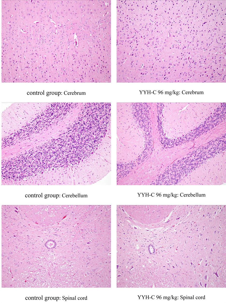
3.5. Histopathology
Histopathology pictures are shown in Appendix.
The histopathological examination revealed no findings related to the treatment. The results of histomorphological examination indicated that oral administration of at the doses up to 96 mg/kg for successive 180 days did not cause toxicologically histomorphological damage in main organs.
Table 6. Blood biochemistry analysis of rats after 28 days withdrawal (n = 5).
Values are presented as mean ± standard variation. *Significantly different from the controls at levels of P < 0.05. **Significantly different from the controls at levels of P < 0.01. ***Significantly different from the controls at levels of P < 0.001.
4. Discussion
The present study was conducted to evaluate the safety of the total flavones of E. leptorrhizum Stearn administered once daily gavage to male and female Sprague-Dawley rats at dose levels of 24, 48, and 96 mg/kg/day for 180 days. Throughout the administration and recovery time, no deaths and no clinical signs were observed. No treatment-related changes were found in body weight, food consumption, hematology parameters, blood biochemistry, organ weights and histopathological examination in male and female in each treatment group.
Our study found that taking YYH-C for a long time didn’t inhibit the growth of animal body weight, but could delay the growth rate for a long time. Changes in food consumption and body weight were corresponding in male rat, taking a long time for the test materials, could lead to reduced food intake, and showed the time-re- lated changes in degree. No above mentioned effect were found both in body weight and food consumption in female.
A dose-depended obvious decrease of LYM% and increase of MID% were seen in male rats of all three dose groups after 90 days administration, moreover, decrease of LYM% and increase of MID% were also seen after 180 days administration with no obviously dose-depended manner. However, an obvious increase of LYM% and decrease of MID% were seen in female rats of 48, 96 mg/kg dose groups after 180 days administration. Some other indicators which showed an obvious changes, including hematology parameters, blood biochemistry and organ weights, were considered as no toxicological significant as the values were within the normal ranges recorded in the experimental facility and no obvious dose effect relationship. It is concluded that the administration of YYH-C at levels up to 96 mg/kg/day for 180 days is well-tolerated for both genders without obvious signs and toxicity. However, leukocyte differential count should be observed carefully.
In Chinese Pharmacopoeia (2010), total flavonoid (calculated by Icariin) in Epimedium should be no less than 5.0% on a dry basis. Icariin content is 1.17% in YYH-C which extract from E. leptorrhizum Stearn, it is far from the criteria in Chinese Pharmacopoeia; Epimendin C content is 8.87%, it is the main composition. Even though Icariin may be safe as a food additive, no studies have been performed systematically to evaluate the effects of long-term intake of YYH-C. The reason why we conducted this study was to confirm the safety of excessive intake of individual YYH-C because during YYH-C efficacy assessment, we found YYH-C have the same efficacy activity as Icariin to prevent osteoporosis. The present study showed that the no-observed-effect level for YYH- C was 96 mg/kg/day for both genders. This dose is 100 times of clinical dose, therefore it is preliminary considered as a safe food additive.
References
- Chinese Pharmacopoeia Commission (2010) Pharmacopoeia of the People’s Republic of China. People’s Medical Publishing House, Beijing, 306.
- Zhang, H.F. and Yang, X.H. (2010) Bioactive Constituents in Herba Epimedii and Their Developmental Strategies. Chinese Traditional and Herbal Drugs, 41, 329.
- Zhang, H.F. and Yang, X.H. (2010) Application of Herba Epmiedii in Food Industry: Current Status and Prospect. Science and Technology of Food Industry, 31, 390.
- Zhang, D.W., Cheng, Y., Wang, N.L., Zhang, J.C., Yang, M.S. and Yao, X.S. (2008) Effects of Total Flavonoids and Flavonol Glycosides from Epimedium koreanum Nakai on the Proliferation and Differentiation of Primary Osteoblasts. Phytomedicine, 15, 55-61. http://dx.doi.org/10.1016/j.phymed.2007.04.002
- Lu, Y., Wang, D.Y., Hu, Y.L., Huang, X.Y. and Wang, J.M. (2008) Sulfated Modification of Epimedium Polysaccharide and Effects of the Modifiers on Cellular Infectivity of IBDV. Carbohydrate Polymers, 71, 180-186. http://dx.doi.org/10.1016/j.carbpol.2007.05.024
- Pan, Y., Kong, L., Li, Y., Xia, X., Kung, H.F. and Jiang, F.X. (2007) Icariin from Epimedium brevicornum Attenuates Chronic Mild Stress-Induced Behavioral and Neuroendocrinological Alterations in Male Wistar Rats. Pharmacology Biochemistry and Behavior, 87, 130-140. http://dx.doi.org/10.1016/j.pbb.2007.04.009
- Ezio, B., Paolo, M., Antonella, R., et al. (2008) Formulations Useful in the Treatment of Male and Female Impotence. US Patent No. 7361370.
- Carsten, S. (2009) Multifaceted Weight Control System. US Patent No. 60572184.
- Susumu, M., Muneaki, T., Ichiro, W., et al. (1982) Polysaccharideps-Aisolation from the Plant of Genus Epimedium Violaceum Morr1 et Decne1, Process for Its Preparation, and Infection-Preventing Agent and Immunostimulating Agent Containing the Polysaccharide as Effective Ingredient. The World Intellectual Property Organization Patent No. WO/1982/003771.
- Cohen, I. (2006) Method of Using Extracts of Epimedium Species. The World Intellectual Property Organization Patent No. WO/2006/065599.
- Koch, E., Erdelmeier, C. and Hauer, H. (2007) Extracts of Epimedium Species Method for Production and Use Thereof. The World Intellectual Property Organization Patent No. WO/2007/031140.
- Li, L. and Wang, X.M. (2008) Advances in Study on Pharmacological Effects of Icariin. China Journal of Chinese Materia Medica, 33, 2727.
- Meng, F.H., Li, Y.B., Xiong, Z.L., Jiang, Z.M. and Li, F.M. (2005) Osteoblastic Proliferative Activity of Epimedium brevicornum Maxim. Phytomedicine, 12, 189-193. http://dx.doi.org/10.1016/j.phymed.2004.03.007
- Iinuma, M., Tanaka, T., Sakakibara, N., Mizuno, M., Matsuda, H., Shiomoto, H. and Kubo, M. (1990) Phagocytic Activity of Leaves of Epimedium Species on Mouse Reticuloendotherial System. Journal of the Pharmaceutical Society of Japan, 110, 179-185.
- Liang, H.R., Vuorela, P., Vuorela, H. and Hiltunen, R. (1997) Isolation and Immunomodulating Effect of Flavonol Glycosides from Epimedium hunanense. Planta Medica, 63, 316-319. http://dx.doi.org/10.1055/s-2006-957690
- Liu, T.Z., Chen, C.Y., Yiin, S.J., Chen, C.H., Cheng, J.T., Shih, M.K., et al. (2006) Molecular Mechanism of Cell Cycle Blockage of Hepatoma SK-Hep-1 Cells by Epimedin C through Suppression of Mitogen-Activated Protein Kinase Activation and Increased Expression of CDK Inhibitors p21Cip1 and p27Kip1. Food and Chemical Toxicology, 44, 227-235. http://dx.doi.org/10.1016/j.fct.2005.07.003
- Deng, W., Zheng, M.Q. and Huang, Y.Q. (2011) Pharmacodynamics Comparative Study of Two Species of Epimedium in Guizhou on the Anti Osteoporosis in Mice. China Journal of Chinese Materia Medica, 36, 939.
- Liu, T., Cao, C.Y., Hao, R. and Hui, L.Q. (2013) Influence of Total Flavones of Epimedium leptorrhizum Stearn on hFOB1.19 Osteoblast Activity. Chinese Journal of Experimental Traditional Medical Formulae, 18, 196.
- Sui, H.X., Gao, P. and Xu, H.B. (2006) The Safety Evaluation of Herba Epimedii Water Extract. Carcinogenesis, Teratogenesis & Mutagenesis, 18, 439.
- Li, D.M., Yin, X.F., Liu, J.H., et al. (2008) Experimental Study on Long-Term Toxicity with Flavones of Epimedium in Rats. Chinese Journal of Experimental Traditional Medical Formulae, 14, 60.
- Li, D.M., Yin, X.F., Cai, D.W., et al. (2007) Experimental Study on Acute Toxicity with Flavones of Epimedium in Rats. China Pharmacist, 10, 1011.
- Han, B., Shen, T., Liu, D. and Yang, J.S. (2002) Study on Chemical Components of Epimedium leptorrhizum. Chinese Pharmaceutical Journal, 37, 740.
Appendix (Histopathology Pictures after 180 Days Administration, HE × 200)


NOTES

*Corresponding author.