Surgical Science, 2011, 2, 140-143
doi:10.4236/ss.2011.23029 Published Online May 2011 (http://www.scirp.org/journal/ss)
Copyright © 2011 SciRes. SS
Clinical and Pathological Studies of Meningioma-Glioma
Mixed Tumor
Junyang Li1, Weixing Hu1, Zhihong Zhang2, Dong Wei1
1Department of Neurosurgery, The First Affiliated Hospital of Nanjing Medical University, Nanjing, China
2Departmen t o f Pathology, The First Af filiated Hospital of Nanjing Medical University, Nanjing, China
E-mail: hwx66@126.com
Received February 20, 2011; revised A pril 2, 2011; accepted April 8, 2011
Abstract
Meningioma-glioma mixed tumor is rare central nervous system tumor. It is necessary to study its clinical
and pathological characteristics as well as its possible genesis. This case was a 54-year-old man who was
readmitted for recurrent glioma. Magnetic resonance imaging showed a big mass in right temporal lobe
which was confirmed as meningioma-glioma by immunohistochemical analysis. Specific immunohisto-
chemical staining is significant in tumor differential diagnosis, and helps to confirm tumor histic origin. By
pathological studies, we found that glioma could stimulate adjacent normal meninges into neoplastic prolif-
eration.
Keywords: Glioma; Immunohistochemical Staining; Meningioma; Mixed Tumor; Neoplastic Proliferation
1. Introduction
The term “meningeal gliomatosis” was firstly adopted to
designate involvement of the leptomeninges by tumors of
neuroectodermal origin by Polmeteer [1]. The authors
primarily did studies abou t metastatic implantatio ns from
gliomas to the leptomeninges of the brain and the spinal
cord and the ependyma of the ventricular system. These
gliomas established themselves in multiple sites via
cerebrospinal fluid (CSF) system and were responsible
for a generalized involvement of the leptomeninges.
Usually, there are no differences in tumor cell morphous
between the meningeal gliomas and intracerebral glio-
mas. We presently report a special case of meningioma-
glioma which possessed meningioma pathological mor-
phous, but its immunohistochemical characteristics be-
long to glioma.
2. Case Report
This 54-year-old man was readmitted for “recurrent
glioma in the right temporal lobe” on October 27, 2008.
In August, 2007, he was admitted with onset of iterative
headache and abduction limitation of the left eyeball.
MRI showed a mass (size 3.0 × 3.0 cm), surrounded by
digitatus type of cerebral edema in the right temporal
lobe. The ventriculus dexter cerebri was severely ex-
truded. The entire mass was resected after the first op-
eration. Pathological diagnosis was glioma (Grade IV)
with glial fibrillary acidic protein (GFAP) positive of
immunohistochemical staining. The patient refused ra-
diotherapy and chemotherapy after the first operation
because of financial limitation. In October 2008, he felt
headache again, accompanied by limbs’ acratia, function
loss of left eyeball abduction, myodynamia decrescence
of limbs and malfunction of equilibrium. MRI showed a
mass (size 5.0 × 6.8 cm) with a bursa located in the right
temporal lobe, and the ventriculus dexter cerebri was
extruded. Enhanced MR images showed the capsule wall
in the superior part and the inferior part of the mass was
significantly enhanced (Figures 1(a) and (b)). The pa-
tient’s second operation was performed on October 29,
2008. About 20 ml intraluminal fluid was drawn from
the cavum inside the mass. After we resected the soft
and fish-flesh-like tissue of its superior part, an elliptic
and smooth hemisphere mass appeared at the bottom of
the middle cranial fossa with a clear and smooth bound-
ary, which involved the anterior part of the tentorium of
cerebellum. In this one solid tumor, there were no inter-
laced tissues found between the two parts, in spite of the
fact that they were closely proximate in his MRI and
during operation. Pathological examination showed the
spherical or elliptic cells with mild heteromorphosis and
plentiful cytoplasm of the superior part tissue. The cells
J. Y. LI ET AL. 141
(a) (b)
Figure 1. MRI for the patient before his second surgery. (a) A mass (size 5.0 × 6.8 cm) with a bursa located in right temporal
lobe, the ventriculus dexter cerebri was extruded. (b) After image contrast processing, the inferior part tumor was markedly
illustrated, which was indicated by arr ows.
Figure 2. Histopathology of specimens at the time of diagnosis of the tumor which was resected in second surgery for the pa-
tient. (a) HE staining shows spherical or elliptic cells of superior part tumor, with mild heteromorphosis and plentiful cyto-
plasm (× 200). (b) EMA staining for superior part tumor cells is negative (× 400). (c) GFAP staining for superior part tumor
cells is positive (× 200). (d) HE staining shows cells of inferior part tumor, composed of spindle cells arranging in whorl pat-
terns (× 200). (e) EMA staining for inferior part tumor cells is positive (× 400). (f) GFAP staining for inferior part tumor cells
is positive (× 200).
C
opyright © 2011 SciRes. SS
J. Y. LI ET AL.
142
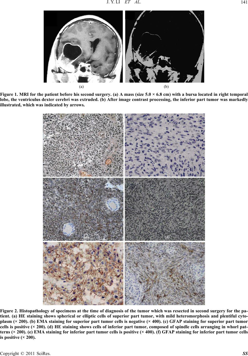
were active in growth and accompanied with abundant
vascular proliferation and extensive tissue necrosis. All
of these indicated glioma (Figure 2(a)). The immuno-
histochemical staining showed epithelial membrane an-
tigen (EMA) negative, GFAP positive (Figures 2(b) and
(c)). However, the inferior part of the tumor was outside
the cerebrum and based at the bottom of the middle fossa.
Microscopically, this part composed of spindle cells ar-
ranged in whorl patterns. The cells grew slowly and
mildly with no karyokinesis. These were characteristics
of meningioma (Figure 2(d)) with EMA positive (Fig-
ure 2(e)) and GFAP positive (Figure 2(f)) of immuno-
histochemical staining. The patient recovered well after
operation, under advice of radiotherapy and chemother-
apy.
3. Discussion
In this case, we found the inferior part, extracerebral
smooth, tough, and firm tumor, which located in the bot-
tom of the middle cranial fossa, had defined and clear
border to the intracerebral upper part tumor. Pathological
findings indicated that the upper part tumor exhibits
morphological characteristics of glioma, as well as
GFAP positive and EMA negative. Meanwhile, inferior
part tumor reflect the morphing characteristic of men-
inges, but its immunohistochemical staining shows
GFAP positive, and EMA positive appeared in cells ar-
ranging in whorl patterns. EMA is specific marker for
normal epithelium or epithelial origin tumor which ex-
pressed in most meningioma, but usually negative in
glioma cells [2]. As a kind of intermediate filaments in
gliocyte, GFAP expressed in most astrocytoma and was
considered as the major evidence for diagnosing glioma
[2,3]. In our case, the inferior part tumor exhibits the
basement of meninges and there was a clear boundary
with the upper part tumor even though they were in one
solid mass in MRI. Positive of EMA indicated that tumor
cells of the inferior part originated from meningothelium,
and positive of GFAP indicated its features of glioma.
We think the term “meningioma-glioma” was more suit-
able than “meningeal glioma” for our case.
Our meningioma-glioma case is different from previ-
ously reported cases of meningeal gliomatosis. The latter
concerned with tumor-like lesions in meninges of the
brain or the spinal cord or the ependyma of the ventricu-
lar system caused by metastatic implantations from in-
tracerebral glioma. Those metastatic glioma cells estab-
lished themselves in multiple sites via CSF system,
caused clinical manifestation like meningitis, and usu ally,
there’re no differences in cell characteristics between
meningeal gliomatosis and intracerebral gliomas [4,5].
Moreover, previous researches showed that glioma cells
which were cultured in normal leptomeningeal extracel-
lular matrix proteins still kept the features of gliocytes
[6]. This suggested th at if glioma cells in vaded mening es
directly and proliferated, their features won’t be changed
by extracellular environment of meningocytes. To our
meningioma-glioma case, it is not only there was a
boundary between the su perior an d infer ior parts but also
the cells of the meningeal glioma possess features like
meningioma cell rather than glioma cell, and immuno-
histochemical staining showed EMA positive. These
indicated that this inferior part tumor was not an invaded
or implanted tumor of intracerebral glioma cells but was
a tumor originated from the meningocytes.
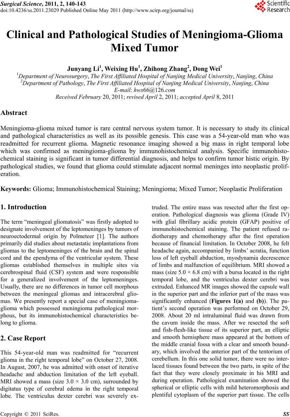
Cooper and Kernohan ha d suggested that primary lep-
tomeningeal glioma originated from dedifferentiation of
heterotopic glial nests [4]. This suggestion seemed un-
suitable for our case, since the tumor divided into two
parts (Figure 1) in one solid mass, the inferior men-
ingeal glioma tight conjuncted the superior glioma. And
there’s no neoplasm in meninges in first surgery for
glioma. We presumed that the recurrence malignant
glioma stimulated the adjacent meninges into neoplastic
proliferation, altho ugh ther e is n o more knowledg e about
the biomechanism or material basis relate to this irritant
action. For our case, this hypothesis seemed more rea-
sonable than “leptomeningeal gliomas originated from
dedifferentiation of heterotopic glial nests” [4]. Davis
had given similar hypothesis after their research of con-
current meningioma and astrocytoma growth [7]. They
considered that meningioma or glioma can stimulate the
adjacent brain parenchyma or arachnoid cells into neo-
plastic proliferation. To our case, we consider that the
irritant action from the malignant glioma not only caused
neoplastic proliferation of meninges but also changed
their phenotypes into expressing both EMA and GFAP.
Further more works are needed to demonstrate the bio-
mechanism and material basis relate to this kind of irri-
tant action.
4. Conclusions
Pathological examinations are necessary for men-
ingioma-glioma mixed tumor diagnosis. Specific immu-
nohistochemical staining should be an important deter-
mination in differential diagnosis and could help confirm
tumor histic origins. This rare tumor which we reported
was recurrent and had meningeal neoplastic proliferation
to primary tumor lesions and was considered as single
and unitary tumor in MRI scans and intraoperative find-
ings. Further pathological studies were necessary to re-
veal the tumor’s real characteristics. Our case demon-
strated that glioma may stimulate adjacent meninges into
neoplastic proliferation, but related biomechanism and
C
opyright © 2011 SciRes. SS
J. Y. LI ET AL. 143
material basis warrants further investigation.
5. References
[1] F. E. Polmeteer and J. W. Kernohan, “Meningeal Glio-
matosis: A Study of 42 Cases,” Archives of Neurology
and Psychiatry, Vol. 57, No. 5, 1947, pp. 593-616.
[2] H. Ikota, S. Kinjo, H. Yokoo and Y. Nakazato, “System-
atic Immunohistochemical Profiling of 378 Brain Tumors
with 37 Antibodies Using Tissue Microarray Technol-
ogy,” Acta Neuropathologica, Vol. 111, No. 5, 2006, pp.
475-482. doi:10.1007/s00401-006-0060-1
[3] E. Bongcam-Rudloff, M. Nistér, C. Betsholtz, J. L. Wang,
G. Stenman, K. Huebner, C. M. Croce and B. Westermark,
“Human Glial Fibrillary Acidic Protein: Complementary
DNA Cloning, Chromosome Localiz a tion, and Messenger
RNA Expression in Human Glioma Cell Lines of Various
Phenotypes,” Cancer Research, Vol. 51, No. 5, 1991, pp.
1553-1560.
[4] I. S. Cooper and J. W. Kernohan, “Heterotopic Nests in
the Subarachnoid Space: Histologic Characteristics,
Mode of Origin and Relation to Meningeal Gliomas,”
Journal of Neuropathology and Experimental Neurology,
Vol. 10, No. 1, 1951, pp. 16-21.
doi:10.1097/00005072-195110010-00002
[5] M. Riva, S. Bacigaluppi, C. Galli, A. Citterio and M.
Collice, “Primary Leptomeningeal Gliomatosis: Case
Report and Review of the Literature,” Neurological Sci-
ences, Vol. 26, No. 2, 2005, pp. 129-134.
doi:10.1007/s10072-005-0446-1
[6] J. T. Rutka, J. R. Giblin, G. Apodaca, S. J. DeArmond, R.
Stern and M. L. Rosenblum, “Inhibition of Growth and
Induction of Differentiation in a Malignant Human
Glioma Cell Line by Normal Leptomeningeal Extracel-
lular Matrix Proteins,” Cancer Research, Vol. 47, No. 13,
1987, pp. 3515-3522.
[7] G. A. Davis, G. C. Fabinyi, R. M. Kalnins, G. A. Brazenor
and M. A. Rogers, “Concurrent Adjacent Meningioma
and Astrocytoma: A Report of Three Cases and Review
of the Literature,” Neurosurgery, Vol. 36, No. 3, 1995, pp.
599-604. doi:10.1227/00006123-199503000-00023
C
opyright © 2011 SciRes. SS