Surgical Science, 2011, 2, 134-136
doi:10.4236/ss.2011.23027 Published Online May 2011 (http://www.scirp.org/journal/ss)
Copyright © 2011 SciRes. SS
Patent Vitellointestinal Duct: A Close Differential
Diagnosis of Umbilical Granuloma: A Case Report and
Review of Literature
Saurabh Piparsaliya1, Milind Joshi2, Nitin Rajput1, Priti Zade1
1Department of pedi at ri cs , Sri Aurobindo Institu te of Medical Sciences, Indore, Indi a
2Department of pediatric surgery, Sri Aurobindo Institute of Medical Sciences, Indore, India
Email: milindj79@yahoo.com
Received January 27, 2011; revised April 2, 2011; accepted April 17 , 20 11
Abstract
Umbilical granuloma is a very common cause of umbilical discharge. It is managed by chemical cauteriza-
tion or simple thread ligation. However, it can be a differential diagnosis of patent vitello intestinal duct and
this should be ruled out before managing such patients. We report a case of a 10-week-old male infant re-
ferred by his General Practitioner for silver nitrate cauterisation, with a diagnosis of suspected umbilical
granuloma (UG). The child underwent subsequent exploratory laparotomy and bowel anastomosis.
Keywords: Umbilical Granuloma, Patent Vitello Intestinal Duct, Umbilical Discharge
1. Introduction
Discharge from umbilicus is a very common presentation
in pediatric age group and umbilical granuloma being the
most common differential diagnosis in such patients. It is
commonly managed by simple thread ligation of the
granuloma or by chemical cauterization. Patent vitel-
lointestinal duct should be ruled out in such patients to
avoid catastrophy. The authors report the case of a 10-
week-old male infant referred by his General Practition er
for silver nitrate cauterisation, with a diagnosis of sus-
pected umbilical granuloma (UG). The child underwent
subsequent exploratory laparotomy and bowel anastomo-
sis was performed after excision of the patent vitello in-
testinal duct.
2. Case Report
A 10-week-old male infant was referred with pink tissue
protruding from the umbilicus. The infant was otherwise
well with no vomiting , abdomin al d istension or any oth er
signs of intestinal obstruction. He was born at term by
emergency caesarean section following failure to pro-
gress, to a primigravida mother. He did not require re-
suscitation and weighed 3.47 kg at birth. The pregnancy
itself had been unremarkable with no history of polyhy-
dramnios and a normal prenatal ultrasound scan.
Both parents had noted the unusual nature of the in-
fant’s umbilicus following cord separation. Its size had
doubled from the time it was first noticed and although
there had been no discharge, some contact bleeding had
been observed. The umbilical tissue did not appear to
increase in size on crying. Following initial reassur-
ances from their health visitor, the infant was referred
by their General Practitioner when the tissue failed to
subside at 10 weeks.
On examination the infant was well grown, comfort-
able and had no abdominal distension. Pink-red, fleshy
tissue, about 1 cm in diameter, protruded from the um-
bilicus. There was no discharge from the umbilicus ei-
Figure 1. Clinical photograph showing patent vitellointes-
tinal duct presenting as umbilical granuloma.
S. PIPARSALIYA ET AL. 135
ther superficially or on gentle palpation (Figure 1). It is
quite understandable how, on cursory examination, this
could have been misdiagnosed as an UG. However, more
detailed inspection suggested that this was not typical
granulation tissue; rather the tissue had the appearance of
intestinal mucosa.
cal team, operative correction has been done for the
patent Vitello Intestinal duct.
3. Discussion
The umbilical cord is fo rmed following the fusion of the
yolk stalk, containing the VID, the body stalk, which
contains the two umbilical arteries and the umbilical
vein and the allantois. Umbilical abnormalities can arise
from any retained umbilical cord elements and may
present clinically with inflammation, discharge, a pal-
pable mass or a hernia, either singly or in combination.
Umbilical disorders can be classified according to em-
bryonic remnants contained in the umbilicus, including
the urachus, the VID, the round ligament of the liver,
An abdominal ultrasound was organised, demonstrat-
ing divarication of the recti at the level of the umbilicus
with protrusion of small bowel through these muscles
(Figure 2). A connection between the umbilicus and the
intestine was also noted and the diagnosis of VID rem-
nant (or umbilical adenoma) was made. The dome of the
urinary bladder was reported as normal and no umbilical
connection was seen.
Following further assessment by the paediatric surgi-
Table 1. All things “umbilical”.
Condition Aetiology
A
.
Si ngle Umbilica l Artery
B.
Ura chal abnormal i ti es
i. Urachal Sinus
ii. Urachal Diverticulum
iii.
Urachal Cyst
iv. V esicoumbilical fist ula
v.
A
lternati ng sinus
C.
VID abnormalitie
s
i. U mb ilical Ade noma / Polyp
ii. V i tell ine Sinus
iii.
M ec kel ’ s Diverticulum
iv. Vi
t
ell i ne Cyst
v. Umbi lical f i s t u l a
vi . Fi brous bands
D. Umbi l i cal r i ng abnorm al i ties
i. Umbi lic a l her n ia
ii. Gastroschisis
iii.
Exomphalos
iv. Omphalocoele
D. Roun d Ligam ent of li ve
r
i. Recanalisation of portal vein.
E.
E xt raper i to neal para ves ical
spaces
i. Flui d coll ect ions
F. Inflamm
a
tor
y
and re l ate d
disorders
i. Umbilical
g
ranuloma
ii. Omphalitis
iii.
N ecrotising fasciitis
iv. Leu coc yt e adhesion
deficiency
G. Neoplastic disorders
Isol ated (m ore c om m on)
ss ociat ed wit h congenit al a nom ali es
Congenital
Distall y pat ent urachus
Proximally patent ura
c
hus
P aten t mi d- porti on of ur achus
Compl et e pat ency of u rac hus
Cyst c om m unicating wi th both um bi l icus and
bladde
r
Congenital
Distal VID remnant
Distall y pat ent VI D
Proximally patent VID
P aten t mi d- porti on of VID
Compl et e pat ency of VI D
Fi brous remnants o
f
VID
A
cquired (A) or Con geni t al (C )
Fai l ure of closure of the umbili cal ring aft er cord
se parati on (A)
Paraumbilical muscular defect in anterior
abdominal wall (C)
Failure of mid-gut to retract f rom umbilical cord
into the abdomen (C)
Defect of anterior abdom i nal wall due t o
incomp le te c losure of umbi li ca l r ing (C)
Portal hypertension
Several
Inflammator
y
p
rocess
Over
g
rowt h of
g
ranul at i on tis sue and lo
w
-grade
infection
Sim pl e infect ion
Polymicrobial infect ion
Neutr ophi l chem otaxi s disorder
Primary and Secondary
Com m on di sor ders are underlined. Ot hers a re relat i vel
y
rare.
-grade
Copyright © 2011 SciRes. SS
S. PIPARSALIYA ET AL.
136
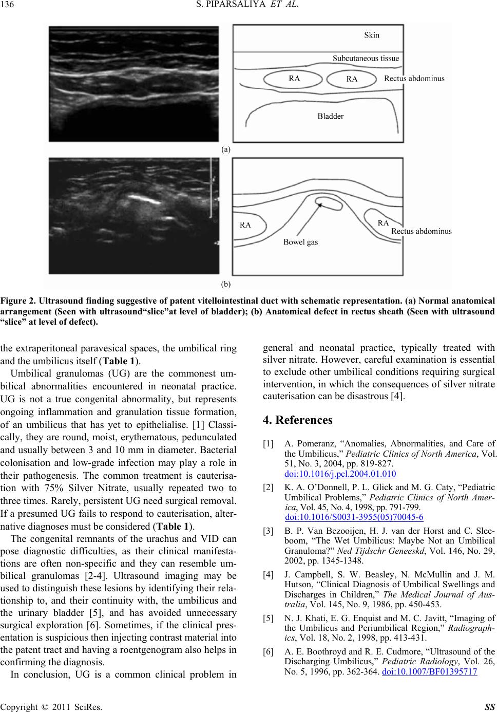
Figure 2. Ultrasound finding suggestive of patent vitellointestinal duct with schematic repr esentation. (a) Normal anatomical
arrangement (Seen with ultrasound“slice”at level of bladder); (b) Anatomical defect in rectus sheath (Seen with ultrasound
“slice” at level of defec t) .
the extraperitoneal paravesical spaces, the umbilical ring
and the umbilicus itself (Table 1).
Umbilical granulomas (UG) are the commonest um-
bilical abnormalities encountered in neonatal practice.
UG is not a true congenital abnormality, but represents
ongoing inflammation and granulation tissue formation,
of an umbilicus that has yet to epithelialise. [1] Classi-
cally, they are round, moist, erythematous, pedunculated
and usually between 3 and 10 mm in diameter. Bacterial
colonisation and low-grade infection may play a role in
their pathogenesis. The common treatment is cauterisa-
tion with 75% Silver Nitrate, usually repeated two to
three times. Rarely, persistent UG need surgical removal.
If a presumed UG fails to respond to cauterisation, alter-
native diagnoses must be considered (Table 1).
The congenital remnants of the urachus and VID can
pose diagnostic difficulties, as their clinical manifesta-
tions are often non-specific and they can resemble um-
bilical granulomas [2-4]. Ultrasound imaging may be
used to distinguish these lesions by identifying their rela-
tionship to, and their continuity with, the umbilicus and
the urinary bladder [5], and has avoided unnecessary
surgical exploration [6]. Sometimes, if the clinical pres-
entation is susp icious then injecting contrast material into
the patent tract and having a roentgenogram also helps in
confirming the diagnosis.
In conclusion, UG is a common clinical problem in
general and neonatal practice, typically treated with
silver nitrate. However, careful examination is essential
to exclude other umbilical conditions requiring surgical
intervention, in wh ich the consequences of silver nitrate
cauterisation ca n be di sast r ous [4] .
4. References
[1] A. Pomeranz, “Anomalies, Abnormalities, and Care of
the Umbilicus,” Pediatric Clinics of North America, Vol.
51, No. 3, 2004, pp. 819-827.
doi:10.1016/j.pcl.2004.01.010
[2] K. A. O’Donnell, P. L. Glick and M. G. Caty, “Pediatric
Umbilical Problems,” Pediatric Clinics of North Amer-
ica, Vol. 4 5, N o. 4, 1 998, p p. 791 -799.
doi:10.1016/S0031-3955(05)70045-6
[3] B. P. Van Bezooijen, H. J. van der Horst and C. Slee-
boom, “The Wet Umbilicus: Maybe Not an Umbilical
Granuloma?” Ned Tijdschr Geneeskd, Vol. 146, No. 29,
2002, pp. 1345-1348.
[4] J. Campbell, S. W. Beasley, N. McMullin and J. M.
Hutson, “Clinical Diagnosis of Umbilical Swellings and
Discharges in Children,” The Medical Journal of Aus-
tralia, Vol. 145, No. 9, 1986, pp. 450-453.
[5] N. J. Khati, E. G. Enquist and M. C. Javitt, “Imaging of
the Umbilicus and Periumbilical Region,” Radiograph-
ics, Vol. 18, No. 2, 1998, pp. 413-431.
[6] A. E. Boothroyd and R. E. Cudmore, “Ultrasound of the
Discharging Umbilicus,” Pediatric Radiology, Vol. 26,
No. 5, 1996, pp. 362-364. doi:10.1007/BF01395717
Copyright © 2011 SciRes. SS