Surgical Science, 2011, 2, 127-129
doi:10.4236/ss.2011.23025 Published Online May 2011 (http://www.scirp.org/journal/ss)
Copyright © 2011 SciRes. SS
127
Osteoblastoma Mimic Failed Back Surgery
Juan F. Blanco1, Susana Gomez-Castro2, Ruth Lopez-Gonzalez2, Juan C. Paniagua3
1Department of Traum a and Orthopedics, Hospital Universitario de Salmanca, Sala manca, Spain
2Department of Rheumatology, Hospital Universitario de Salmanca, Salamanca, Spain
3Department o Ra di ol o gy , Hospital Universitario de Salmanca, Salamanca, Spain
E-mail: juanfblanco@telefonica.net; susigom888@yahoo.es; ruthielg@hotmail.com; ruthielg@hotmail.com
Received October 25, 2010; revised January 10, 2011; accepted April 21, 2011
Abstract
A 44 year-old women with persistent back and irradiate pain after discectomy in L5 - S1 lumbar level pre-
sented a osteoblastoma in L4 - L5 level. The osteoblastoma mimic a failed back surgery. She was success-
fully treated by removing the tumour with instrumented posteroalateral fusion. This case warns people that a
new disease (such as a tumour in this case), irrelevant to the original one, may happen simultaneously even if
the original one has been successfully treated and should be differentiated from the previous disease or sur-
gical failure and complications.
Keywords: Osteoblastoma, Lumber Spine, Surgery, Back Pain, Disc Herniation
1. Introduction
Osteoblastoma of the spine is a rare benign bone tumour
accounting for less than 1% of all primary bone tumours
[1]. Generally, it affects persons 10 to 40 years of age,
with a male to female ratio of approximately 2 to 1 [2,3].
Spinal involvement occurs in 35% to 45% of cases.
Lumbar localization is frequent. The majority arise from
posterior spinal elements: pedicles, lamina and facets
[4,5]. Clinically the patients with osteoblastoma have
pain and stiffness [6,7]. This case is presented because of
is rarity and also it happened after spine surgery; this one
was the reason of the delay in the diagnosis. The symp-
toms were attributed to a syndrome of failed back sur-
gery.
2. Case Report
44-year-old women were admitted in June 2006 with low
back and irradiate pain. The past medical history in-
cluded: apendectomy, histerectomy and depression. In
2004 a lumbar discectomy was performed because of a
slipped disc in L5 - S1 level (Figure 1). After surgery
the patients continued with lumbar and irradiate pain.
Because of the suspicion of periradicular fibrosis, the
patient underwent a Magnetic Resonance Imaging (MRI)
(Figure 2). The disc degeneration in the L4 - L5 and L5 -
S1 levels was considered the aetiology of pain. The
symptoms assumed to a failed back surgery and she was
remitted to the pain un it. In 2006 when she was admitted
to our unit, she has back and irradiate pain. Physical and
neurological examination found no abnormalities. Plain
radiographs showed degenerative disc disease in L4 - L5
and L5 - S1 levels. A MRI was performed and revealed
an inflated focal lesion with a 1.5 cm diameter located in
the right lamina of L4, heterogenic, isointensive relation-
ship to the muscle that through contrast administration
presents a discrete highlight signs of paravertebral mus-
cles oedema, adjacent to the lesion with notable contrast
(Figure 3). Technetium bone scan showing increased
focal uptake in the right side of the L4 - L5 level (Figure
4). Before surgery a computed tomography (CT) was
performed and showed a well limited expansible litic
lesion in the inferior right lamina of L4 vertebra (Figure
5). Routine laboratory examinations were within normal
limits. In a restrospective way, little alterations of the
signal in MRI can be seen previous to the appearance of
the tumour (Figure 2). Posterior approached was per-
formed with complete excision of tumour. Simultane-
ously a posterolateral fusion with pedicular screws was
performed. Pathologic examination of surgical specimen
from patient revealed osteoblastoma (Figure 6). After
the surgery the symptoms were relieved.
3. Discussion
Osteoblastoma is a uncommon neoplasm that accounts
for approximately 3% of benign and 1% of all primary
J. F. BLANCO ET AL.
128
Figure 1. Sagital T2 weighted MRI. Signs of posterocentral
slipped disc protude at the L5 - S1 level in the year 2003.
(a)
(b)
Figure 2. Axial T2 weighted MRI showing, surgical changes
in L5 - S1 level (a) and slight high signal intensity in the
facet and lamine of the L4 vertebra on the right side (b).
Yellow arrow. (w here it wasn’t noticed or found important).
Year 2004.
Figure 3. Axial T2 MRI (year 2006) showing expansive le-
sion and anormal intensity changes in the L4 - L5 right
facet. This lesion is heterogenic, isointensive relationship to
the muscle that through contrast administration presents a
discrete highlight signs of paravertebral muscles oedema,
adjacent to the lesion with notable contrast.
Figure 4. Increase focal uptake in bone scan at the L4 - L5
right level.
Figure 5. CT showing well-defined litic lesion parcial calci-
fied matrix with a 1.5 cm diameter located in the right lam-
ina of L4 and that it is associated with a notable sclerosis
and periostotic enlargement of the adjacent bones struc-
tures.
Copyright © 2011 SciRes. SS
J. F. BLANCO ET AL.
Copyright © 2011 SciRes. SS
129
4. Conclusions
We report a rare case of spinal osteoblastoma after sur-
gery in adjacent vertebral level. The osteoblastoma
mimic a syndrome of failed back surgery which can warn
people that a new disease (such as a tumor in this case),
irrelevant to the original one, may happen simultane-
ously even if the original one has been successfully
treated and should be differentiated from the previous
disease or surgical failure and complications.
5. References
[1] D. R. Lucas, K. K. Unni, R. A. McLeod, M. I. O’Connor
and F. H. Sim, “Osteoblastoma: Clinicopathologic Study
of 306 Cases,” Human Pathology, Vol. 25, No. 2, 1994,
pp. 117-134. doi:10.1016/0046-8177(94)90267-4
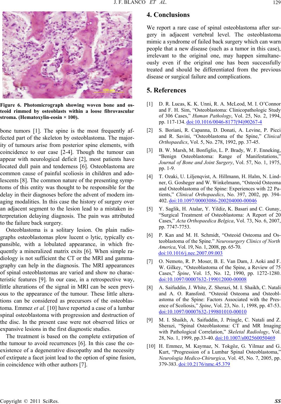
Figure 6. Photomicrograph showing woven bone and os-
teoid rimmed by osteoblasts within a loose fibrovascular
stroma. (Hematoxylin-eosin × 100).
[2] S. Boriani, R. Capanna, D. Donati, A. Levine, P. Picci
and R. Savini, “Osteoblastoma of the Spine,” Clinical
Orthopaedics, Vol. 5, No. 278, 1992, pp. 37-45.
bone tumors [1]. The spine is the most frequently af-
fected part of the skeleton by osteoblastoma. The major-
ity of tumours arise from posterior spine elements, with
coincidence to our case [2-4]. Though the tumour can
appear with neurological deficit [2], most patients have
located dull pain and tenderness [6]. Osteoblastoma are
common cause of painful scoliosis in children and ado-
lescents [8]. The common nature of the presenting symp-
toms of this entity was thought to be responsible for the
delay in their diagnoses before the advent of modern im-
aging modalities. In this case the history of surgery over
an adjacent segment to the lesion lead to a mistaken in-
terpretation delaying diagnosis. The pain was attributed
to the failure back surgery.
[3] B. W. Marsh, M. Bonfiglio, L. P. Brady, W. F. Enneking,
“Benign Osteoblastoma: Range of Manifestations,”
Journal of Bone and Joint Surgery, Vol. 57, No. 1, 1975,
pp. 1-9.
[4] T. Ozaki, U. Liljenqvist, A. Hillmann, H. Halm, N. Lind-
ner, G. Gosheger and W. Winkelmann, “Osteoid Osteoma
and Osteoblastoma of the Spine: Experiences with 22 Pa-
tients,” Clinical Orthopaedics, No. 397, 2002, pp. 394-
402. doi:10.1097/00003086-200204000-00046
[5] Y. Saglik, H. Atalar, Y. Yildiz, K. Basari and C. Gunay,
“Surgical Treatment of Osteoblastoma: A Report of 20
Cases,” Acta Orthopaedica Belgica, Vol. 73, No. 6, 2007,
pp. 7747-7753.
Osteoblastoma is a solitary lesion. On plain radio-
graphs osteoblastomas plow lucent o lytic, typically ex-
pansible, with a lobulated appearance, in which fre-
quently a mineralized matrix exits [6]. When simple ra-
diology is not sufficient the CT or the MRI and gamma-
graphy can help in the diagnosis. The MRI appearances
of spinal osteoblastomas are varied and show no charac-
teristic features [9]. In our case, in a retrospective way,
little alterations of the signal in MRI can be seen previ-
ous to the appearance of the tumour. These little altera-
tions can be considered as precursors of the osteoblas-
toma. Emmez et al. [10] have reported a case of a lumbar
spinal osteoblastoma with progression and destruction of
the disc. In the present case were not observed litics or
expansive lesions in the first diagnostic studies.
[6] P. Kan and M. H. Schmidt, “Osteoid Osteoma and Os-
teoblastoma of the Spine.” Neurosurgery Clinics of North
America, Vol. 19, No. 1, 2008, pp. 65-7 0.
doi:10.1016/j.nec.2007.09.003
[7] O. Nemoto, R. P. Moser, B. E. Van Dam, J. Aoki and F.
W. Gilkey, “Osteoblastoma of the Spine, a Review of 75
Cases,” Spine, Vol. 15, No. 12, 1990, pp. 1272-1280.
doi:10.1097/00007632-199012000-00008
[8] A. Saifuddin, J. White, Z. Sherazi, M. I. Shaikh, C. Natali
and A. O. Ransford. “Osteoid Osteoma and Osteobl-
astoma of the Spine: Factors Associated with the Pres-
ence of Scoliosis,” Spine, Vol. 23, No. 1, 1998, pp. 47-53.
doi:10.1097/00007632-199801010-00010
[9] M. I. Shaikh, A. Saifuddin, J. Pringle, C. Natali and Z.
Sherazi, “Spinal Osteoblastoma: CT and MR Imaging
with Pathological Correlation,” Skeletal Radiology, Vol.
28, No. 1, 1999, pp.33-40. doi:10.1007/s002560050469
The treatment is based on the complete extirpation of
the tumour to avoid recurrences [6]. In this case the co-
existence of a degenerative discopathy and the necessity
of extirpate a facet joint lead to the option of spine fusion,
in coincidence with other authors [7].
[10] H. Emmez, M. Kaymaz, N. Tokgöz, G. Yilmaz and G.
Kurt, “Progression of a Lumbar Spinal Osteoblastoma,”
Neurologia Medico-Chirurgica, Vol. 45, No. 7, 2005, pp.
379-383. doi:10.2176/nmc.45.379