Paper Menu >>
Journal Menu >>
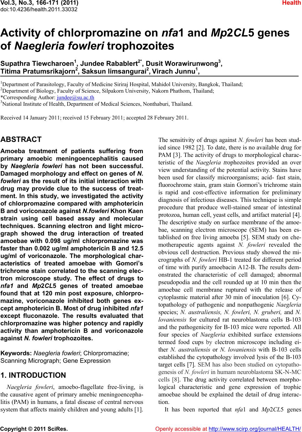 Vol.3, No.3, 166-171 (2011) Health doi:10.4236/health.2011.33032 Copyright © 2011 SciRes. Openly accessible at http://www.scirp.org/journal/HEALTH/ Activity of chlorpromazine on nfa1 and Mp2CL5 genes of Naegleria fowleri trophozoites Supathra Tiewcharoen1, Jundee Rabablert2*, Dusit Worawirunwong3, Titima Pratumsrikajorn2, Saksun limsangurai2, Virach Junnu1, 1Department of Parasitology, Faculty of Medicine Siriraj Hospital, Mahidol University, Bangkok, Thailand; 2Department of Biology, Faculty of Science, Silpakorn University, Nakorn Phathom, Thailand; *Corresponding Author: jundee@su.ac.th 3National Institute of Health, Department of Medical Sciences, Nonthaburi, Thailand. Received 14 January 2011; received 15 February 2011; accepted 28 February 2011. ABSTRACT Amoeba treatment of patients suffering from primary amoebic meningoencephalitis caused by Naegleria fowleri has not been successful. Damaged morphology and effect on genes of N. fowleri as the result of its initial interaction with drug may provide clue to the success of treat- ment. In this study, we investigated the activity of chlorpromazine compared with amphotericin B and voriconazole against N.fowleri Khon Kaen strain using cell based assay and molecular techniques. Scanning electron and light micro- graph showed the drug interaction of treated amoebae with 0.098 ug/ml chlorpromazine was faster than 0.002 ug/ml amphotericin B and 12.5 ug/ml of voriconazole. The morphological char- acteristics of treated amoebae with Gomori’s trichrome stain correlated to the scanning elec- tron microscope study. The effect of drugs to nfa1 and Mp2CL5 genes of treated amoebae found that at 120 min post exposure, chlorpro- mazine, voriconazole inhibited both genes ex- cept amphotericin B. Most of drug inhibited nfa1 except fluconazole. The results evaluated that chlorpromazine was higher potency and rapidly activity than amphotericin B and voriconazole against N. fowleri trophozoites. Keywords: Naegleria fowleri; Chlorpromazine; Scanning Micrograph; Gene Expression 1. INTRODUCTION Naegleria fowleri, amoebo-flagellate free-living, is the causative agent of primary amebic meningoencepha- litis (PAM) in humans, a fatal disease of central nervous system that affects mainly children and young adu lts [1]. The sensitivity of drugs against N. fowleri has been stu d- ied since 1982 [2]. To date, there is no available drug for PAM [3]. The activity o f drugs to morphological charac- teristic of the Naegleria trophozoites provided an over view understanding of the potential activity. Stains have been used for classify microorganisms; acid- fast stain, fluorochrome stain, gram stain Gormori’s trichrome stain is rapid and cost-effective information for preliminary diagnosis of infectious diseases. This technique is simple procedure that produce well-stained smear of intestinal protozoa, human cell, yeast cells, and artifact material [4]. The descriptive study on surface membrane of the amoe- bae, scanning electron microscope (SEM) has been es- tablished on free living amoeba [5]. SEM study on che- motherapeutic agents against N. fowleri revealed the obvious cell destruction. Previous study showed the mi- crographs of N. fowleri HB-1 treated for different period of time with purify amoebacin A12-B. The results dem- onstrated the characteristic of cell damaged; abnormal pseudopodia and the cell rounded up at 10 min then the amoebae cell membrane ruptured with the release of cytoplasmic material after 30 min of inoculation [6]. Cy- topathology of pathogenic and nonpathogenic Naegleria species; N. australiensis, N. fowleri, N. gruberi, and N. lovaniensis for cultured rat neuroblastoma cells B-103 and the pathogenicity for B-103 mice were reported. All four species of Naegleria exhibited surface extensions termed food cups by electron microscope including ei- ther N. australiensis or N. lovaniensis with B-103 cells established the cytopathology involved lysis of the B-103 target cells [7]. SEM has also been studied on cytopatho- genesis of N. fowleri in humam neuroblastoma SK-N - MC cells [8]. The drug activity correlated between morpho- logical characteristic and gene expression of trophic amoebae should be explained the detail of drug interac- tion. It has been reported that nfa1 and Mp2CL5 genes 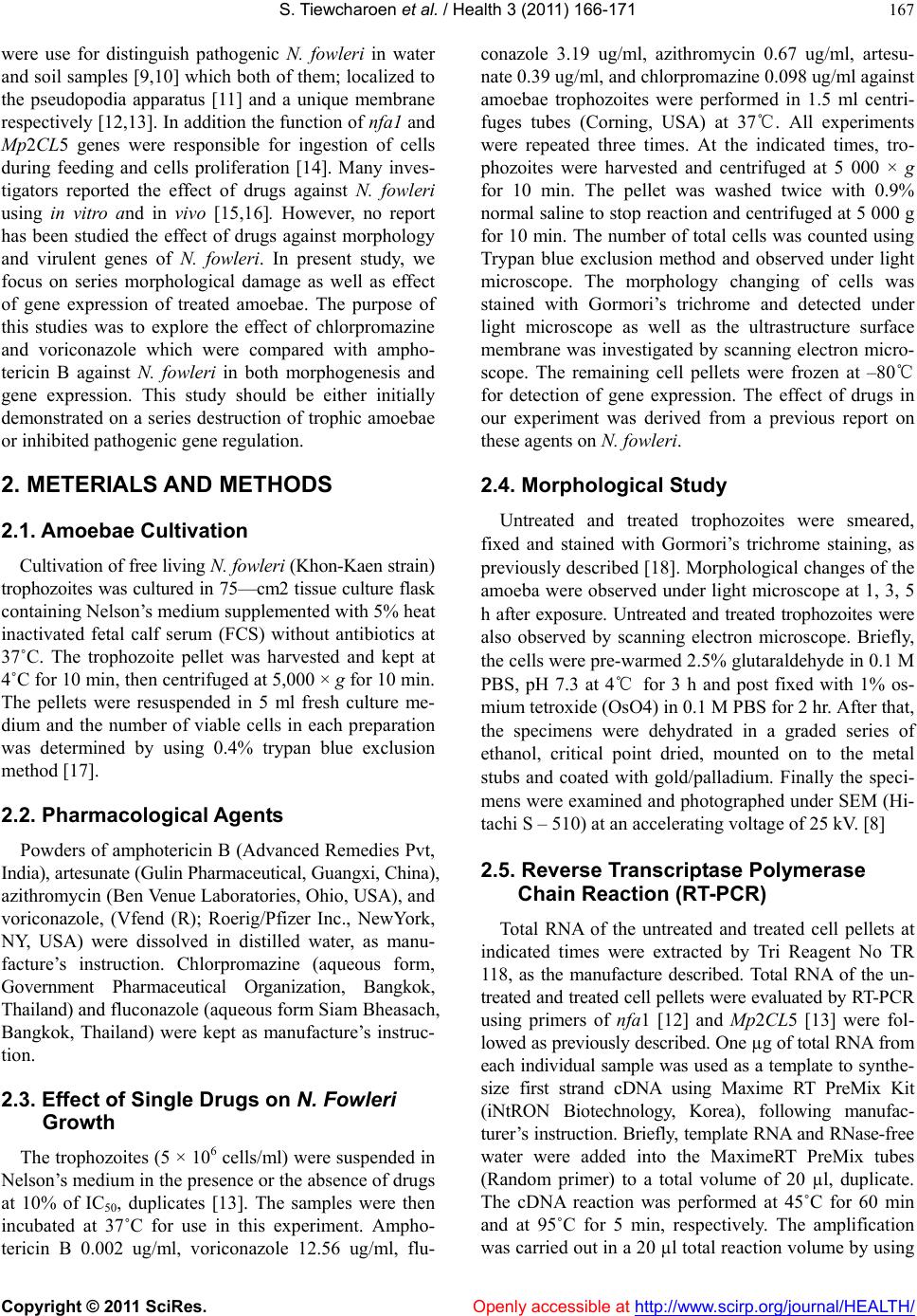 S. T iewcharoen et al. / Health 3 (2011) 166-171 Copyright © 2011 SciRes. Openly accessible at http://www.scirp.org/journal/HEALTH/ 167 were use for distinguish pathogenic N. fowleri in water and soil samples [9,10] which both of them; lo calized to the pseudopodia apparatus [11] and a unique membrane respectively [12,13]. In additio n the function of nfa1 and Mp2CL5 genes were responsible for ingestion of cells during feeding and cells proliferation [14]. Many inves- tigators reported the effect of drugs against N. fowleri using in vitro and in vivo [15,16]. However, no report has been studied the effect of drugs against morphology and virulent genes of N. fowleri. In present study, we focus on series morphological damage as well as effect of gene expression of treated amoebae. The purpose of this studies was to explore the effect of chlorpromazine and voriconazole which were compared with ampho- tericin B against N. fowleri in both morphogenesis and gene expression. This study should be either initially demonstrated on a series destruction of trophic amoebae or inhibited pathogenic gene regulation. 2. METERIALS AND METHODS 2.1. Amoebae Cultivation Cultivatio n of free liv ing N. fowleri (Khon-Kaen strain) trophozoites was cultured in 75—cm2 tissue culture flask containing Nelson’s medium supplemented with 5% heat inactivated fetal calf serum (FCS) without antibiotics at 37˚C. The trophozoite pellet was harvested and kept at 4˚C for 10 min, then centr ifuged at 5,00 0 × g fo r 10 min. The pellets were resuspended in 5 ml fresh culture me- dium and the number of viable cells in each preparation was determined by using 0.4% trypan blue exclusion method [17]. 2.2. Pharmacological Agents Powders of amphotericin B (Advanced Remedies Pvt, India), artesunate (Gulin Pharmaceutical, Guangxi, China), azithromycin (Ben Venue Laboratories, Ohio, USA) , and voriconazole, (Vfend (R); Roerig/Pfizer Inc., NewYork, NY, USA) were dissolved in distilled water, as manu- facture’s instruction. Chlorpromazine (aqueous form, Government Pharmaceutical Organization, Bangkok, Thailand) and fluconazole (aqueous form Siam Bheasach, Bangkok, Thailand) were kept as manufacture’s instruc- tion. 2.3. Effect of Single Drugs on N. Fowleri Growth The trophozoites (5 × 10⁶ cells/ml) were suspended in Nelson’s mediu m in the presence or the absence of drugs at 10% of IC50, duplicates [13]. The samples were then incubated at 37˚C for use in this experiment. Ampho- tericin B 0.002 ug/ml, voriconazole 12.56 ug/ml, flu- conazole 3.19 ug/ml, azithromycin 0.67 ug/ml, artesu- nate 0.39 ug/ml, and chlorpromazine 0.098 ug/ml against amoebae trophozoites were performed in 1.5 ml centri- fuges tubes (Corning, USA) at 37℃. All experiments were repeated three times. At the indicated times, tro- phozoites were harvested and centrifuged at 5 000 × g for 10 min. The pellet was washed twice with 0.9% normal saline to stop reaction and centrifuged at 5 000 g for 10 min. The number of total cells was counted using Trypan blue exclusion method and observed under light microscope. The morphology changing of cells was stained with Gormori’s trichrome and detected under light microscope as well as the ultrastructure surface membrane was investigated by scanning electron micro- scope. The remaining cell pellets were frozen at –80℃ for detection of gene expression. The effect of drugs in our experiment was derived from a previous report on these agents on N. fowleri. 2.4. Morphological Study Untreated and treated trophozoites were smeared, fixed and stained with Gormori’s trichrome staining, as previously described [18]. Morphological changes of the amoeba were observed under light microscope at 1, 3, 5 h after exposure. Untreated and treated trophozoites were also observed by scanning electron microscope. Briefly, the cells were pre-warmed 2.5% glutaraldehyde in 0.1 M PBS, pH 7.3 at 4℃ for 3 h and post fixed with 1% os- mium tetroxide (OsO4) in 0.1 M PBS for 2 hr. After that, the specimens were dehydrated in a graded series of ethanol, critical point dried, mounted on to the metal stubs and coated with gold/palladium. Finally the speci- mens were ex amined and photograph ed under SEM (Hi- tachi S – 510) at an accelerating voltage of 25 kV. [8] 2.5. Reverse Transcriptase Polymerase Chain Reaction (RT-PCR) Total RNA of the untreated and treated cell pellets at indicated times were extracted by Tri Reagent No TR 118, as the manufacture described. Total RNA of the un- treated and treated cell pellets were evaluated by RT-PCR using primers of nfa1 [12] and Mp2CL5 [13] were fol- lowed as previously described. One µg of total RNA fro m each individual sample was used as a template to synthe- size first strand cDNA using Maxime RT PreMix Kit (iNtRON Biotechnology, Korea), following manufac- turer ’s instruction. Briefly, template RNA and RNase-fre e water were added into the MaximeRT PreMix tubes (Random primer) to a total volume of 20 µl, duplicate. The cDNA reaction was performed at 45˚C for 60 min and at 95˚C for 5 min, respectively. The amplification was carried out in a 20 µl total reaction volume by using 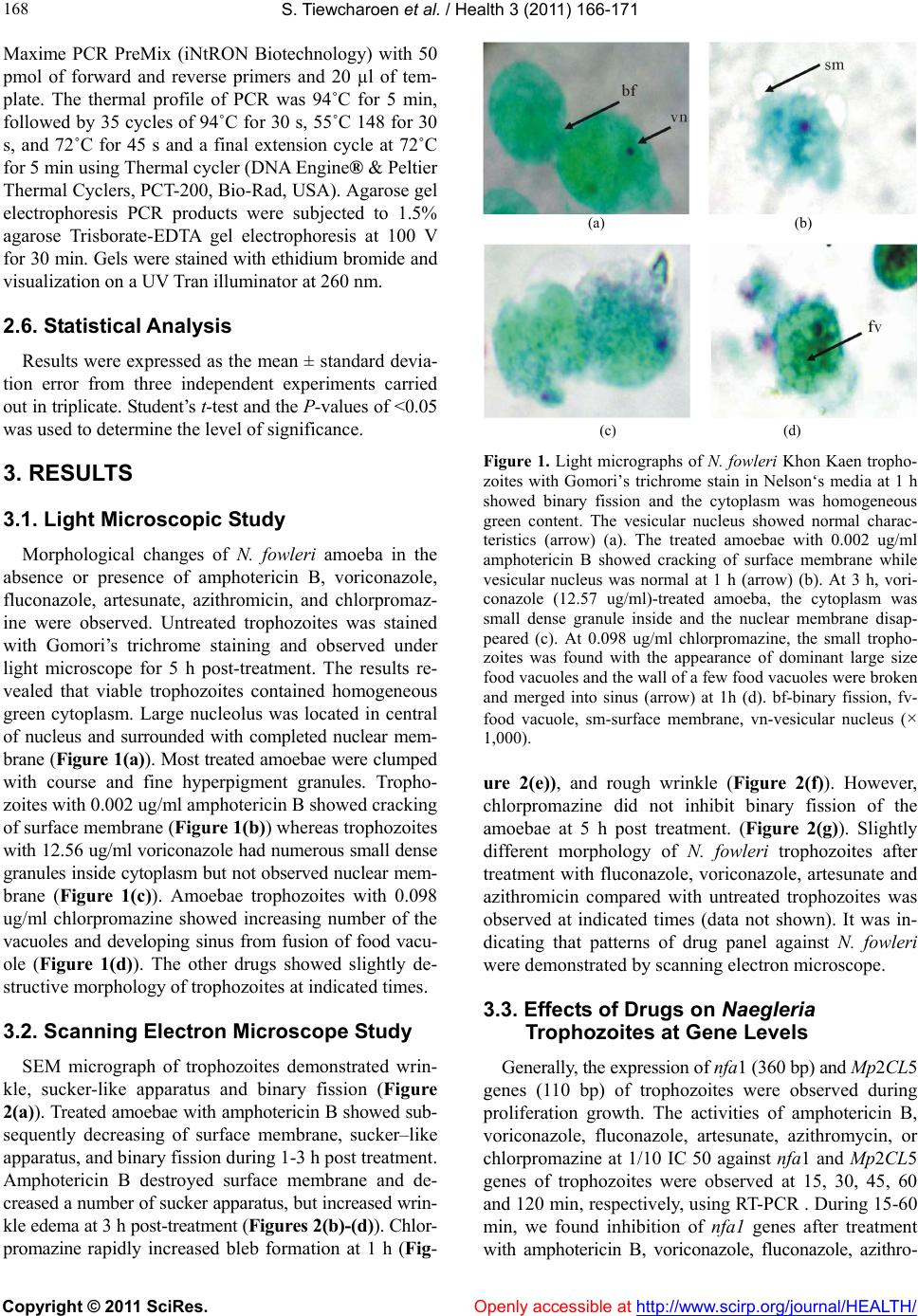 S. T iewcharoen et al. / Health 3 (2011) 166-171 Copyright © 2011 SciRes. Openly accessible at http://www.scirp.org/journal/HEALTH/ 168 Maxime PCR PreMix (iNtRON Biotechnology) with 50 pmol of forward and reverse primers and 20 µl of tem- plate. The thermal profile of PCR was 94˚C for 5 min, followed by 35 cycles of 94˚C for 30 s, 55˚C 148 for 30 s, and 72˚C for 45 s and a final extension cycle at 72˚C for 5 min using Thermal cycler (DNA Engine® & Peltier Thermal Cyclers, PCT-200, Bio-Rad, USA). Agarose gel electrophoresis PCR products were subjected to 1.5% agarose Trisborate-EDTA gel electrophoresis at 100 V for 30 min. Gels were stained with ethidium bromide and visualization on a UV Tran illuminator at 260 nm. 2.6. Statistical Analysis Results were expressed as the mean ± standard devia- tion error from three independent experiments carried out in tripli cate. Student’s t-test and the P-values of <0.05 was used to determine the level of significance. 3. RESULTS 3.1. Light Microscopic Study Morphological changes of N. fowleri amoeba in the absence or presence of amphotericin B, voriconazole, fluconazole, artesunate, azithromicin, and chlorpromaz- ine were observed. Untreated trophozoites was stained with Gomori’s trichrome staining and observed under light microscope for 5 h post-treatment. The results re- vealed that viable trophozoites contained homogeneous green cytoplasm. Large nucleolus was located in central of nucleus and surrounded with completed nuclear mem- brane (Figure 1(a)). Most treated amoebae were clumped with course and fine hyperpigment granules. Tropho- zoites with 0.002 ug/ml amphotericin B showed cracking of surface membrane (Figure 1(b)) whereas trophozoites with 12.56 ug/ml voriconazole had numerous small dense granules inside cytoplasm but not observed nuclear mem- brane (Figure 1(c)). Amoebae trophozoites with 0.098 ug/ml chlorpromazine showed increasing number of the vacuoles and developing sinus from fusion of food vacu- ole (Figure 1(d)). The other drugs showed slightly de- structive morphology of trophozoites at indicated times. 3.2. Scanning Electron Microscope Study SEM micrograph of trophozoites demonstrated wrin- kle, sucker-like apparatus and binary fission (Figure 2(a)). Treated amoebae with amphotericin B showed sub- sequently decreasing of surface membrane, sucker–like apparatus, and bi nary fission duri n g 1- 3 h p ost treatment. Amphotericin B destroyed surface membrane and de- creased a number of sucker apparatus, but increased wrin- kle edema at 3 h post-treatment (Figures 2(b)-(d)). Chlor- promazine rapidly increased bleb formation at 1 h (Fig- (a) (b) (c) (d) Figure 1. Light micrographs of N. fowleri Khon Kaen tropho- zoites with Gomori’s trichrome stain in Nelson‘s media at 1 h showed binary fission and the cytoplasm was homogeneous green content. The vesicular nucleus showed normal charac- teristics (arrow) (a). The treated amoebae with 0.002 ug/ml amphotericin B showed cracking of surface membrane while vesicular nucleus was normal at 1 h (arrow) (b). At 3 h, vori- conazole (12.57 ug/ml)-treated amoeba, the cytoplasm was small dense granule inside and the nuclear membrane disap- peared (c). At 0.098 ug/ml chlorpromazine, the small tropho- zoites was found with the appearance of dominant large size food vacuoles and the wall of a few food vacuoles were broken and merged into sinus (arrow) at 1h (d). bf-binary fission, fv- food vacuole, sm-surface membrane, vn-vesicular nucleus (× 1,000). ure 2(e)), and rough wrinkle (Figure 2(f)). However, chlorpromazine did not inhibit binary fission of the amoebae at 5 h post treatment. (Figure 2(g)). Slightly different morphology of N. fowleri trophozoites after treatment with fluconazole, voriconazole, artesunate and azithromicin compared with untreated trophozoites was observed at indicated times (data not shown). It was in- dicating that patterns of drug panel against N. fowleri were demonstrated by scanning electron microscope. 3.3. Effects of Drugs on Naegleria Trophozoites at Gene Levels Generally, the expression of nfa1 (360 bp) and Mp2CL5 genes (110 bp) of trophozoites were observed during proliferation growth. The activities of amphotericin B, voriconazole, fluconazole, artesunate, azithromycin, or chlorpromazine at 1/10 IC 50 against nfa1 and Mp2CL5 genes of trophozoites were observed at 15, 30, 45, 60 and 120 min, respectively, using RT-PCR . During 15-60 min, we found inhibition of nfa1 genes after treatment with amphotericin B, voriconazole, fluconazole, azithro-  S. T iewcharoen et al. / Health 3 (2011) 166-171 Copyright © 2011 SciRes. Openly accessible at http://www.scirp.org/journal/HEALTH/ 169 (a) (b) (c) (d) (e) (f) (g) Figure 2. Scanning electron micrograph of N. fowleri trophozoite in the absence (a) or presence of amphotericin B (0.002 ug/ml) (b)-(d) or chlorpromazine at 0.098 ug/ml (e-g). Untreated trophozoites showed normal characteristics; equal binary fission and sucker apparatus (arrow) (a). Amphotericin B-treated amoebae showed sucker apparatus at 1 h (b). Treated amoebae showed the destructive surface membrane started at 3 h; disappearance of sucker apparatus and edematous wrinkles surface (c). The series damage continued until wrinkles surface was not found (d). Chlorpromazine -treated amoebae showed the surface membrane was rapidly damage and the bleb formations were found (arrow) at 1 h (e). The destruction of amoebae cell was continued; sucker and pseudopodia formation were disappeared including the surface membrane showed edematous change at 3h (arrow) (f). The unequal binary fission of the amoeba was seen at 5 h (arrow) (g). Bars represent 10 μm: b-bleb formation, bf-binary fission, sa-sucker apparatus, sm-surface mem- brane, vn-vesicular nucleus. mycin, artesunate, and chlorpromazine (data not shown). Furthermore, all drugs, except fluconazole inhibited nfa1 genes at 120 min (Figure 3(a)). During 15-30 min, all drugs inhibited mp2c15 gene expression of trophozoites (data not shown). From 30 min to 120 min, voriconazole, fluconazole, and chlorpromazine also inhibited Mp2CL5 gene expression (Figure 3(b)), there by amphotericin B, azithromycin, and artesunate did not inhibit Mp2CL5 gene. It was indicating that the activities of drugs against N. fowleri trophozoites were time-dependent manner. 4. DISCUSSIONS PAM is a severe infection of human that typically leads to death during 1 to 2 weeks from the onset of symptoms [19]. Amphotericin B is only drug of choice for PAM treatment [1]. However, it was a very toxic antibiotic and it caused renal toxicity, electrolyte distur- bances, hematopoietic effects, and other organ damage, as well as chills, fever, nausea, vomiting, and headache [20]. In our study, we investigated the patterns of drugs against the trophozoites using light microscope and scanning electron microscope. Furthermore, we studied the activities of drugs against viru lent nfa1 and Mp 2CL5 gene expression. This is the first report which demon- strated the characterization activity of drugs against amoeba trophozoites in both cellular and molecular lev- els. In the present study, light micrographs of N. fowleri Khon Kaen trophozoites showed binary fission and showed normal characteristics of the vesicular nucleus. SEM micrograph of trophozoites demonstrated wrinkle, sucker-like apparatus and binary fission. Morphological characteristic of N. fowleri in this study was consistent with previous reports [8,21]. Morphology changing of trophozoites after drug treatment was revealed. Among these drugs, we found that the activity of chlorpromazine was higher potency than amphotericin B and voricona- zole due to increasing number of the vacuoles, bleb for- mation and rough wrinkle at 1 h. As previously de- scribed, our study correlated to the mechanism of chlor- promazine that acted on lipophilic action which effected on the plasma membrane and reflected sensitivity of amoeba calcium regulatory protein. Many researches revealed that nfa1 and Mp2CL5 genes were found only pathogenic N. fowleri [10,11]. Nfa1 protein expressed from nfa1 11 gene was located in pseudopodia and around food vacuole [22]. Nfa1 protein was specifically localized food cups which are involved in phagocytic activity [11]. In this study, we found that most of drugs inhibited nfa1 gene which represented that the amoebae should be loss of phagocytic activity Reve iller et al., de- monstrated that the expression of native Mp2CL5 protein in N. flowleri appeared to be growth phase regul at ed [12]. The increased expression of the protein in the stationary phase of growth when the cells are experiencing nutrient  S. T iewcharoen et al. / Health 3 (2011) 166-171 Copyright © 2011 SciRes. Openly accessible at http://www.scirp.org/journal/HEALTH/ 170 1 2 3 4 5 6 7 (a) 1 2 3 4 5 6 7 (b) Figure 3. The nfa1 (a) and Mp2CL5 (b) genes of N. fowleri trophozoites expressed in the absence or presence of drugs at 120 min post-treatment. Untreated N. fowleri (lane 1). N. fowl- eri treated with amphotericin B (lane 2), voriconazole (lane 3), fluconazole (lane 4), azithromycin (lane 5), artesunate (lane 6), and chlorpromazine (lane 7), respectively. deprivation is consistent with our hypothesis that the protein may be involved in nutrient or environment sensing. In our study, we found that chlorpromazine and voriconazole inhibited Mp2CL5 gene, suggesting that activity of both drugs invo lved losing of cell recogn ition , sensing of environment and growth of amoeba. However, previously our report showed that at MIC100 ampho- tericin B and chlorpromazine inhibited trophic amoeba in 3 days but voriconazole did not [15]. Therefore, chlorpromazine should be proposed for treatment p ri mary amoebic meningoencephalitis. 5. CONCLUSIONS Our studies presented the correlation activity of drugs at ultrastructure and gene expressions of N. fowleri tro- phozoites. Chlorpromazine was rapidly damage surface membrane due to bleb formation occurred at 1 hour whereas the activity of amphotericin B was slow re- sponse at the same time in contrast it showed completely destruction during 5 hours. However both drugs could inhibit nfa1 and Mp2CL5 gene of N. fowleri 6. ACKNOWLEDGMENTS We are thank Associate Professor Dr. Darawan Wana- chiwanawin, head of Department of Parasitology, Fac- ulty of Medicine, Siriraj hospital, Mah idol University for providing the research facility. We also thank to Dr. Pathom awanpanyalert Director, National Institute of Health, Department of Medical Sciences. This work was supported by with Grant Number 017(II)50 from Faculty of Medicine, Siriraj Hospital, Mahidol University Foun- dation in 2007-2009 and partial fund from Grant Num- ber RGP 2551/01 from Department of Biology, Faculty of Science, Silpakorn University at Sanamchan Palace, Nakhon Pathom, Thailand. REFERENCES [1] Schuster, F.L. and Visvesvara, G.S. (2004) Free-living amoebae as opportunistic and nonopportunistic patho- gens of humans and animals. International Journal for Parasitology, 34, 1001-1027. doi:10.1016/j.ijpara.2004.06.004 [2] Ferrante, A. (1981) Comparative sensitivity of Naegleria fowleri to amphotericin B and amphotericin B methyl ester. Transactions of the Royal Society of Tropical Medicine and Hygiene, 76, 476-478. doi:10.1016/0035-9203(82)90141-9 [3] Soltow, S.M. and Brenner, G.M. (2007) Synergistic ac- tivities of azithromycin and amphotericin B against Nae- gleria fowleri in vitro and in a mouse model of primary amebic meningoencephalitis. Antimicrobial Agents and Chemotherapy, 51, 23-27. doi:10.1016/0035-9203(82)90141-9 [4] Madison, B.M. (2001) Application of stains in clinical microbiology. Biotechnic & Histochemistry, 76, 119-125. doi:10.1016/j.exppara.2009.03.016 [5] González-Robles, A., Cristóbal-Ramos, A.R, González- Lázaro, M., Omaña-Molina, M. and Martínez-Palomo, A. (2009) Naegleria fowleri: Light and electron microscopy study. Experimental Parasitology, 122, 212-217. [6] Gálvez, A., Valdivia, E., González-Segura, A., Lebbadi, M., Martínez-Bueno, M. and Maqueda, M. (1993) Puri- fication, characterization, and lytic activity against Nae- gleria fowleri of two amoebicins produced by Bacillus licheniformis A12. Applied and Environmental Microbi- ology, 59, 1480-1486. [7] Marciano-Cabral, F. and John, D. T. (1983) Cytopatho- genicity of Naegleria fowleri for rat neuroblastoma cell cultures: scanning electron microscopy study. Infect Im- munity, 40, 1214-1217. [8] Tiewcharoen, S., Rabablert, J., Chetanachan, P., Junnu, V., Worawirounwong, D. and Malainual, N. (2008) Scanning electron microscope of Naegleria fowleri Thai strain during cytopathogenesis in human neuroblastoma cells.  S. T iewcharoen et al. / Health 3 (2011) 166-171 Copyright © 2011 SciRes. Openly accessible at http://www.scirp.org/journal/HEALTH/ 171 Parasitology Research, 103, 1119-1123. doi:10.1007/s00436-008-1103-4 [9] Maclean, R.C., Richardson, D.J., LePardo, R. and Marciano-Cabral, F. (2004) The identification of Naegle- ria fowleri from water and soil samples by nested PCR. Parasitology Research, 93, 211-217. doi:10.1007/s00436-004-1104-x [10] Reveiller, F.L., Cabanes, P.A. and Marciano-Cabral, F. (2002) Development of a nested PCR assay to detect the pathogenic free-living amoeba Naegleria fowleri. Para- sitology Research, 88, 443-450. doi:10.1007/s00436-002-0591-x [11] Kang, S.Y., Song, K.J., Jeong, S.R., Kim, J.H., Park, S., Kim, K., Kwon, M.H. and Shin, H.J. (2005) Role of the Nfa1 protein in pathogenic Naegleria fowleri cocultured with CHO target cells. Clinical and Diagnostic Labora- tory, 12, 873- 876. [12] Reveiller, F. L., Suh, S., Sullivan, K., Cabanes, P.A and Marciano-Cabral, F. (2001) Isolation of a unique mem- brane protein from Naegleria fowleri. Journal of Eu- karyotic Microbiology, 48, 676-682. doi:10.1111/j.1550-7408.2001.tb00208.x [13] Madarova, L., Trnkova, K., Feiková, S., Klement, C. and Obernauerová, M. (2010) A real-time PCR diagnostic method for detection of Naegleria fowleri. Experimental Parasitology, 126, 37-41. doi:10.1016/j.exppara.2009.11.001 [14] Song, K.J., Jeong, S.R., Park, S., Kim, K., Kwon, M.H., Im, K., Pak, J. H. and Shin, H.J. (2006) Naegleria fowleri: Functional expression of the Nfa1 protein in transfected Naegleria gruberi by promoter modification. Experi- mental Parasitology, 112, 115-20. doi:10.1016/j.exppara.2005.10.004 [15] Tiewcharoen, S., Rabablert, J. and Junnu, V. (2009) In vitro susceptibility of Naegleria fowleri trophozoites to amphotericin B-combined chlorpromazine. Research Journal of Microbiology, 4, 320-333. doi:10.3923/jm.2009.320.333 [16] Kim, J.H., Jung, S.Y., Lee,Y.J., Song, K.J., Kwon, D., Park, S., Im, K.I. and Shin, H.J. (2008) Effect of thera- peutic chemical agents in vitro and on experimental meningo-encephalitis due to Naegleria fowleri. Antim- icrobial Agents Chemother, 52, 4010-4016. doi:10.1128/AAC.00197-08 [17] Tiewchreon, S., Malainual, N., Junnu, V., Chetanachan, P. and Rabablert, J. (2008) Cytopathogenesis of Naegleria fowleri Thai strains for cultured human neuroblastoma cells. Parasitology Research, 102, 997-1000. doi:10.1007/s00436-007-0866-3 [18] Gormori, G. (1950) A rapid one-step trichrome stain American Journal of Clinical Pathology, 20, 661-664. [19] Shin, H. J. and Im, K. I. (2004) Pathogenic free-living amoebae in Korea. Korean Journal of Parasitology, 42, 93-119. doi:10.3347/kjp.2004.42.3.93 [20] Mandell, G. L. and Petri W. A. Jr (1996) Antimicrobial Agents. In: Hardman, J.G. and Limbird, L.E. eds., Good- man and Gilman's—The Pharmacological Basis of Therapeutics. McGraw-Hill, New York, US, 1165-1168. [21] Antonios, S. N. (2010) Scanning electron microscopic study of trophozoite and cyst stages of Naegleria fowleri. Journal of the Egyptian Society of Parasitology, 40, 271-276. [22] Shin, H.J., Cho, M.S., Jung, S.Y., Kim, H.I, Park, S. and Im, K.I. (2001) Molecular cloning and characterization of a gene encoding a 13·1 kDa antigenic protein of Nae- gleria fowleri. Journal of Eukaryotic Microbiology, 48, 713-717. doi:10.1111/j.1550-7408.2001.tb00211.x |