K. A. KHALIL ET AL.
OPEN ACCESS MSCE
nanofibers solutions. M. A. Kanjwal et al. [14] investi-
gated the influence of the silver content and the mor-
phology of nanofibers on the photocatalytic activity of
silver-grafted titanium dioxide. Titanium dioxide con-
taining different weight percentages of silver was pre-
pared in nanofibrous and nanoparticulate forms. Sil-
ver-grafted titanium dioxide nanofibers were synthesized
by the electro spinning process. The prepared nanostruc-
tures were utilized as a photocatalyst to degrade two dyes.
The obtained results endorse the use of this composite in
a nanofibrous form. Y. Li et al. reported a Ag-TiO2 na no-
particles were prepared by a miniemulsion method using
Ti (O B un) 4 and Ag(NO3) as starting materials. The results
show that Ag doping showed a controlling effect on the
transformation of titania from anatase to rutile. The spe-
cific surface area increased with the Ag-doped amount to
reach a maximum (86.3 m2·g−1) at Ag/Ti molar ratio of
0.8% and then decreased with further increase of the
Ag-doped amount. The applications of nanoparticulate in
waste water treatment, for example; Ag (I) and silver
compounds have been used as antimicrobial compounds
for coliform found in waste water. Nanoscale silver par-
ticles are typically 1 - 40 nanometers (nm) with an aver-
age particle size of 2 - 10 micron range with a specific
surface area of approximately 1 m2·g−1. Applications for
silver nanocrystals include as an anti-microbial, anti-
biotic and anti-fungal agent when incorporated in coat-
ings, nanofibers, first aid bandages, plastics, soap and
textiles, in treatment of certain viruses, in self cleaning
fabrics, as conductive filler and in nanowire and certain
catalyst applications. It has been reported that Ag
nanoparticles were active biocides against Gram-positive
Gram-negative bacteria [15]. H. Bai et al. [16] reported a
novel kind of multifunctional membrane was fabricated
via integrating the advantages of conventional polymer
membrane as supporting layer like hierarchically struc-
tured TiO2/ZnO nanomaterial as functional layer. This
novel membrane possesses the common advantages of
polymer membrane and multifunctional properties of the
hierarchical TiO2 nanofibers/ZnO nanorod materials,
which demonstrated to be able to produce clean water at
a constant high flux with no membrane fouling problem
and energy saving manner.
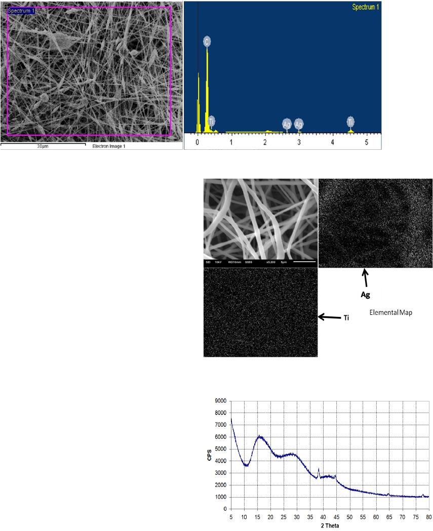
In this paper, polyacrylonitrile solution containing
Ag/TiO2 was directly electrospun to obtain nanofibers
films containing Ag Ag/TiO2, and the Ag/TiO2 of rsuling
composite nanofibers were reduced to Ag/TiO2 nnoparti-
cles. Then, we treated PAN/Ag/TiO2 composite nanofi-
bers at different temperatures. The PAN/ Ag/TiO2 nano-
composite film was characterized by scanning eletron
microscopy (SEM), X-ray diffraction (XRD) paterns and
surface-enhanced Raman scattering (SERS) spectros-
copy.
2. Experimental Work
PANNF film was prepared by electrospinning. PAN (7
wt.%) was dissolved in DMF, and stirred until homoge-
nous at room temperature. 0.03 gm AgNO3 was dis-
solved in 70 ml DMF with stirring at 30 min (UV/vis
spectrometer used to obtain the optimum weight per-
centage and the time of reduction for AgNO3), then 0.01
gm of polyethylene glycol was added as stabilizer and
reduction agent. This solution was stirred for 30 min be-
fore analyze using UV spectra. 0.5 ml of acetic acid and
2 ml of titanium is opropoxide were added into 20 ml
DMF. The solution was stirred for 15 min, then 5 wt%
PAN was added to the solution. The two solutions were
mixed together by adding the first (TIP/PAN) to the sec-
ond one (Ag/PAN) gradually with continuous stirring
until homogenous. The solution containing silver and
titanium isopropoxide salt with PAN were stirred for 24
h at room temperature. After that, the solution obtained
was added into a plastic syringe, the internal diameter of
plastic was 20 cm, the pinhead was connected to a
(20-kV) high-voltage, and aluminum foil served as
counter electrode. TCD was (21 cm), the feed rate of the
solution was adjusted to (0.1 ml/h) through a syringe
pump. The electrospinning was performed at room tem-
perature. The nanofibers were stabilized in an air atmos-
phere at 270˚C for 2 h (at a heating rate of 2˚C/min) and
followed by carbonization at 1000˚C for 1 h (at a heating
rate of 4.5˚C/min) under an inert nitrogen atmosphere to
yield carbonized. The resulting carbon nanofibres were
cooled down to room temperature in an inert gas atmos-
phere before they were taken out of the furnace. Full-
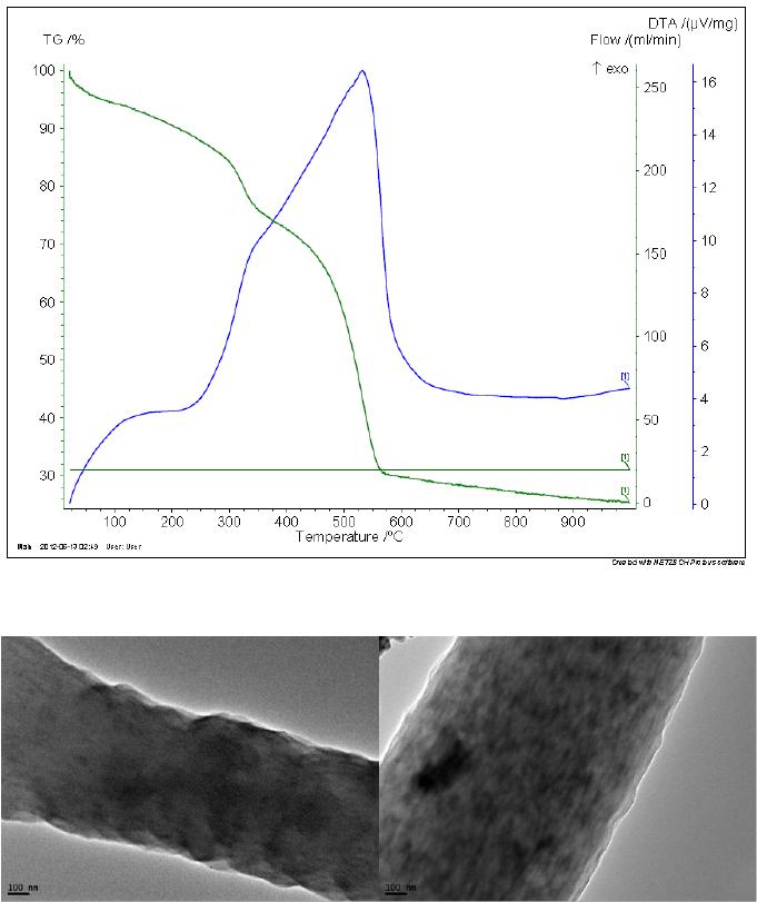
stained ultra-thin sections were examined using the field
e mission transmission electron microscope (JEOL-JEM-
2100F, Japan). Thermal properties of electrospinning
nanofibers were examined using thermogravimetric anal-
ysis (TGA) carried on TA-Q500 System of TA; samples
of 5 - 10 mg were heated in the temperature range 30˚C -
800˚C at a scanning rate of 10˚C·mi n−1 under nitrogen
atmosphere, and by using TG-DTA: NETZSCH Ger-
many(Model: STA 449 F3). The bonding configurations
of the samples of carbon nanofibers were recorded by
Four ie r -transformer infrared (FT-IR) Spectra using
TENSOR 27. Tube furnace (Model: T2F-16/610, carbo-
lite-England) was used in treatment nanofibers to convert
them into carbon nanofibers. Thermoanalytical technique
in which the difference in the amount of heat required to
increase the temperature of sample and reference is
measured as a function of temperature. (DSC Q 20-TA
National scientific company USA). Counter ions are
stored in the electrical double layers which form at the
solution interface inside the porous electrodes, with the
ions of cations stored in the negatively charged electrode,
and anions stored in the positively charged electrode
(anode). Plimmer test unit.